. 2016 Jul 22:6:30122.
doi: 10.1038/srep30122.

miR-877-3p targets Smad7 and is associated with myofibroblast differentiation and bleomycin-induced lung fibrosis

Affiliations

miR-877-3p targets Smad7 and is associated with myofibroblast differentiation and bleomycin-induced lung fibrosis

Cong Wang et al. Sci Rep. .

Abstract

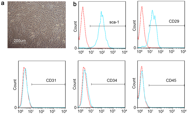
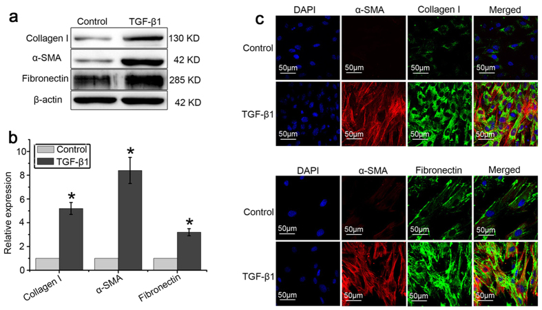
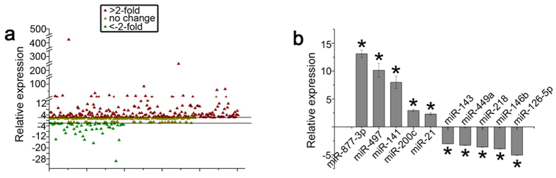
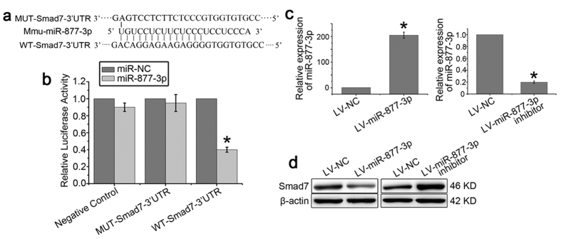
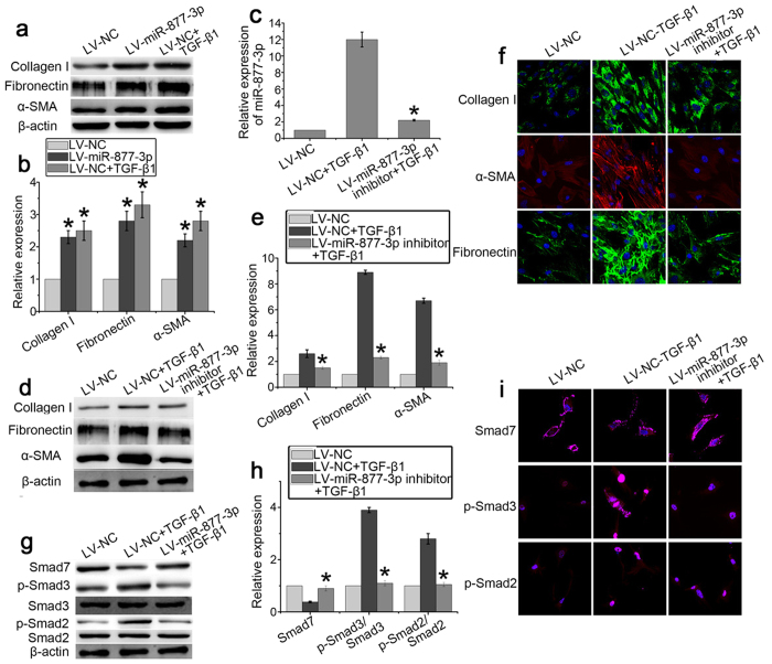
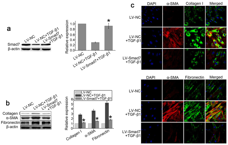
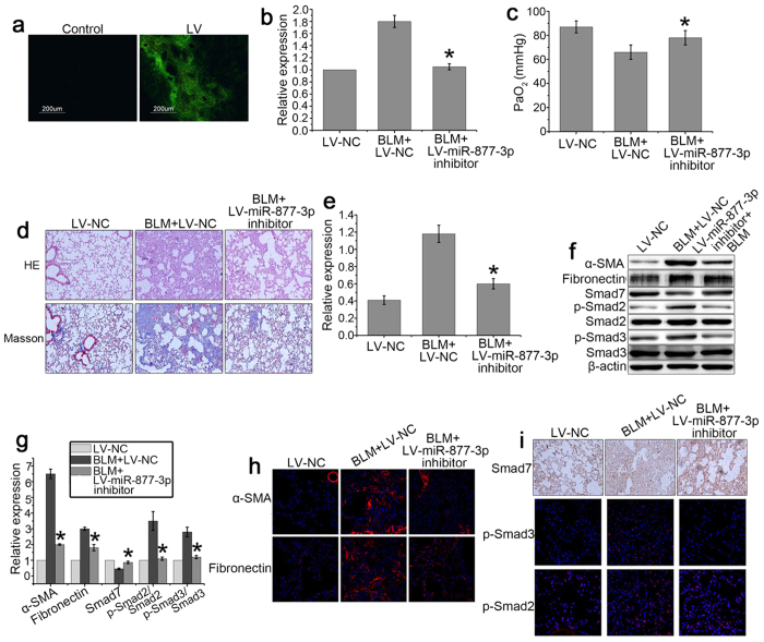
Myofibroblast differentiation of lung resident mesenchymal stem cells (LR-MSC) plays an important role in idiopathic pulmonary fibrosis. By comparing the expression profiles of miRNAs before and after myofibroblast differentiation of LR-MSC, we identified miR-877-3p as a fibrosis-related miRNA. We found that miR-877-3p sequestration inhibited the myofibroblast differentiation of LR-MSC and attenuates bleomycin-induced lung fibrosis by targeting Smad7. Smad7, as an inhibitory smad in the TGF-β1 signaling pathway, was decreased in the myofibroblast differentiation of LR-MSC and up-regulation of Smad7 could inhibit the differentiation process. Our data implicates a potential application of miR-877-3p as a fibrosis suppressor for pulmonary fibrosis therapy and also as a fibrosis marker for predicting prognosis.


Figures

Figure 1
Figure 1. The phenotypes of the lung resident mesenchymal stem cells (LR-MSCs) were confirmed by microscopy and flow cytometry.
Mouse LR-MSCs were collected and cultured for 7 days. (a) Cell morphology was examined by a standard light microscope. (b) Representative flow cytometric analysis of the cell surface markers of LR-MSCs is shown. The negative control (red) and indicated surface marker (blue) was merged, 1 × 104 cells were counted per experiment.
Figure 2
Figure 2. TGF-β1 induced LR-MSCs to differentiate into myofibroblasts.
LR-MSCs were cultured with or without TGF-β1 (10 ng/ml) for 7 days followed by measurement of myofibroblast markers on LR-MSCs. (a,b): Expression of collagen I, α-SMA, and fibronectin on LR-MSCs was examined by western blotting. Representative gel electrophoresis bands are shown (a), and protein expression levels were quantified by densitometry and normalized to the expression of β-actin (b). Densitometry data are shown as mean ± SD. Statistical analysis was performed using one sample Student t-test. *P < 0.05 vs. control. (c) Expression of collagen I, α-SMA and fibronectin in LR-MSCs was measured by immunofluorescent microscopy (×600).
Figure 3
Figure 3. Expression of miRNAs in LR-MSCs is regulated by TGF-β1 (10 ng/ml).
(a) MiRNA expression in LR-MSCs treated with TGF-β1 for 7 days is shown in scatter plots. Black lines indicate the 2-fold threshold. All values are expressed as fold-changes. (b) Altered expression of miRNAs was confirmed by quantitative real-time polymerase chain reaction (Q-PCR). *P < 0.05 vs. Control.
Figure 4
Figure 4. Smad7 is directly regulated by miR-877-3p.
(a) Smad7 3′UTR fragment containing wild-type (WT) or mutated (MUT) miR-877-3p–binding sequence. (b) Luciferase reporter assays were carried out in 293T cells following cotransfection of negative control (NC), WT-Smad7-3′UTR or MUT-Smad7-3′UTR and miR-877-3p or miR-NC as indicated. Firefly luciferase activity was normalized by the Renilla luciferase activity. *P<0.05 vs. miR-NC. (c,d) LR-MSCs were transiently transfected with LV-miR-877-3p, LV-control, or LV-miR-877-3p inhibitor, followed by incubation for 7 days. The expression of miR-877-3p was measured by Q-PCR (c). The expression of Smad7 in LR-MSCs was examined by western blotting (d).
Figure 5
Figure 5. miR-877-3p is increased and Smad7 is downregulated in pulmonary fibrosis.
(a,b) LR-MSCs were cultured with or without TGF-β1 (10 ng/ml) for 7 days. The expression levels of Smad7 during the myofibroblast differentiation of LR-MSCs were measured by Q-PCR (a) and western blotting (b). (cf) Pulmonary fibrosis was induced in bleomycin-treated mice (BLM). Mice (n = 10 in each group) received either saline or bleomycin (5 mg/kg body wt in 50 μl saline) intratracheally. Mice were killed on days 7 and 14. The expression of miR-877-3p and Smad7 was assessed by Q-PCR. *P < 0.05 vs. Control (c,d). The levels of Smad7 were examined by western blotting (n = 10 per group). Representative gel electrophoresis bands are shown (e). The expression levels of proteins were quantified by densitometry and normalized to the expression of β-actin (f). Three repeats were performed. *P < 0.05 vs. Control.
Figure 6
Figure 6. miR-877-3p regulates the differentiation of LR-MSCs into myofibroblasts.
LR-MSCs were transiently transfected with LV-miR-877-3p, LV-negative control (NC), or LV-miR-877-3p inhibitor in the presence or absence of TGF-β1 (10 ng/ml) followed by incubation for 7 days. (a,b) The expression of Collagen I, fibronectin, and α-SMA regulated by LV-miR-877-3p were examined by western blotting. Representative gel electrophoresis bands are shown (a). The expression levels of proteins were quantified by densitometry and normalized to the expression of β-actin (b). Three repeats were performed. *P < 0.05 vs. LV-NC. (c) The expression of miR-877-3p regulated by LV-miR-877-3p inhibitor was measured by Q-PCR. *P < 0.05 vs. LV-NC + TGF-β1. (d,e) The expression of collagen I, α-SMA, and fibronectin regulated by LV-miR-877-3p inhibitor on LR-MSCs was examined by western blotting. Representative gel electrophoresis bands are shown (d). The expression levels of proteins were quantified by densitometry and normalized to the expression of β-actin (e). Three repeats were performed. *P < 0.05 vs. LV-NC + TGF-β1. (f) The expression of collagen I, α-SMA, and fibronectin regulated by LV-miR-877-3p inhibitor on LR-MSCs was examined by immunofluorescent microscopy (×600). (g,h) Expression of Smad7, p-Smad2, Smad2, p-Smad3, and Smad3 in LR-MSCs was examined by western blotting. Representative gel electrophoresis bands are shown (g). The expression levels of proteins were quantified by densitometry and normalized to the expression of β-actin (h). Three repeats were performed. *P < 0.05 vs. LV-NC + TGF-β1. (i) Expression of Smad7, p-Smad2, Smad2, p-Smad3, and Smad3 in LR-MSCs was examined by immunofluorescent microscopy (×600).
Figure 7
Figure 7. Smad7 inhibits myofibroblast differentiation of LR-MSCs.
LR-MSCs were transiently transfected with LV-Smad7 or LV-control in the presence or absence of TGF-β1 (10 ng/ml) followed by incubation for 7 days. (a) Expression of Smad7 was measured by western blotting. Representative gel electrophoresis bands are shown (left panel). The expression levels of proteins were quantified by densitometry and normalized to the expression of β-actin (right panel). Three repeats were performed. *P < 0.05 vs. LV-NC + TGF-β1. (b) Expression of collagen I, α-SMA, and fibronectin on LR-MSCs was examined by western blotting. Representative gel electrophoresis bands are shown (left panel). The expression levels of proteins were quantified by densitometry and normalized to the expression of β-actin (right panel). Three repeats were performed. *P < 0.05 vs. LV-NC + TGF-β1. (c) Expression of collagen I, α-SMA, and fibronectin on LR-MSCs was examined immunofluorescent microscopy (×600).
Figure 8
Figure 8. Downregulation of miR-877-3p inhibits the development of bleomycin-induced pulmonary fibrosis.
Mice (n = 10 in each group) received either LV-negative control (NC) or LV-miR-877-3p inhibitor (2 × 108 TU in 50 μl saline) intratracheally on three days before intratracheal instillation of bleomycin (BLM) (5 mg/kg body wt in 50 μl saline) or saline. (a) mice were killed 14 days after bleomycin instillation. The distribution of lentivirus was detected by fluorescent microscopy. (b) Expression of miR-877-3p was measured by Q-PCR. (c) PaO2 was assessed by blood gas analysis. Statistical analysis was performed using pairwise t-test. *P < 0.05 vs. BLM + LV-NC. (d) Bleomycin-induced pulmonary fibrotic lesions were determined by H&E staining (×100) and Masson’s trichrome stain (×200). (e) The effect of LV-miR-877-3p inhibitor on the level of collagen was determined by hydroxyproline measurement. *P < 0.05 vs. BLM + LV-NC. (f,g) Expression of α-SMA, fibronectin, Smad7, p-Smad2, Smad2, p-Smad3, and Smad3 in the injured lungs was analyzed by western blotting. Representative gel electrophoresis bands are shown (f), and the expression levels of the proteins were quantified by densitometry and normalized to the expression of β-actin (g). Densitometry data are shown as mean ± SD. *P < 0.05 vs. BLM + LV-NC. (h) Expression of α-SMA and fibronectin in the injured lungs was examined by immunofluorescent microscopy (×600). (i) Expression of Smad7, p-Smad2, and p-Smad3 in the injured lungs was examined by immunofluorescent microscopy (×600) or immunohistochemistry (×200).

Similar articles

Cited by

References

    1. Shinde A. et al.. Effects of the bifunctional sulfoxide MMS350, a radiation mitigator, on hematopoiesis in long-term bone marrow cultures and on radioresistance of marrow stromal cell lines. In Vivo 28, 457–465 (2014). - PMC - PubMed
    1. King T. E. Jr., Pardo A. & Selman M. Idiopathic pulmonary fibrosis. Lancet 378, 1949–1961 (2011). - PubMed
    1. Phan S. H. The myofibroblast in pulmonary fibrosis. Chest 122, 286s–289s (2002). - PubMed
    1. Phan S. H. Biology of fibroblasts and myofibroblasts. Proceedings of the American Thoracic Society 5, 334–337 (2008). - PMC - PubMed
    1. Rastegar F. et al.. Mesenchymal stem cells: Molecular characteristics and clinical applications. World journal of stem cells 2, 67–80 (2010). - PMC - PubMed

Publication types

MeSH terms

LinkOut - more resources