. 2016 Sep;75(9):855-67.
doi: 10.1093/jnen/nlw061. Epub 2016 Jul 21.

Fibrosis of the Choroid Plexus Filtration Membrane

Affiliations

Fibrosis of the Choroid Plexus Filtration Membrane

John W Prineas et al. J Neuropathol Exp Neurol. 2016 Sep.

Abstract

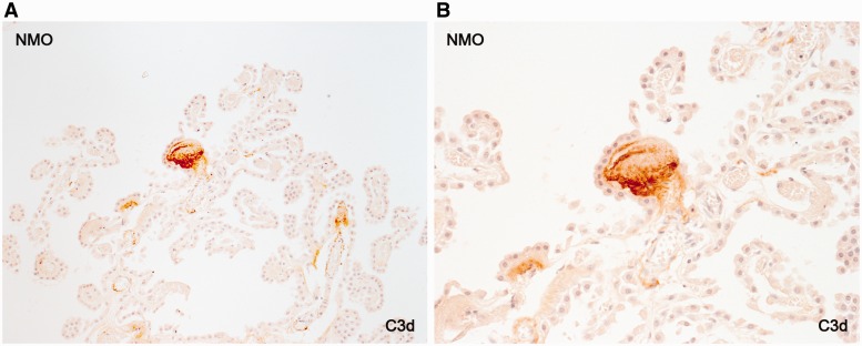
We report a previously undescribed inflammatory lesion consisting of deposition of activated complement (C3d and C9neo) in association with major histocompatibility complex type II (MHC2)-positive activated microglia in choroid plexus villi exhibiting classical fibrous thickening of the pericapillary filtration membrane. The proportion of villi affected ranged from 5% to 90% in 56 adult subjects with diseases of the CNS and 11 subjects with no preexisting disease of the CNS. In 3 of the 4 children studied, 2% or less of examined villi showed stromal thickening, complement deposition, and the presence of MHC2-positive microglia; in adults, the proportion of villi affected increased with age. Other features of the lesion included loss of capillaries and failure by macrophages to clear extracellular particulate electron-dense material by clathrin-mediated phagocytosis. This choroid plexus lesion may relate pathogenetically to age-related macular degeneration and to Alzheimer disease, 2 other conditions with no known risk factors other than increasing age. All 3 conditions are characterized by the presence of damaged capillaries, inflammatory extracellular aggregates of mixed molecular composition and defective clearance of the deposits by macrophages.

Keywords: Alzheimer disease; Choroid plexus; Complement; Macular degeneration; Multiple sclerosis..


Figures

FIGURE 1
FIGURE 1
Choroid plexus (A) A choroid plexus in the roof of the fourth ventricle immunostained using a labeled lectin that stains vascular endothelium a brownish color. The staining pattern reflects the fact that choroid plexuses are composed largely of massed capillaries. (B) A choroid plexus capillary located within a normal-appearing choroid plexus villus. The asterisk is located within the pericapillary space of the filtration membrane. Vacuoles at the base of each epithelial cell represent extracellular fluid-filled cisterns located amongst epithelial cell foot processes as shown in Figure 2. Human tissue: A, RCA-1, ×5; B, Semi-thin section, toluidine blue, ×900.
FIGURE 2
FIGURE 2
The filtration membrane. The asterisk is located in the pericapillary space of the filtration membrane. Other components of this complex structure are seen below and above the asterisk. Below is the fenestrated endothelium of the capillary and its very thin basal lamina. Above is the basal lamina of the epithelium together with the tips of epithelial cell foot processes and large extracellular fluid-filled cisterns that bulge into the base of each of the 2 epithelial cells. Human tissue: electron micrograph, ×7000).
FIGURE 3
FIGURE 3
Complement deposition in the choroid plexus of a 45-year-old male patient with MS. (A, B) A lateral ventricle choroid plexus stains positively for activated complement. A, H&E; B, C3d. Magnifications: A, B, ×25.
FIGURE 4
FIGURE 4
Interstitial fibrosis. (A) The pericapillary space in each of the 3 villi is occupied by a thick layer of connective tissue. The capillaries have reduced diameters. (B) The relatively narrow perivascular space of 3 villi and the connecting portion of the frond is occupied by a thin layer of connective tissue that is immunostained positively for C9neo. A, Mallory trichrome, ×460; B, C9neo, ×520.
FIGURE 5
FIGURE 5
Early deposition of complement in the choroid plexus of a patient with NMO. (A, B) Complement immunoreactivity (C3d) is restricted to a single large villous nodule. A, ×90; B, ×180.
FIGURE 6
FIGURE 6
Complement and IgG deposits. Choroid plexus perivascular nodules (stromal fibrosis) in patients with cerebral infarction (A) and NMO (B). Immunoreactivity is restricted to material located within the fibrosed pericapillary space. A, C9neo, ×560; B, IgG, ×570.
FIGURE 7
FIGURE 7
Rat and human choroid plexus villi. (A) The choroid plexus of a young rat showing each villous entirely occupied by a large capillary. The pericapillary space is narrow and without discernible contents. (B) A choroid plexus in a 71-year-old male patient with MS in which more than half the number of villi present exhibit narrowing or loss of capillaries together with marked stromal fibrosis. A, Frozen section, H&E, ×430; B, paraffin section, H&E, ×200.
FIGURE 8
FIGURE 8
Stromal fibrosis nodules in choroid plexus of MS and NMO patients. Nodules occupy most of the space normally occupied by a capillary. A, MS, Luxol fast blue-PAS, ×460; B, MS, Bodian’s silver stain, ×210; C, NMO, IgG immunohistochemistry, ×450.
FIGURE 9
FIGURE 9
Inflammatory cells. In the choroid plexus of a patient with MS, a villous nodule contains a mineral inclusion (A). CD68-positive putative macrophages (B), although present close to complement-positive material, are unreactive for complement (C). (D) UCHL1-positive lymphocytes are numerous in the choroid plexus of a patient with MS and perivenous encephalomyelitis; the latter followed injections of pig brain given as a treatment for MS. (E, F) MHC2-positive cells are present throughout pericapillary spaces in a patient with NMO. A, C9neo; B, CD68; C, C3d; D, UCHL1; E, F, MHC2. Magnifications: A–C, E, F, ×320; D, ×400; F, ×180.
FIGURE 10
FIGURE 10
Electron photomicrographs of particulate electron-dense material in choroid plexus villi. (A) A cell located in the pericapillary space invests extracellular fragments of electron-dense material. (B) An area within the cell illustrated in panel A showing cell processes contacting but not endocytosing dark material. (C, D) Extracellular dark material contacts but is not endocytosed by perivascular cells. Arrows indicate points of contact with what appear to be clathrin-coated pits. (E) An elongated fragment of dark material roughly the size of adjacent collagen fibers. (F) Cell processes surround extracellular dark material located close to a villous capillary. A, ×2,900; B, ×2,100; C, ×68,200; D, ×29,000; E, ×64,400; F, ×4,050.
FIGURE 11
FIGURE 11
Interstitial fibrosis - capillary and epithelial damage in the choroid plexus of a 39-year-old woman with MS. (A) A semi-thin section of a choroid plexus villous in which the several components of the filtration membrane appear normal. The capillary endothelium is intact; there is a narrowly patent pericapillary space, and vacuoles corresponding to fluid-filled cistern located amongst epithelial cell foot processes are intact. (B) A semi-thin section of a villous in which the filtration membrane is replaced by marked stromal fibrosis. There is no centrally located capillary, the pericapillary space is gone, and the line of fluid cisterns at the base of the epithelial monolayer is no longer evident. There are 8 cells (a mixture presumably of fibroblasts and CD68-positive, MHC2-positive cells [nuclei only visible]) located within and around the nodule. A, B, toluidine blue, ×800.
FIGURE 12
FIGURE 12
Interstitial fibrosis. There is widening of the pericapillary space in an affected choroid plexus villous. Commencing fibrosis is accompanied by thickening of the subepithelial basal lamina together with crowding and elongation of epithelial cell foot processes. Note the absence of evidence of immune complex deposition as seen in the kidney in Figure 15. F Cap, fenestrated capillary. Electron micrograph, ×3200.
FIGURE 13
FIGURE 13
Filtration membranes. Semi-thin sections of retinal (left) and choroid plexus (right) filtration membranes showing fenestrated capillaries separated by a narrow pericapillary space from the absorptive zone of epithelial cell foot processes. Expanding inflammatory lesions within this space damage the overlying epithelial monolayer in AMD and in inflammation of choroid plexus villi. F cap, fenestrated capillary; BM, Bruch's membrane; Foot Proc, foot processes; SBL, subepithelial basement membrane. Toluidine blue; Magnifications: left, ×2200; right, ×1500.
FIGURE 14
FIGURE 14
Age-related macular degeneration. The retinal filtration membrane in a patient with pathologically defined AMD. Nodules of drusen capped with a pale band of thickened basement membrane impinge on and displace upwards the retinal pigmentary epithelial cell layer (arrows). When compared with normal regions of the retinal filtration membrane (Fig. 13A), it is apparent that there is also some loss of the vacuolated absorptive foot process zone at the base of each epithelial cell. Such appearances indicate possible injury both to the pigmentary cell layer and to the epithelial monolayer. F Cap, fenestrated capillary. Semi-thin section, toluidine blue, ×2100.
FIGURE 15
FIGURE 15
Renal immune complex disease. Immune complex deposition (asterisks) is associated with a thickly ridged renal glomerular basement membrane in a patient with membranous glomerulonephritis (stage 2). Electron micrograph, ×6200.

References

    1. Parratt JD, Prineas JW. Neuromyelitis optica: a demyelinating disease characterized by acute destruction and regeneration of perivascular astrocytes. Mult Scler 2010;16:1156–72 - PubMed
    1. Misu T, Fujihara K, Kakita A, et al. Loss of aquaporin 4 in lesions of neuromyelitis optica: distinction from multiple sclerosis. Brain 2007;130:1224–34 - PubMed
    1. Roemer SF, Parisi JE, Lennon VA, et al. Pattern-specific loss of aquaporin-4 immunoreactivity distinguishes neuromyelitis optica from multiple sclerosis. Brain 2007;130:1194–205 - PubMed
    1. Prineas JW. Multiple sclerosis: presence of lymphatic capillaries and lymphoid tissue in the brain and spinal cord. Science 1979;203:1123–5 - PubMed
    1. Adams RD, Oksche A, Haymaker W. Meninges, choroid plexuses, ependyma and their reactions. Part 2: Age-related changes and pathology In: Haymaker W, Adams RD, eds. Histology and Histopathology of the Nervous System. Springfield, IL: Charles C. Thomas; 1982:641–713

Publication types