

Surgical tissue handling methods to optimize ex vivo fluorescence with the activatable optical probe γ-glutamyl hydroxymethyl rhodamine green
- PMID: 27444370
- PMCID: PMC6467211
- DOI: 10.1002/cmmi.1705
Surgical tissue handling methods to optimize ex vivo fluorescence with the activatable optical probe γ-glutamyl hydroxymethyl rhodamine green
Abstract
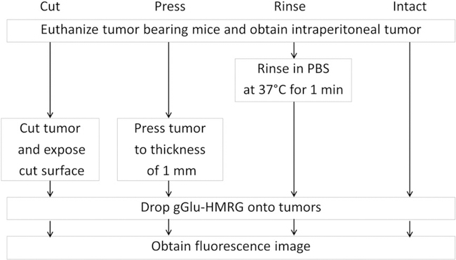
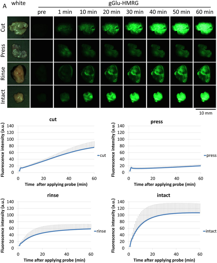
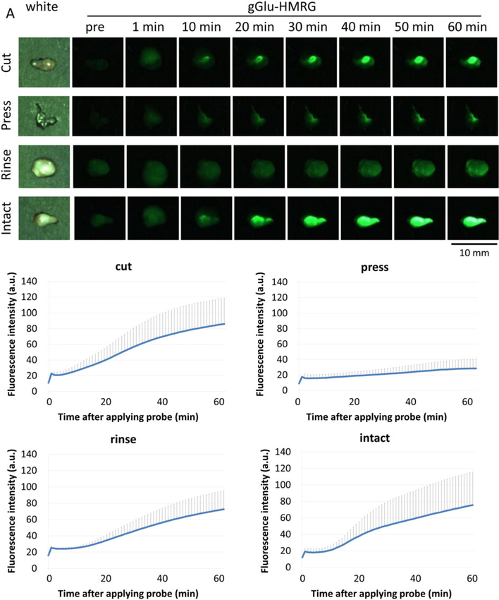
Optical fluorescence imaging has been developed as an aid to intraoperative diagnosis to improve surgical and endoscopic procedures. Compared with other intraoperative imaging methods, it is lower in cost, has a high safety margin, is portable and easy to use. γ-glutamyl hydroxymethyl rhodamine green (gGlu-HMRG) is a recently developed activatable fluorescence probe that emits strong fluorescence in the presence of the enzyme γ-glutamyl transpeptidase (GGT), which is overexpressed in many cancers, including ovarian cancer. Ex vivo testing is important for clinical approval of such probes. The diagnostic performance of gGlu-HMRG in fresh excised surgical specimens has been reported; however, details of tissue handling have not been optimized. In this study, we investigated four different tissue handling procedures to optimize imaging in excised tumor specimens. The fluorescence intensity time courses after the different tissue handling methods were compared. Additionally, the fluorescence positive areas were correlated with the presence of red fluorescent protein (RFP) in an RFP positive cell line as the standard of reference for cancer location. In the 'intact' groups, tumors yielded quick and homogeneous activation of gGlu-HMRG. In the 'rinse' and 'cut' groups, the fluorescence intensity of the tumor was a little lower than that in the intact group. In the 'pressed' groups, however, fluorescence intensity from gGlu-HMRG was lower over the entire time course, suggesting a decrease or relocation of excreted GGT. In conclusion, we demonstrate that the method of tissue handling prior to ex vivo imaging with the activatable probe gGlu-HMRG has a strong influence on the signal derived from the specimen. Published 2016. This article is a U.S. Government work and is in the public domain in the USA.
Keywords: fluorescent probe; optical navigation surgery; ovarian cancer; surgical specimen; γ-glutamyl transpeptidase.
Published 2016. This article is a U.S. Government work and is in the public domain in the USA.
Figures

Similar articles
-
Exploit the γ-Glutamyl hydroxymethyl rhodamine green fluorescence in surgical resection: A systematic literature review on clinical indications, fields of application and outcomes.Neurosurg Rev. 2025 Mar 28;48(1):335. doi: 10.1007/s10143-025-03484-3. Neurosurg Rev. 2025. PMID: 40148698
-
Dynamic fluorescent imaging with the activatable probe, γ-glutamyl hydroxymethyl rhodamine green in the detection of peritoneal cancer metastases: Overcoming the problem of dilution when using a sprayable optical probe.Oncotarget. 2016 Aug 9;7(32):51124-51137. doi: 10.18632/oncotarget.9898. Oncotarget. 2016. PMID: 27286461 Free PMC article.
-
Rapid visualization of mammary gland tumor lesions of dogs using the enzyme-activated fluorogenic probe; γ-glutamyl hydroxymethyl rhodamine green.J Vet Med Sci. 2022 Apr 15;84(4):593-599. doi: 10.1292/jvms.22-0003. Epub 2022 Mar 7. J Vet Med Sci. 2022. PMID: 35249908 Free PMC article.
-
Rapid intraoperative visualization of breast lesions with γ-glutamyl hydroxymethyl rhodamine green.Sci Rep. 2015 Jul 13;5:12080. doi: 10.1038/srep12080. Sci Rep. 2015. PMID: 26165706 Free PMC article.
-
Recent Advances in the Development of Optical Imaging Probes for γ-Glutamyltranspeptidase.Chembiochem. 2019 Feb 15;20(4):474-487. doi: 10.1002/cbic.201800370. Epub 2018 Sep 19. Chembiochem. 2019. PMID: 30062708 Review.
Cited by
-
Novel pH-activatable NIR fluorogenic spray mediated near-instant and precise tumor margins identification in human cancer tissues for surgical resection.Theranostics. 2023 Aug 15;13(13):4497-4511. doi: 10.7150/thno.85651. eCollection 2023. Theranostics. 2023. PMID: 37649597 Free PMC article.
-
Association between serum γ-glutamyl transferase and advanced colorectal adenoma among inpatients: a case-control study.Front Oncol. 2024 Jan 12;13:1188017. doi: 10.3389/fonc.2023.1188017. eCollection 2023. Front Oncol. 2024. PMID: 38282678 Free PMC article.
-
A novel method for rapid detection of a Helicobacter pylori infection using a γ-glutamyltranspeptidase-activatable fluorescent probe.Sci Rep. 2019 Jul 1;9(1):9467. doi: 10.1038/s41598-019-45768-x. Sci Rep. 2019. PMID: 31263136 Free PMC article.
-
Rapid detection of papillary thyroid carcinoma by fluorescence imaging using a γ-glutamyltranspeptidase-specific probe: a pilot study.Thyroid Res. 2018 Nov 21;11:16. doi: 10.1186/s13044-018-0060-y. eCollection 2018. Thyroid Res. 2018. PMID: 30479665 Free PMC article.
-
Exploit the γ-Glutamyl hydroxymethyl rhodamine green fluorescence in surgical resection: A systematic literature review on clinical indications, fields of application and outcomes.Neurosurg Rev. 2025 Mar 28;48(1):335. doi: 10.1007/s10143-025-03484-3. Neurosurg Rev. 2025. PMID: 40148698
References
-
- Keereweer S, Kerrebijn JD, van Driel PB, Xie B, Kaijzel EL, Snoeks TJ, Que I, Hutteman M, van der Vorst JR, Mieog JS, Vahrmeijer AL, van de Velde CJ, Baatenburg de Jong RJ, Lowik CW. Optical image-guided surgery - where do we stand? Mol Imaging Biol 2011; 13: 199–207. doi:10.1007/s11307-010-0373-2. - DOI - PMC - PubMed
-
- Ueo H, Shinden Y, Tobo T, Gamachi A, Udo M, Komatsu H, Nambara S, Saito T, Ueda M, Hirata H, Sakimura S, Takano Y, Uchi R, Kurashige J, Akiyoshi S, Iguchi T, Eguchi H, Sugimachi K, Kubota Y, Kai Y, Shibuta K, Kijima Y, Yoshinaka H, Natsugoe S, Mori M, Maehara Y, Sakabe M, Kamiya M, Kakareka JW, Pohida TJ, Choyke PL, Kobayashi H, Urano Y, Mimori K. Rapid intraoperative visualization of breast lesions with gamma-glutamyl hydroxymethyl rhodamine green. Sci Rep 2015; 5: 12080. doi:10.1038/srep12080. - DOI - PMC - PubMed
MeSH terms
Substances
Grants and funding
LinkOut - more resources
Full Text Sources
Other Literature Sources
Miscellaneous
