. 2016 Jul 22:6:30196.
doi: 10.1038/srep30196.

Role of Zn doping in oxidative stress mediated cytotoxicity of TiO2 nanoparticles in human breast cancer MCF-7 cells

Affiliations

Role of Zn doping in oxidative stress mediated cytotoxicity of TiO2 nanoparticles in human breast cancer MCF-7 cells

Maqusood Ahamed et al. Sci Rep. .

Abstract

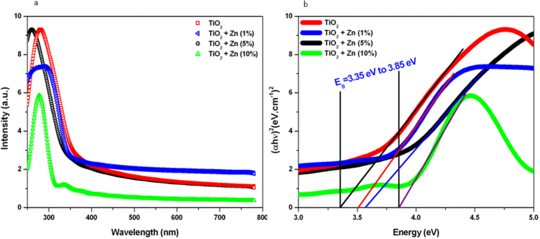
We investigated the effect of Zn-doping on structural and optical properties as well as cellular response of TiO2 nanoparticles (NPs) in human breast cancer MCF-7 cells. A library of Zn-doped (1-10 at wt%) TiO2 NPs was prepared. Characterization data indicated that dopant Zn was incorporated into the lattice of host TiO2. The average particle size of TiO2 NPs was decreases (38 to 28 nm) while the band gap energy was increases (3.35 eV-3.85 eV) with increasing the amount of Zn-doping. Cellular data demonstrated that Zn-doped TiO2 NPs induced cytotoxicity (cell viability reduction, membrane damage and cell cycle arrest) and oxidative stress (reactive oxygen species generation &glutathione depletion) in MCF-7 cells and toxic intensity was increases with increasing the concentration of Zn-doping. Molecular data revealed that Zn-doped TiO2 NPs induced the down-regulation of super oxide dismutase gene while the up-regulation of heme oxygenase-1 gene in MCF-7 cells. Cytotoxicity induced by Zn-doped TiO2 NPs was efficiently prevented by N-acetyl-cysteine suggesting that oxidative stress might be the primarily cause of toxicity. In conclusion, our data indicated that Zn-doping decreases the particle size and increases the band gap energy as well the oxidative stress-mediated toxicity of TiO2 NPs in MCF-7 cells.


Figures

Figure 1
Figure 1. Transmission electron microscopy characterization of pure and Zn-doped TiO2 NPs.
Upper & middle panels represent the low resolution images while lower panel shows the high resolution images.
Figure 2
Figure 2. XRD and Raman characterization of pure and Zn-doped TiO2 NPs.
(a) XRD spectra and (b) Raman spectra.
Figure 3
Figure 3. Optical characterization of pure and Zn-doped TiO2 NPs.
(a) UV-visible absorption spectra and (b) (αhν)2 vs photon energy plots of the corresponding sample used to determine their band gap energy levels.
Figure 4
Figure 4. Cytotoxic activity of pure and Zn-doped TiO2 NPs in MCF-7 cells.
(a) Cell viability determined by MTT assay. This assay measures the mitochondrial function by determining the ability of living cells to reduce MTT into blue formazon product. Cells were treated with 50, 100 & 200 μg/ml of pure and Zn-doped TiO2 NPs as well as pure Zn NPs for 24 h. Cells not exposed to NPs served as a negative control. (b) Lactate dehydrogenase (LDH) assay. LDH is an enzyme widely present in the cytosol that converts lactate into pyruvate. When plasma membrane integrity is disrupted, LDH leaks into culture media and its extracellular level is elevated. Exposure of NPs to cells was similar as in MTT assay. Data represented are mean ± SD of three identical experiments made in three replicate. *Significant difference as compared to control (p < 0.05).
Figure 5
Figure 5. Oxidant ROS and antioxidant GSH levels in MCF-7 cells after exposure to pure and Zn-doped TiO2 NPs.
(a) Intracellular generation of ROS was measured by 2,7-dichlorofluorescin diacetate (DCFH-DA) fluorescence-based assay. Cells were treated with 50, 100 & 200 μg/ml of pure and Zn-doped TiO2 NPs as well as pure Zn NPs for 6 h. Cells not exposed to NPs served as a negative control. (b) Intracellular ROS levels in the presence or absence of N-acetyl cysteine (NAC). (c) GSH level was determined by Ellman’s reagent. Exposure of NPs to cells was similar as in ROS assay. (d) Intracellular GSH levels in the presence or absence of buthionine sulphoximine (BSO) and NAC. The BSO is an inhibitor of GSH biosynthesis. GSH levels were expressed in terms of nmole/mg protein. Data represented are mean ± SD of three identical experiments made in three replicate. *Significant difference as compared to the control (p < 0.05). #Significant inhibitory effect of NAC on ROS generation and GSH depletion (p < 0.05).
Figure 6
Figure 6. Effect of pure and Zn-doped TiO2 NPs on superoxide dismutase (SOD) and heme oxygenase 1 (HO-1) genes in MCF-7 cells.
(a) Western blot analysis of SOD1 (CuZn-SOD), SOD2 (Mn-SOD) and HO-1 protein levels. Cells were treated with 200 μg/ml pure and Zn-doped TiO2 NPs for 6 h. Cells not exposed to NPs served as a negative control. The treated and untreated cells were lysed in RIPA buffer and cell extract subjected to western blots with anti-SOD1, anti-SOD2 & anti-HO-1 antibodies. The β-actin blot is a loading control. (b) Protein levels were also analyzed by desitometric analysis using AlphaEase TM FC StandAlone V.4.0.0 software. Results are expressed as a fold change over the control group. (c) SOD enzyme activity in MCF-7 cells after exposure to pure and Zn-doped TiO2 NPs. Cells were treated with 200 μg/ml of pure and Zn-doped TiO2 NPs as well as pure Zn NPs for 6 h. SOD activity was expressed in terms of U/ml. (d) SOD enzyme extract attenuates Zn-doped TiO2 NPs induced cytotoxicity. Cells were treated with 200 μg/ml of Zn-doped TiO2 NPs in the presence or absence of SOD enzyme extract. Data represented are mean ± SD of three identical experiments made in three replicate. *Significant difference as compared to the control (p < 0.05). #Significant inhibitory effect of SOD on cell viability reduction (p < 0.05).
Figure 7
Figure 7. Possible mechanism of cytotoxicity caused by Zn-doped TiO2 NPs. Pure TiO2 NPs was not able to elicit cellular response because of lack of electrons (e) or holes (h+) on the surface of NPs.
However, integration of Zn impurities contributes electron energy levels high in semiconductor band gap so that electrons can be easily excited into conduction band, which causes Fermi level to be shifted towards conductions band. Movement of electrons (e) across the band gap to conduction band creates a hole (h+) in valence band. The free holes (h+) can generate hydroxyl radicals (OH), while the free electrons (e) could lead to formation of superoxide radicals (O2•−). These free oxygen radicals might be responsible for oxidant injury to cellular systems.

References

    1. Nel A., Xia T., Madler L. & Li N. Toxic potential of materials at the nanolevel. Science 311, 622–627 (2006). - PubMed
    1. Jiang W., Kim B. S., Rutka J. T. & Chan W. W. Nanoparticle-mediated cellular response is size-dependent. Nat. Nanotechnol. 3, 145–150 (2008). - PubMed
    1. Xia T. et al.. Comparison of the abilities of ambient and manufactured nanoparticles to induce cellular toxicity according to an oxidative stress paradigm. Nano Lett. 6, 1794–1807 (2006). - PubMed
    1. Pujalte I. et al.. Cytotoxicity and oxidative stress induced by different metallic nanoparticles on human kidney cells. Part. Fibre Toxicol. 8, 10 (2011). - PMC - PubMed
    1. Setyawati M. I., Tay C. Y. & Leong D. T. Mechanistic investigation of the biological effects of SiO2, TiO2, and ZnO nanoparticles on intestinal cells. Small 11, 3458–3468 (2015). - PubMed

Publication types