. 2016 Sep 16;119(7):853-64.
doi: 10.1161/CIRCRESAHA.116.309001. Epub 2016 Jul 21.

Proliferation and Recruitment Contribute to Myocardial Macrophage Expansion in Chronic Heart Failure

Affiliations

Proliferation and Recruitment Contribute to Myocardial Macrophage Expansion in Chronic Heart Failure

Hendrik B Sager et al. Circ Res. .

Abstract

Rationale: Macrophages reside in the healthy myocardium, participate in ischemic heart disease, and modulate myocardial infarction (MI) healing. Their origin and roles in post-MI remodeling of nonischemic remote myocardium, however, remain unclear.

Objective: This study investigated the number, origin, phenotype, and function of remote cardiac macrophages residing in the nonischemic myocardium in mice with chronic heart failure after coronary ligation.

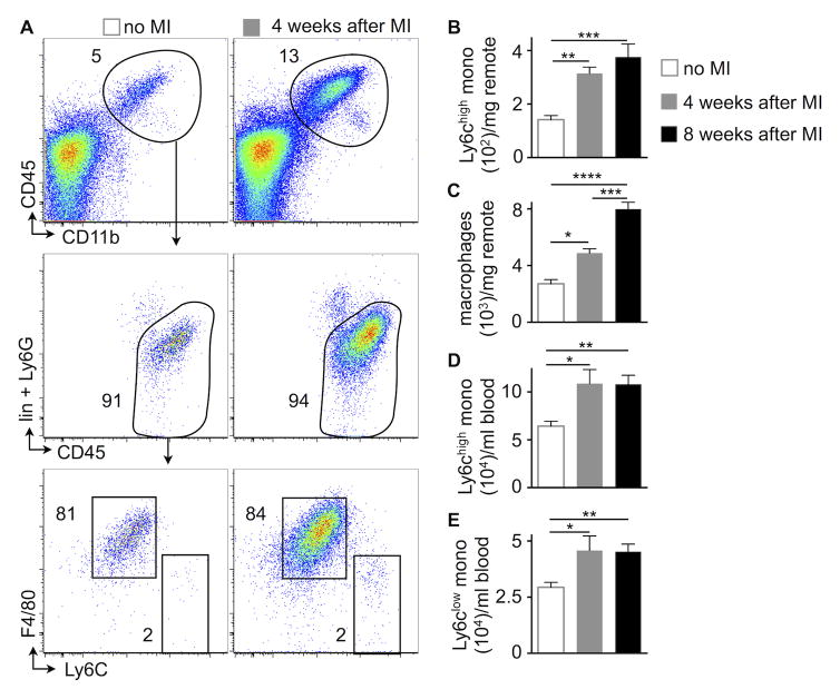
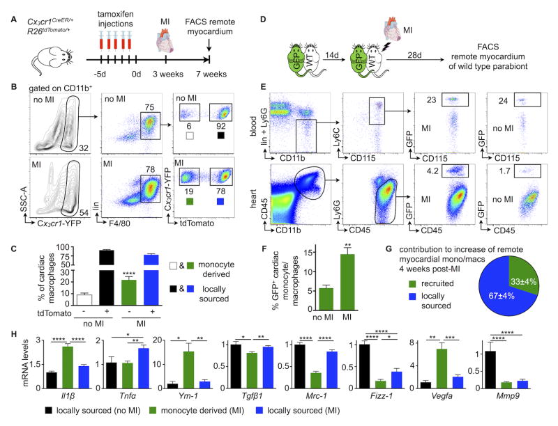
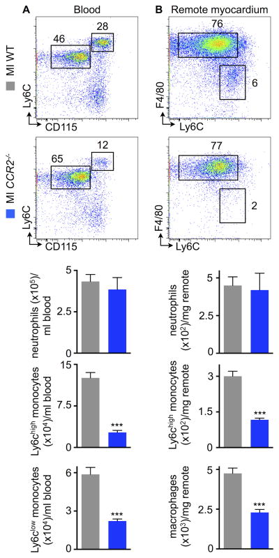
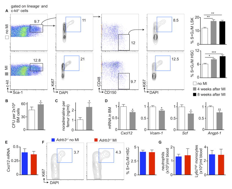
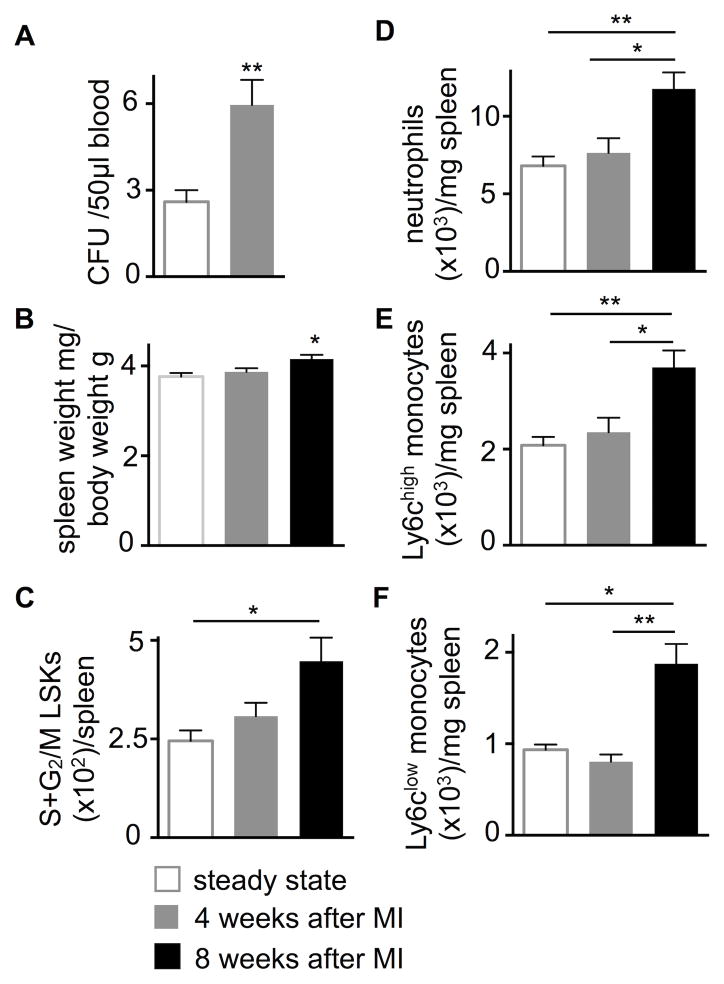
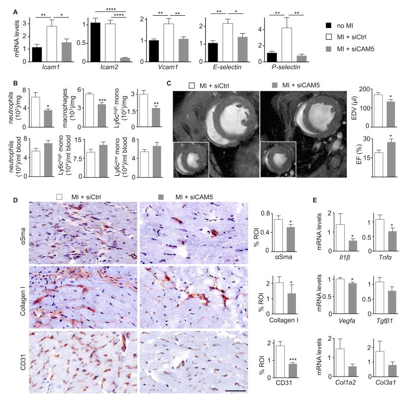
Methods and results: Eight weeks post MI, fate mapping and flow cytometry revealed that a 2.9-fold increase in remote macrophages results from both increased local macrophage proliferation and monocyte recruitment. Heart failure produced by extensive MI, through activation of the sympathetic nervous system, expanded medullary and extramedullary hematopoiesis. Circulating Ly6C(high) monocytes rose from 64±5 to 108±9 per microliter of blood (P<0.05). Cardiac monocyte recruitment declined in Ccr2(-/-) mice, reducing macrophage numbers in the failing myocardium. Mechanical strain of primary murine and human macrophage cultures promoted cell cycle entry, suggesting that the increased wall tension in post-MI heart failure stimulates local macrophage proliferation. Strained cells activated the mitogen-activated protein kinase pathway, whereas specific inhibitors of this pathway reduced macrophage proliferation in strained cell cultures and in the failing myocardium (P<0.05). Steady-state cardiac macrophages, monocyte-derived macrophages, and locally sourced macrophages isolated from failing myocardium expressed different genes in a pattern distinct from the M1/M2 macrophage polarization paradigm. In vivo silencing of endothelial cell adhesion molecules curbed post-MI monocyte recruitment to the remote myocardium and preserved ejection fraction (27.4±2.4 versus 19.1±2%; P<0.05).

Conclusions: Myocardial failure is influenced by an altered myeloid cell repertoire.

Keywords: heart failure; hypertrophy; macrophage; monocyte; myocardial infarction.


Conflict of interest statement

Disclosures

J.E.D., and D.G.A. have filed intellectual property protection related to 7C1 nanoparticles. The authors declare that they have no further competing interests.

Figures

Figure 1
Figure 1. Expansion of cardiac macrophages in HFrEF
A–C, Gating and quantification of myeloid cells in steady-state versus 4 and 8 weeks after MI in the remote area (i.e. the myocardium that was never ischemic), n=8–23 WT mice per group, mean±SEM, *p<0.05, **p<0.01, ***p<0.001, ****p<0.0001. D and E, Blood monocytes in steady-state versus 4 and 8 weeks after MI, n=8–23 WT mice per group, mean±SEM, *p<0.05, **p<0.01.
Figure 2
Figure 2. Contribution of recruitment to cardiac macrophage expansion in HFrEF
A, Experimental design. B and C, Gating and quantification of resident versus bone marrow-derived cardiac macrophages in steady-state versus 4 weeks after MI, n=4–8 per group, mean±SEM, ****p<0.0001. D, Experimental design. E and F, Gating and quantification of chimerism for blood monocytes and cardiac monocytes and macrophages in steady-state versus 4 weeks after MI, n=4–10 pairs per group, mean±SEM, **p<0.01. G, Relative contribution of monocyte-derived versus locally sourced macrophages to total remote monocyte/macrophage population 4 weeks after MI, n=4–10 pairs per group, mean±SEM. H, Phenotyping of resident versus bone marrow-derived cardiac macrophages using fate mapping outlined in 2A (4 weeks after MI, n=4–8 per group, mean±SEM, *p<0.05, **p<0.01, ***p<0.001, ****p<0.0001).
Figure 3
Figure 3. Ccr2 dependent monocyte recruitment contributes to cardiac macrophage expansion
A, Gating and quantification of blood myeloid cells in WT vs. Ccr2−/− mice, 4 weeks after MI, n=6–8 per group, mean±SEM, ***p<0.001. B, Gating and quantification of cardiac myeloid cells in WT vs. Ccr2−/− mice, 4 weeks after MI, n=6–8 per group, mean±SEM, ***p<0.001.
Figure 4
Figure 4. In situ macrophage proliferation and the role of biomechanical strain
A, Gating and quantification of cardiac macrophage proliferation in steady-state versus 4 weeks after MI, n=6–9 per group, mean±SEM, **p<0.01. B and C, Gating, quantification of cell numbers and proliferation with Ki67 (B) and BrdU (C) in stretched vs. non-stretched cultured murine peritoneal macrophages (n=6–8 dishes per group, mean±SEM, **p<0.01, ***p<0.001). D, In-dish confocal microscopy, macrophage numbers and macrophage proliferation in stretched versus non-stretched murine cultured peritoneal macrophages (n=5 per group, mean±SEM, *p<0.01). E, PhosphoErk1/2 (pT202/Y204) to total Erk1/2 ratio in stretched versus non-stretched cultured peritoneal murine macrophages by ELISA (n=6 per group, mean±SEM, *p<0.05). F, Cell numbers and BrdU incorporation in stretched cultured peritoneal murine macrophages that were treated with Mek inhibitor (n=6 per group, mean±SEM, *p<0.05). G and H, Gating and quantification of cardiac macrophage proliferation and numbers in mice with HFrEF, treated with a Mek inhibitor (4 weeks after MI, n=7–8 per group, mean±SEM, *p<0.05, ***p<0.001). I, Blood monocytes in mice with HFrEF, treated with a Mek inhibitor (n=7–8 per group, mean±SEM).
Figure 5
Figure 5. Strain enhances proliferation of human macrophages
A, Strain exposure of human macrophages. Gating and quantification of stretched versus non-stretched human primary macrophages (n=12 per group). B, Histological evaluation of heart tissue obtained from patients with ischemic cardiomyopathy undergoing left ventricular assist device implantation. Controls are unused donor hearts (n=8–11 per group, mean±SEM, **p<0.01).
Figure 6
Figure 6. HFrEF activates bone marrow hematopoiesis
A, Gating and quantification of bone marrow hematopoietic stem and progenitor cell proliferation in steady-state versus 4 and 8 weeks after MI, n=9–11 per group, mean±SEM, *p<0.05, **p<0.01, ***p<0.001. B, Bone marrow colony forming unit (CFU) assay in steady-state versus HFrEF (n=5 per group, mean±SEM, *p<0.05). C, Bone marrow noradrenaline in steady-state versus 4 weeks after MI, n=5–7 per group, mean±SEM, *p<0.05. D, mRNA of bone marrow hematopoietic stem cell (HSC) retention factors (Cxcl12, chemokine (C-X-C motif) ligand 12; Vcam-1, vascular cell adhesion molecule 1; Scf, stem cell factor; Angpt1, angiopoietin-1) in bone marrow in steady state versus 4 weeks after MI, n=10 per group, mean±SEM, *p<0.05, **p<0.01. E, mRNA of HSC retention factor Cxcl12 in steady-state Adrb3−/− versus Adrb3−/− mice 4 weeks after MI, n=5–6 per group, mean±SEM. F, Gating and quantification of bone marrow hematopoietic stem and progenitor cell proliferation in steady-state Adrb3−/− versus Adrb3−/− mice 4 weeks after MI, n=5–6 per group, mean±SEM. G, Quantification of blood neutrophils and monocytes in steady-state Adrb3−/− versus Adrb3−/− mice 4 weeks after MI, n=5–6 per group, mean±SEM.
Figure 7
Figure 7. HFrEF activates splenic myelopoiesis
A, Blood colony forming unit (CFU) assay in steady-state versus 4 weeks after MI, n=5–12 per group, mean±SEM, **p<0.01. B, Spleen weight in steady-state versus 4 and 8 weeks after MI, n=9–20 per group, mean±SEM, *p<0.05. C, Splenic hematopoietic stem and progenitor cell proliferation in steady state versus 4 and 8 weeks after MI, n=7–16 per group, mean±SEM, *p<0.05. D–F, Splenic myeloid cells in steady-state versus 4 and 8 weeks after MI, n=7–16 per group, mean±SEM, *p<0.05, **p<0.01.
Figure 8
Figure 8. Recruited macrophages contribute to HFrEF development
A, Endothelial cell adhesion molecule mRNA levels in remote myocardium, values normalized to Gapdh (treatment with either siCtrl or siCAM5 for three weeks starting one week after MI, n=9–11 per group, mean±SEM, *p<0.05, ***p<0.001, ****p<0.0001). B, Blood and cardiac myeloid cells in mice with HFrEF that received RNAi treatment with either siCtrl or siCAM5 for three weeks starting one week after MI (n=9–11 per group, mean±SEM, *p<0.05, **p<0.01, ***p<0.001). C, Evaluation of post-MI remodeling by cardiac MRI. Each panel shows the mid-ventricular short axis view at end-diastole and end-systole (inset). End-diastolic volumes (EDV) and left ventricular ejection fraction (EF) were measured on day 28 after MI (n=9–11 per group, mean±SEM, *p<0.05). D, Immunohistochemical evaluation of remote myocardium in mice with HFrEF for myofibroblasts (α-smooth muscle actin, αSMA), collagen (collagen-1), and vessels (CD31). Bar graphs show percentage of positive staining per region of interest (ROI) or number of vessels per high-power field (hpf). Scale bar, 50 μm (n=9–11 per group). E, mRNA levels in remote myocardium, values normalized to Gapdh (treatment with either siCtrl or siCAM5 for three weeks starting one week after MI (n=9–11 per group, mean±SEM, *p<0.05).

Comment in

  • Macrophages in the Remodeling Failing Heart.
    Chen B, Frangogiannis NG. Chen B, et al. Circ Res. 2016 Sep 16;119(7):776-8. doi: 10.1161/CIRCRESAHA.116.309624. Circ Res. 2016. PMID: 27635078 Free PMC article. No abstract available.

References

    1. Yancy CW, Jessup M, Bozkurt B, et al. 2013 ACCF/AHA guideline for the management of heart failure: a report of the American College of Cardiology Foundation/American Heart Association Task Force on practice guidelines. Circulation. 2013;128:e240–327. - PubMed
    1. McMurray JJ, Adamopoulos S, Anker SD, et al. ESC Guidelines for the diagnosis and treatment of acute and chronic heart failure 2012: The Task Force for the Diagnosis and Treatment of Acute and Chronic Heart Failure 2012 of the European Society of Cardiology. Developed in collaboration with the Heart Failure Association (HFA) of the ESC. Eur Heart J. 2012;33:1787–1847. - PubMed
    1. Landmesser U, Drexler H. Chronic heart failure: an overview of conventional treatment versus novel approaches. Nat Clin Pract Cardiovasc Med. 2005;2:628–638. - PubMed
    1. McMurray JJ. Clinical practice. Systolic heart failure. N Engl J Med. 2010;362:228–238. - PubMed
    1. Burchfield JS, Xie M, Hill JA. Pathological ventricular remodeling: mechanisms: part 1 of 2. Circulation. 2013;128:388–400. - PMC - PubMed