Review
. 2016 Nov;150(5):1141-1157.
doi: 10.1016/j.chest.2016.07.003. Epub 2016 Jul 19.

Plugs of the Air Passages: A Clinicopathologic Review

Affiliations
Review

Plugs of the Air Passages: A Clinicopathologic Review

Tanmay S Panchabhai et al. Chest. 2016 Nov.

Abstract

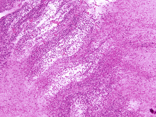
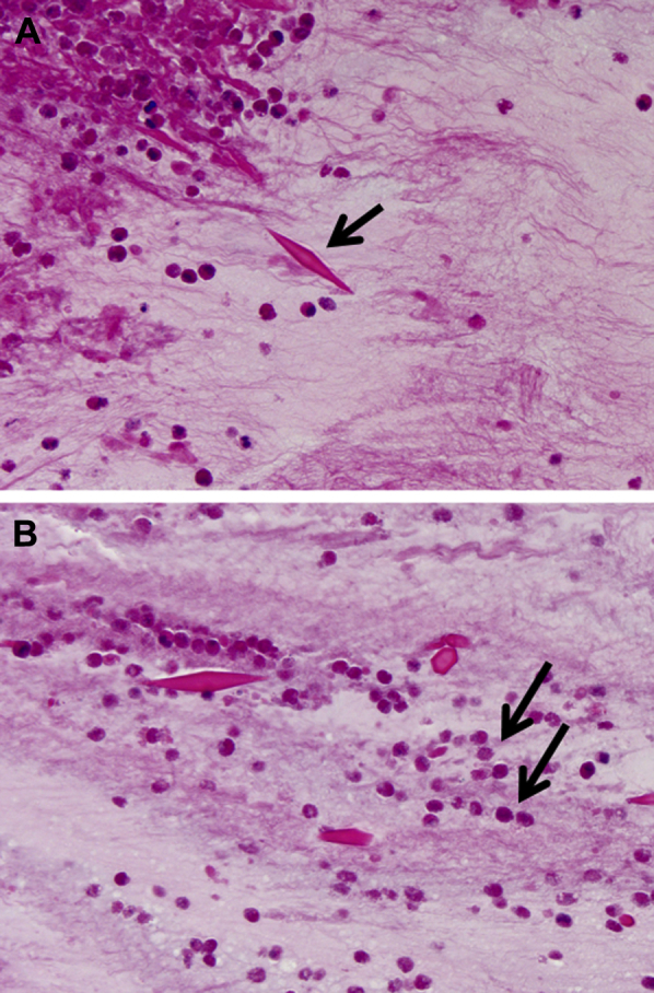
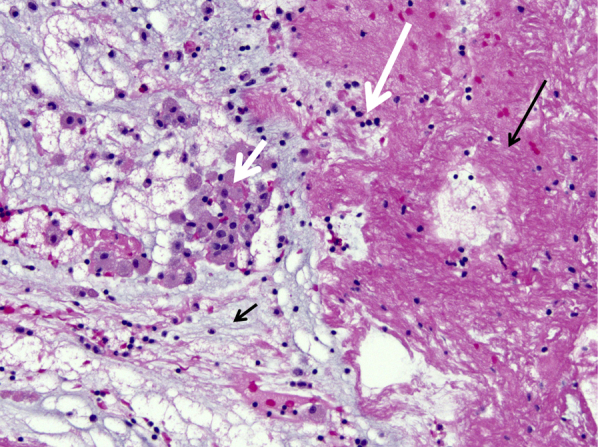
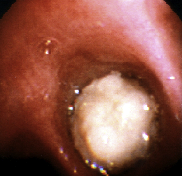
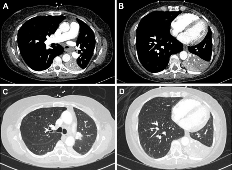
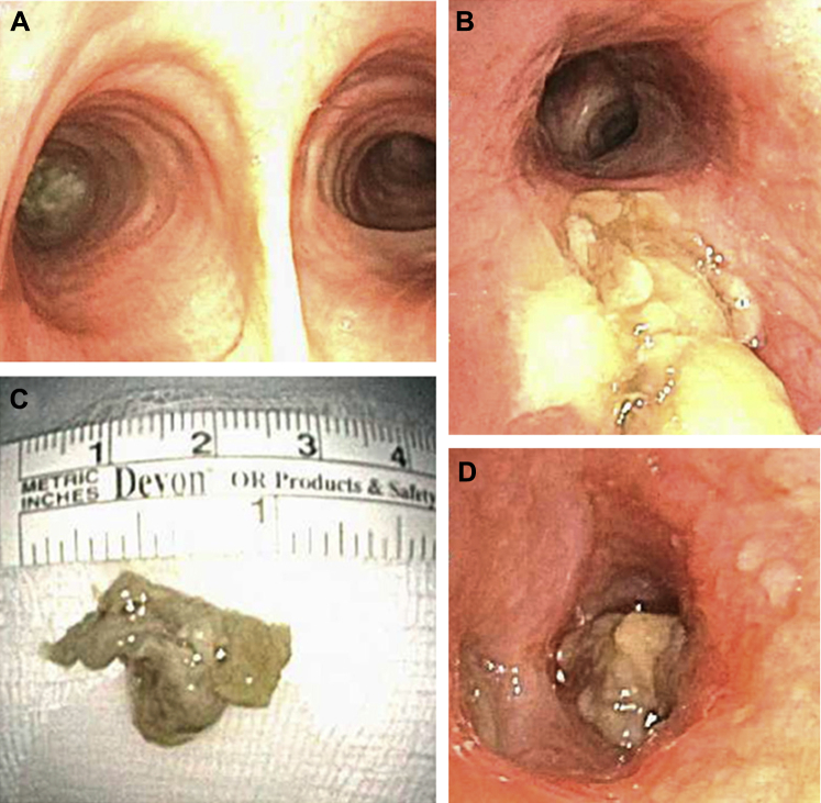
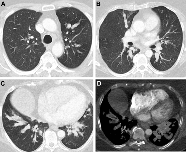
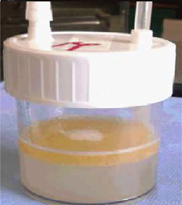
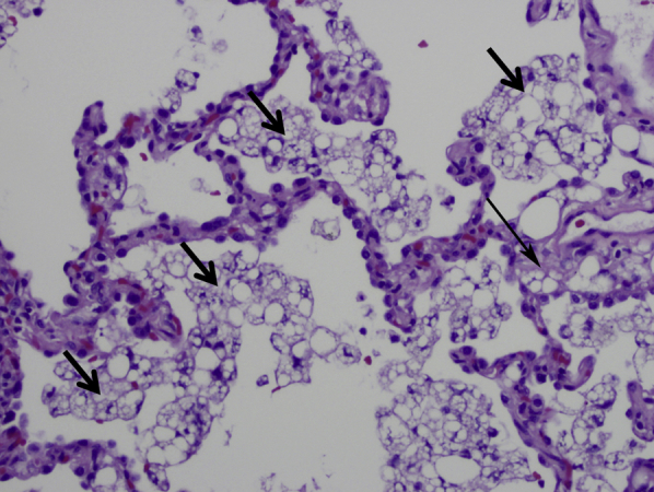
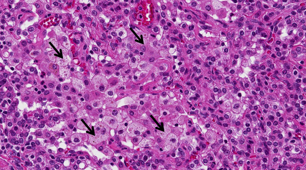
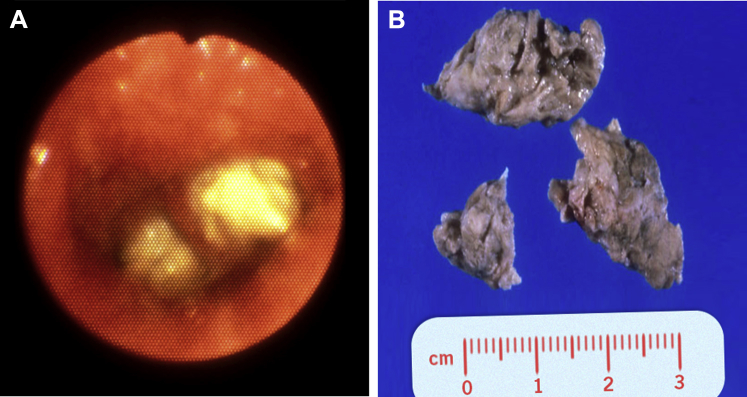
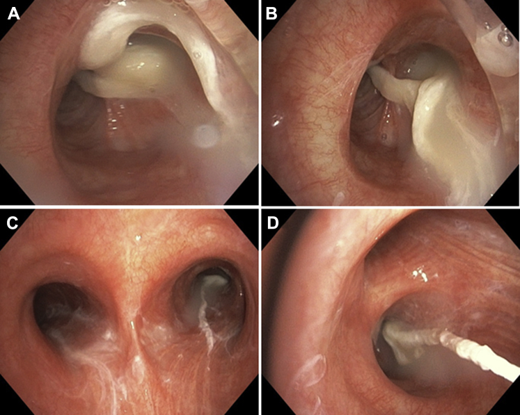
Although mucus is a normal product of the tracheobronchial tree, some diseases of the respiratory tract are characterized by unusually thick (inspissated) forms of mucus that accumulate within the airways. These are known as mucus plugs. The pathologic composition of these plugs is surprisingly diverse and, in many cases, correlates with distinctive clinical, radiologic, and bronchoscopic findings. The best-known conditions that involve mucus plugs are allergic bronchopulmonary aspergillosis, plastic bronchitis, and asthma. Other lung diseases occasionally associated with plugs within the airways include Aspergillus tracheobronchitis, hyper-IgE syndrome, exogenous lipoid pneumonia, pulmonary alveolar proteinosis, and chronic eosinophilic pneumonia. In this review, we describe and illustrate the bronchoscopic, pathologic, and imaging findings in respiratory disorders characterized by mucus plugs or plugs composed of other similar materials. Recognition of the characteristic appearance and differential diagnosis of mucus plugs will hopefully facilitate diagnosis and management of these diseases.

Keywords: airway and airspace diseases; bronchoscopic appearance; bronchoscopy; mucoid pseudotumor; mucus plugging.


Figures

Figure 1
Figure 1
A, B, High attenuation mucus (arrows) seen on CT scan of the chest in (A) mediastinal and (B) lung windows.
Figure 2
Figure 2
Chest radiograph shows finger-in-glove sign (arrows) from left upper lobe mucus impaction in a patient with allergic bronchopulmonary aspergillosis.
Figure 3
Figure 3
A, B, Chest radiograph shows (A) collapsed right upper lobe from mucus impaction in a patient with allergic bronchopulmonary aspergillosis (pretreatment) and (B) resolution of right upper lobe collapse (postbronchoscopic treatment).
Figure 4
Figure 4
Microscopic appearance of mucus plugs in mucoid impaction of bronchi and allergic bronchopulmonary aspergillosis (allergic mucin), showing characteristic lamellated appearance under low magnification (40×).
Figure 5
Figure 5
Microscopic appearance of mucus plugs in mucoid impaction of bronchi and allergic bronchopulmonary aspergillosis (allergic mucin) under high magnification, showing alternating layers of mucin (pale) and fibrin (dark). Numerous eosinophils and Charcot-Leyden crystals are present in both layers (magnification, 200×).
Figure 6
Figure 6
A, B, Charcot-Leyden crystals within allergic mucin in allergic bronchopulmonary aspergillosis. Bipyramidal appearance (arrow). B, Degenerating eosinophils are present in the background (arrows). Charcot-Leyden crystals can be found in any inflammatory process that contains numerous eosinophils (magnification, 400×).
Figure 7
Figure 7
Low magnification of mucus plug in plastic bronchitis containing mucin (short black arrow), fibrin (long black arrow), macrophages (short white arrow), and a few lymphocytes (long white arrow) (magnification, 200×).
Figure 8
Figure 8
Bronchoscopic view of complete obstruction of the right main bronchus with a large cast in a patient with plastic bronchitis.
Figure 9
Figure 9
A, Left-sided airway cast removed via rigid bronchoscopy from a patient with plastic bronchitis. B, Airway cast resembling the entire bronchial tree, removed via rigid bronchoscopy from a patient with plastic bronchitis.
Figure 10
Figure 10
A, B, Autopsy specimen from a patient with fatal asthma. A, Bronchial wall is characterized by thick mucoid secretions within the lumen, marked eosinophilic infiltration, smooth muscle hypertrophy, and thickening of the bronchial basement membrane. B, Gross specimen shows the mucus plugging in the airway (arrow).
Figure 11
Figure 11
A-C, Mucus plug obstructing the right lower and middle lobe bronchi (A) after bronchial thermoplasty in a patient with asthma. B, Histologic examination showed fibrin mixed with some mucus, scattered inflammatory cells and (C) gross examination showed a bronchus-shaped mucus plug.
Figure 12
Figure 12
Gross autopsy specimen of the trachea reveals inflammatory exudates in a patient with aspergillus tracheobronchitis.
Figure 13
Figure 13
A-D, CT scan of the chest in a liver transplant recipient with Aspergillus tracheobronchitis shows complete collapse of the left lower lobe in the (A, B) mediastinal and (C, D) lung windows.
Figure 14
Figure 14
A, Bronchoscopic view of completely obstructed left main bronchus from large mucus plug. B, C, The plug was subsequently removed. D, Distal mucus plugs were still visible in the lobar bronchi after initial removal and were later aspirated.
Figure 15
Figure 15
A-D, CT scan of the chest shows mucus plugging resulting in obstruction of multiple airways and distal consolidation in a patient with hyper-IgE syndrome.
Figure 16
Figure 16
Gross appearance of BAL fluid in exogenous lipoid pneumonia in a patient taking Lorenzo’s oil.
Figure 17
Figure 17
Microscopic appearance of exogenous lipoid pneumonia. Macrophages containing coarse vacuoles of varying sizes fill the airspaces (short arrows). Similar macrophages are focally present within the interstitium (long arrow). This appearance is pathognomonic (magnification, 200×).
Figure 18
Figure 18
Microscopic appearance of endogenous lipoid pneumonia. Macrophages containing fine uniform vacuoles fill the airspaces (arrows). This is a common and nonspecific pathologic finding in lung specimens (magnification, 200×).
Figure 19
Figure 19
A, Bronchoscopic view of exogenous lipoid pneumonia with airway plugs. B, Photograph of airway plugs removed from a patient with exogenous lipoid pneumonia.
Figure 20
Figure 20
A-D, Bronchoscopic images show mucoid plugs within the airways in a patient with pulmonary alveolar proteinosis. Note the white-yellow, thick appearance.
Figure 21
Figure 21
Microscopic findings in a patient with pulmonary alveolar proteinosis. Granular eosinophilic material fills the alveoli (black arrows). Alveolar septa (white arrows) are normal (magnification, 40×).
Figure 22
Figure 22
A, Tracheal cast in situ from a patient who died from complications of mucus plug after insertion of a transtracheal oxygen catheter. Arrows indicate the upper and lower edges of the cast, which filled approximately 90% of the lumen. B, Microscopic image of the tracheal cast showing a distinct marbled pattern of homogenous proteinaceous zones alternating with dark zones of necrotic epithelial cellular nuclei and inflammatory cells.
Figure 24
Figure 23
A-C, Bronchoscopic images show necrotic debris and plugs in the (A) left main, (B) left upper, and (C) left lower lobe bronchi in a patient with community-acquired methicillin-resistant Staphylococcus aureus pneumonia.

References

    1. Rose M.C., Voynow J.A. Respiratory tract mucin genes and mucin glycoproteins in health and disease. Physiol Rev. 2006;86(1):245–278. - PubMed
    1. Karasick D., Karasick S., Lally J.F. Mucoid pseudotumors of the tracheobronchial tree in two cases. AJR Am J Roentgenol. 1979;132(3):459–460. - PubMed
    1. Bosken C.H., Myers J.L., Greenberger P.A., Katzenstein A.L. Pathologic features of allergic bronchopulmonary aspergillosis. Am J Surg Pathol. 1988;12(3):216–222. - PubMed
    1. Mukhopadhyay S., Gal A.A. Granulomatous lung disease: an approach to the differential diagnosis. Arch Pathol Lab Med. 2010;134(5):667–690. - PubMed
    1. Katzenstein A.L., Liebow A.A., Friedman P.J. Bronchocentric granulomatosis, mucoid impaction, and hypersensitivity reactions to fungi. Am Rev Respir Dis. 1975;111(4):497–537. - PubMed