. 2016 Aug:97:556-567.
doi: 10.1016/j.freeradbiomed.2016.07.013. Epub 2016 Jul 19.

NOX4-derived reactive oxygen species limit fibrosis and inhibit proliferation of vascular smooth muscle cells in diabetic atherosclerosis

Affiliations

NOX4-derived reactive oxygen species limit fibrosis and inhibit proliferation of vascular smooth muscle cells in diabetic atherosclerosis

Elyse Di Marco et al. Free Radic Biol Med. 2016 Aug.

Abstract

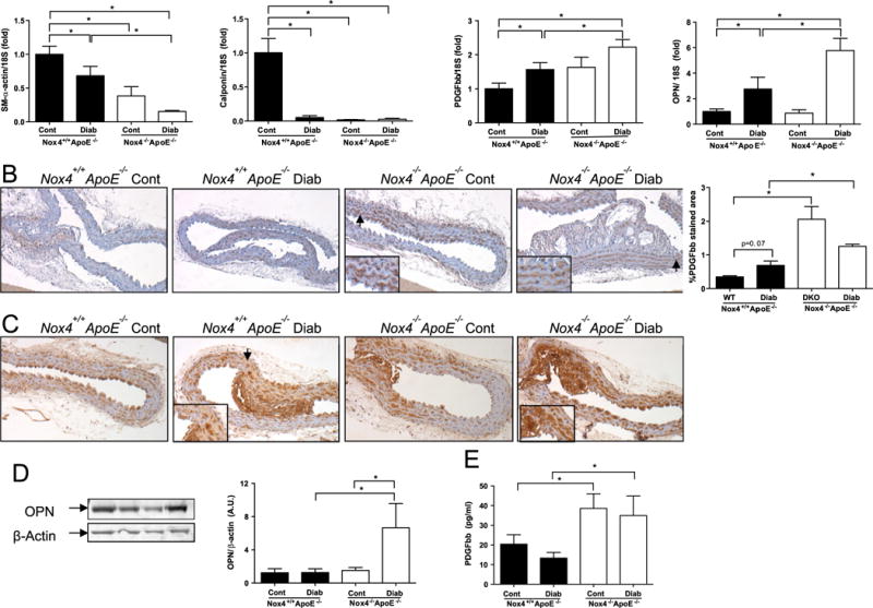
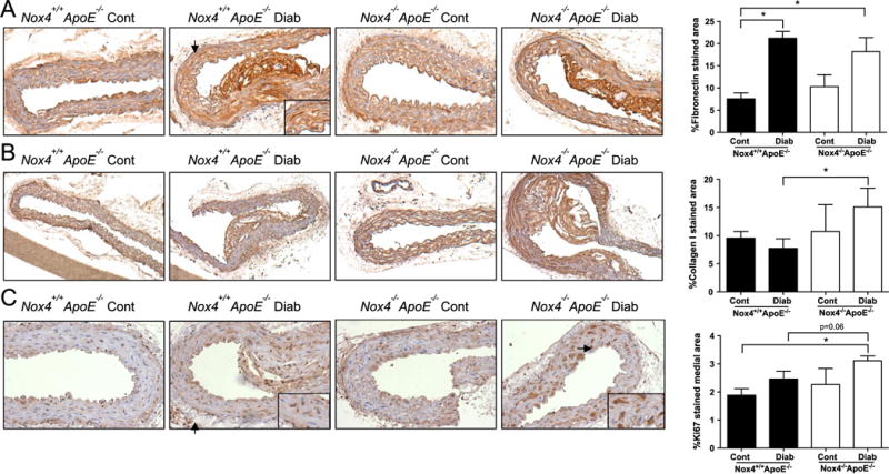
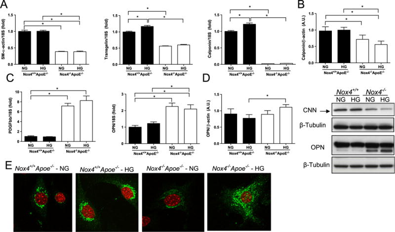
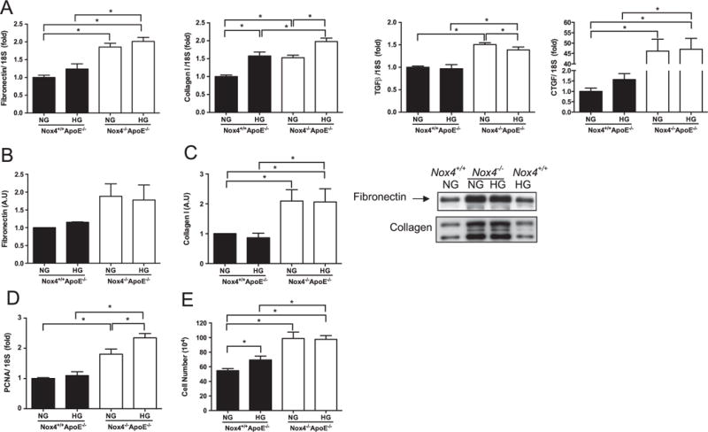
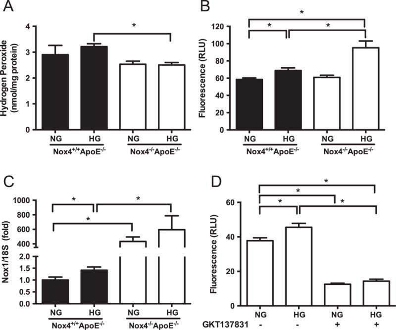
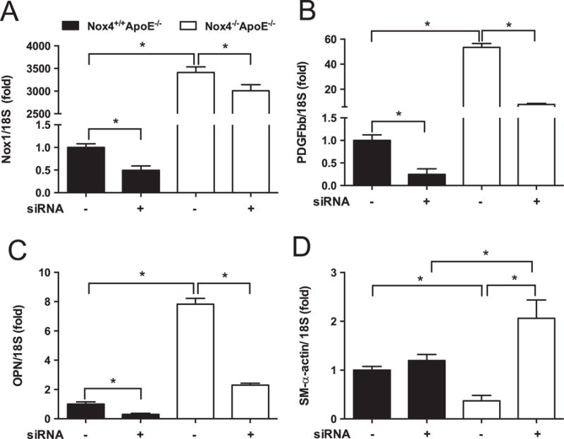
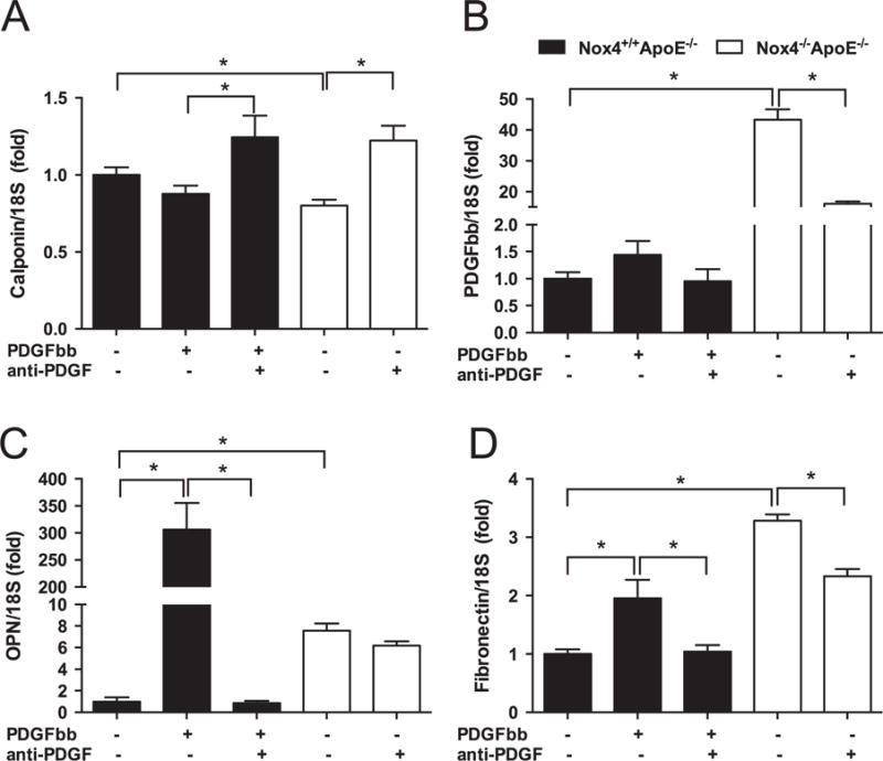
Smooth muscle cell (SMC) proliferation and fibrosis contribute to the development of advanced atherosclerotic lesions. Oxidative stress caused by increased production or unphysiological location of reactive oxygen species (ROS) is a known major pathomechanism. However, in atherosclerosis, in particular under hyperglycaemic/diabetic conditions, the hydrogen peroxide-producing NADPH oxidase type 4 (NOX4) is protective. Here we aim to elucidate the mechanisms underlying this paradoxical atheroprotection of vascular smooth muscle NOX4 under conditions of normo- and hyperglycaemia both in vivo and ex vivo. Following 20-weeks of streptozotocin-induced diabetes, Apoe(-/-) mice showed a reduction in SM-alpha-actin and calponin gene expression with concomitant increases in platelet-derived growth factor (PDGF), osteopontin (OPN) and the extracellular matrix (ECM) protein fibronectin when compared to non-diabetic controls. Genetic deletion of Nox4 (Nox4(-/)(-)Apoe(-/-)) exacerbated diabetes-induced expression of PDGF, OPN, collagen I, and proliferation marker Ki67. Aortic SMCs isolated from NOX4-deficient mice exhibited a dedifferentiated phenotype including loss of contractile gene expression, increased proliferation and ECM production as well as elevated levels of NOX1-associated ROS. Mechanistic studies revealed that elevated PDGF signalling in NOX4-deficient SMCs mediated the loss of calponin and increase in fibronectin, while the upregulation of NOX1 was associated with the increased expression of OPN and markers of proliferation. These findings demonstrate that NOX4 actively regulates SMC pathophysiological responses in diabetic Apoe(-/-) mice and in primary mouse SMCs through the activities of PDGF and NOX1.

Keywords: Atherosclerosis; Diabetes; NOX4; Vascular smooth muscle cell.


Conflict of interest statement

Disclosure

CS is a paid employee and own shares in Genkyotex SA, Geneva, Switzerland. The other authors report no conflicts.

Figures

Fig. 1
Fig. 1
Downregulation of contractile SMC markers with concomitant upregulation of PDGF-BB and osteopontin (OPN) in aortas from diabetic NOX4-deficient mice. (A) mRNA levels of SM-α-actin, calponin, PDGF-BB, and OPN relative to 18S in aortas from NOX4-deficient (Nox4+/+ Apoe−/− , filled bars) and control (NOX4−/− Apoe−/− , open bars) mice measured by quantitative real time polymerase chain reaction (qRT-PCR). (B) Representative immunohistochemistry images and analysis showing PDGF-BB staining in medial layer of aortas from Nox4+/+ Apoe−/− and NOX4−/− Apoe−/− mice; Brown: PDGF-BB, blue: nuclear counterstaining with Hematoxylin. OPN protein expression in aortas from Nox4+/+ Apoe−/− and NOX4−/− Apoe−/− mice as assessed by (C) immunohistochemistry and (D) immunoblotting with fold changes normalised to the β-actin level. (E) Circulating levels of PDGF-BB protein in the serum from NOX4-deficient mice compared to control mice. Values are mean±SEM (n=6–8). *p<0.05.
Fig. 2
Fig. 2
Elevated expression of fibrosis and proliferation markers in the aortas from diabetic NOX4-deficient mice. Representative images and analysis (right) of immunohistochemical staining for (A) Fibronectin, (B) Collagen I and (C) Ki67 in the aortas of Nox4+/+ Apoe−/− (filled bars) and Nox4−/− Apoe−/− (open bars) mice. Values are mean positively stained area±SEM (n=6). *p<0.05.
Fig. 3
Fig. 3
Lack of NOX4 decreases expression of SMC contractility markers in association with marked increases in PDGF-BB and osteopontin (OPN) in mouse aorta SMCs. (A) Relative SM-α-actin, transgelin and calponin mRNA levels in SMCs derived from Nox4+/+ Apoe−/− (filled bars) and Nox4−/− Apoe−/− (open bars) mouse aortas cultured in normal (NG) and high glucose (HG) conditions. (B) Immunoblotting of calponin protein in Nox4+/+ Apoe−/− (denoted Nox4+/+ ) and Nox4−/− Apoe−/− (denoted Nox4+/+) SMCs. (C) PDGF-BB and OPN gene expression as well as OPN protein expression as shown by immunoblotting (D) and immunocytochemistry (E). Analysis of immunoblots represents fold changes from Nox4+/+ Apoe−/− SMCs cultured under NG conditions normalised to the β-tubulin level (n=4). Immunocytochemistry images show Red: nuclear DAPI staining and Green: OPN, magnification ×40. *p<0.05.
Fig. 4
Fig. 4
Upregulation of fibrosis and proliferation markers in SMCs from NOX4-deficient mouse aortas. (A) Relative mRNA levels of extracellular matrix proteins fibronectin and collagen I as well as profibrotic mediators transforming growth factor (TGF)-β and connective tissue growth factor (CTGF) in SMCs derived from Nox4−/− Apoe−/− (filled bars) and Nox4−/− Apoe−/− (open bars) aortas cultured in normal (NG) and high glucose (HG) conditions. Extracellular fibronectin (B) and collagen I (C) protein expression measured by immunoblotting culture media of Nox4−/− Apoe−/− (denoted Nox4−/− ) and Nox4−/− Apoe−/− (denoted Nox4−/− ) SMCs with fold changes normalised to the NG Nox4−/− Apoe−/− SMCs. (D) Expression of proliferation genes PCNA and (E) growth curve assessment of proliferation. Values are mean±SEM (n=4–6). *p<0.05.
Fig. 5
Fig. 5
Reduced hydrogen peroxide and increased superoxide production associated with elevated Nox1 gene expression in high glucose treated NOX4-deficient SMCs. (A) Hydrogen peroxide (A) and superoxide (B) production in SMCs from Nox4+/+ Apoe−/− (filled bars) and Nox4−/− Apoe−/− (open bars) aortas cultured under normal (NG) and high glucose (HG) conditions as measured by Amplex Red and L012 chemiluminescence, respectively. (C) Expression of Nox1 gene relative to 18S. (D) Superoxide levels in DKO cells treated with or without the NOX1/NOX4 dual inhibitor GKT137831. Values are mean±SEM (n=6). *p<0.05.
Fig. 6
Fig. 6
Silencing of NOX1 attenuates the aberrant expression of NOX1, PDGF-BB, osteopontin (OPN), and SM-α-actin genes in NOX4-deficient SMCs. (A) NOX1, (B) PDGF-BB, (C) OPN, and (D) SM-α-actin mRNA levels relative to 18S in Nox4+/+ Apoe−/− (filled bars) and Nox4−/− Apoe−/− (open bars) SMCs following knockdown with siRNA against NOX1. Values are mean±SEM (n=6). *p<0.05.
Fig. 7
Fig. 7
Inhibition of PDGF reverses aberrant expression of calponin, PDGF-BB and fibronectin in NOX4-deficient SMCs. Gene expression of (A) Calponin, (B) PDGF-BB, (C) osteopontin (OPN), and (D) Fibronectin in SMCs derived from Nox4+/+ Apoe−/− (filled bars) and Nox4−/− Apoe−/− (open bars) following treatment with PDGF-BB (10 ng/ml for 24 h, Nox4+/+ Apoe−/− SMCs only) or PDGF neutralising antibody (anti-PDGF; 67 μg/ml for 24 h). Values are mean±SEM (n=3). *p<0.05.

References

    1. Lassègue B, Clempus RE. Vascular NAD(P)H oxidases: specific features, expression, and regulation. Am J Physiol – Regul Integr Comp Physiol. 2003;285(254–2):R277–R297. - PubMed
    1. Clempus RE, Sorescu D, Dikalova AE, Pounkova L, Jo P, Sorescu GP, Schmidt HH, Lassegue B, Griendling KK. Nox4 is required for maintenance of the differentiated vascular smooth muscle cell phenotype. Arterioscler Thromb Vasc Biol. 2007;27(1):42–48. - PMC - PubMed
    1. Schroder K, Wandzioch K, Helmcke I, Brandes RP. Nox4 acts as a switch between differentiation and proliferation in preadipocytes. Arterioscler Thromb Vasc Biol. 2009;29(2):239–245. - PubMed
    1. Schroder K, Zhang M, Benkhoff S, Mieth A, Pliquett R, Kosowski J, Kruse C, Luedike P, Michaelis UR, Weissmann N, Dimmeler S, Shah AM, Brandes RP. Nox4 is a protective reactive oxygen species generating vascular NADPH oxidase. Circ Res. 2012;110(9):1217–1225. - PubMed
    1. Goettsch C, Babelova A, Trummer O, Erben RG, Rauner M, Rammelt S, Weissmann N, Weinberger V, Benkhoff S, Kampschulte M, Obermayer-Pietsch B, Hofbauer LC, Brandes RP, Schroder K. NADPH oxidase 4 limits bone mass by promoting osteoclastogenesis. J Clin Investig. 2013;123(11):4731–4738. - PMC - PubMed

MeSH terms