. 2016 Jul 20;36(29):7663-75.
doi: 10.1523/JNEUROSCI.0839-16.2016.

Supratrigeminal Bilaterally Projecting Neurons Maintain Basal Tone and Enable Bilateral Phasic Activation of Jaw-Closing Muscles

Affiliations

Supratrigeminal Bilaterally Projecting Neurons Maintain Basal Tone and Enable Bilateral Phasic Activation of Jaw-Closing Muscles

Edward Stanek 4th et al. J Neurosci. .

Abstract

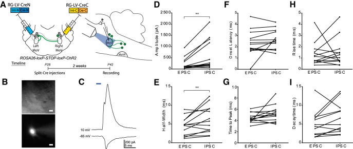
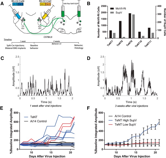
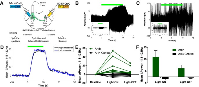
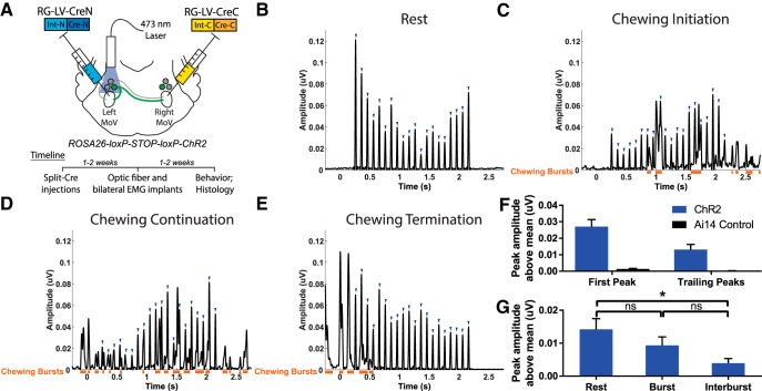
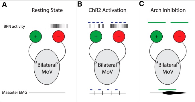
Anatomical studies have identified brainstem neurons that project bilaterally to left and right oromotor pools, which could potentially mediate bilateral muscle coordination. We use retrograde lentiviruses combined with a split-intein-mediated split-Cre-recombinase system in mice to isolate, characterize, and manipulate a population of neurons projecting to both the left and right jaw-closing trigeminal motoneurons. We find that these bilaterally projecting premotor neurons (BPNs) reside primarily in the supratrigeminal nucleus (SupV) and the parvicellular and intermediate reticular regions dorsal to the facial motor nucleus. These BPNs also project to multiple midbrain and brainstem targets implicated in orofacial sensorimotor control, and consist of a mix of glutamatergic, GABAergic, and glycinergic neurons, which can drive both excitatory and inhibitory inputs to trigeminal motoneurons when optogenetically activated in slice. Silencing BPNs with tetanus toxin light chain (TeNT) increases bilateral masseter activation during chewing, an effect driven by the expression of TeNT in SupV BPNs. Acute unilateral optogenetic inhibition of SupV BPNs identifies a group of tonically active neurons that function to lower masseter muscle tone, whereas unilateral optogenetic activation of SupV BPNs is sufficient to induce bilateral masseter activation both during resting state and during chewing. These results provide evidence for SupV BPNs in tonically modulating jaw-closing muscle tone and in mediating bilateral jaw closing.

Significance statement: We developed a method that combines retrograde lentiviruses with the split-intein-split-Cre system in mice to isolate, characterize, and manipulate neurons that project to both left and right jaw-closing motoneurons. We show that these bilaterally projecting premotor neurons (BPNs) reside primarily in the supratrigeminal nucleus and the rostral parvicellular and intermediate reticular nuclei. BPNs consist of both excitatory and inhibitory populations, and also project to multiple brainstem nuclei implicated in orofacial sensorimotor control. Manipulation of the supratrigeminal BPNs during natural jaw-closing behavior reveals a dual role for these neurons in eliciting phasic muscle activation and in maintaining basal muscle tone. The retrograde lentivirus carrying the split-intein-split-Cre system can be applied to study any neurons with bifurcating axons innervating two brain regions.

Keywords: bilaterally projecting neurons; optogenetics; retrograde lentivirus; split-intein-split-Cre system; supratrigeminal nucleus; trigeminal motor nucleus.


Figures

Figure 1.
Figure 1.
Labeling of BPNs innervating both left and right trigeminal motor nucleus with retrograde lentiviruses expressing split-Cre recombinase. A, RG-LV expressing either Cre-N-intein-N or intein-C-Cre-C was injected into the left or right trigeminal MoV, which resulted in the expression of full-length Cre-recombinase selectively in neurons projecting bilaterally to both MoV nuclei. B, Injection of only the retrograde lentivirus expressing Cre-N-intein-N into the MoV did not result in any labeled neurons in the brainstem. Similar results were found after injection of only the retrograde lentivirus expressing Cre-C-intein-C into the MoV. C, D, When these viruses were injected into an Ai14 Cre-reporter line, BPNs were identified primarily in the supratrigeminal nucleus dorsal to MoV (C), and the reticular nucleus dorsal to the facial motor nucleus (D). EI, Axons of labeled BPNs were also observed projecting bilaterally in the peri-trigeminal region (PeriV; D), the central MoVII (E), the MoXII and caudal reticular regions (IRt, PCRt, Gi; F), the vestibular nucleus (G), the oral pontine reticular formation (PnO) and reticulotegmental nucleus (RtTg; H), and the mesencephalic reticular formation (MRF), dorsal raphé (DRN), and lateral periaqueductal gray (lPAG; I). Scale bar, 100 μm.
Figure 2.
Figure 2.
Neurotransmitter phenotypes of SupV and Rt-MoVII BPNs. A–F, Representative images of in situ hybridization (red) analyses of BPNs labeled with the split-Cre retrograde lentivirus (green). BPNs in Rt-MoVII and SupV consisted of GABAergic (A, D), glycinergic (B, E), and glutamatergic neurons (C, F). G, The distributions of neuronal subtypes were similar between Rt-MoVII and SupV. In both cases, the majority of neurons were glutamatergic, while approximately equal proportions of GABAergic and glycinergic neurons were identified. N = 8 sections of each region per probe from four mice. All values shown are ±SEM. Scale bar, 100 μm.
Figure 3.
Figure 3.
Characterization of trigeminal motoneuron responses to ChR2-assisted stimulation of BPNs in acute slices. A, We expressed ChR2 in BPNs by injecting RG-LV-Cre-N and RG-LV-Cre-C into the left and right trigeminal motor nucleus, respectively, in 4-week-old Ai32 mice. Sagittal slices (350 μm) were collected for whole-cell patch-clamp physiology. B, Cell fill showing typical MoV motoneuron morphology. Scale bar, 20 μm. C, Representative traces of excitatory and inhibitory currents from an MoV motoneuron to a 2 ms full-field blue light stimulation of BPNs. MoV motoneuron inhibitory currents showed a significantly larger peak amplitude (D) and half-width (E) than excitatory currents. FI, There was no significant difference between inhibitory and excitatory currents in onset latency (F), time to peak (G), rise time (H), or decay time (I). **p < 0.02. N = 13 cells from six mice.
Figure 4.
Figure 4.
TeNT-mediated chronic silencing of BPN outputs induces bilateral increases in masseter muscle activation amplitude. A, RG-LV-Cre-N and RG-LV-Cre-C were injected into the left and right trigeminal motor nucleus, respectively, to express Cre in bilaterally projecting neurons. Bilateral injections of a Cre-dependent TeNT were targeted for infection of neurons in both Rt-MoVII and SupV. Immediately following injections, EMG implants were inserted into the left and right masseter. B, Variations in the numbers of TeNT-infected BPNs in Rt-MoVII and SupV across all five mice. CE, Representative results of masseter EMG during chewing revealed that both burst amplitude and integrated amplitude increased between 1 week (C) and 3 weeks (D) after viral infection, and this increase occurred to a varying extent across experimental mice (E, red lines indicate left and right masseter integrated amplitude values for TeNT11). F, A median split based on the numbers of TeNT-infected SupV BPNs revealed that only mice with higher numbers of TeNT-expressing SupV BPNs had increased masseter amplitude. All values shown are ±SEM.
Figure 5.
Figure 5.
Acute optogenetic inactivation of SupV BPNs reveals a tonic inhibitory function in setting bilateral muscle tone. A, We expressed Arch in BPNs by injecting RG-LV-Cre-N and RG-LV-Cre-C into the left and right trigeminal motor nucleus, respectively, in Ai35 mice. An optic fiber was implanted dorsal to SupV unilaterally, and EMG electrodes were implanted bilaterally into the left and right masseter muscles. BE, Ten second Arch-mediated silencing of SupV BPNs resulted in an increase in masseter muscle tone both during quiet (B) and active (C) EMG epochs in Ai35 mice (n = 5) versus control Ai14 mice (n = 4). This effect occurred bilaterally in the 118–122 Hz band (D) in all experimental mice (E). F, An aftereffect of increased bilateral muscle tone was also observed post-optogenetic silencing of SupV BPNs, which slowly diminished. All values shown are ±SEM.
Figure 6.
Figure 6.
Acute unilateral optogenetic activation of SupV BPNs is sufficient to induce bilateral masseter activation. A, We expressed ChR2 in BPNs by injecting RG-LV-Cre-N and RG-LV-Cre-C into the left and right trigeminal motor nucleus, respectively, in Ai32 mice. An optic fiber was implanted dorsal to SupV unilaterally, and EMG electrodes were implanted bilaterally into the left and right masseter muscles. BE, Twenty 20 ms pulses of blue light administered at a frequency of 10 Hz induced reliable time-locked bilateral masseter activations (blue arrowheads) in quiet EMG epochs (B), and during the initiation (C), continuation (D), and termination (E) of chewing behavior. Orange bars denote spontaneous jaw-closing bursts. All sample traces are from the right masseter of the same mouse. F, Light stimulation induced EMG amplitudes in Ai32 mice (n = 5) were significantly greater than that observed in control Ai14 mice (n = 4). The first peak was approximately twice the size of trailing peaks across all mice. G, Average trailing peak amplitudes induced by light during resting state, naturally occurring jaw bursts, or during the interburst period. *p < 0.05; ns, not statistically significant. All values shown are ±SEM.
Figure 7.
Figure 7.
A simple model for SupV BPN functions in regulating masseter motoneuron activity. A, During the resting state, excitatory SupV BPNs are inactive while inhibitory neurons are tonically active to maintain a balanced low level masseter muscle tone bilaterally. B, During ChR2-mediated activation, excitatory SupV BPNs are activated to induce phasic bilateral masseter EMG responses, whereas inhibitory SupV BPNs remain active to balance muscle tone. C, During Arch-mediated inhibition, excitatory SupV BPNs remain inactive, whereas inhibitory SupV BPNs are silenced, thereby disinhibiting MoV motoneurons resulting in an increase in bilateral masseter muscle tone.

Similar articles

Cited by

References

    1. Balasubramaniam R, Ram S. Orofacial movement disorders. Oral Maxillofac Surg Clin North Am. 2008;20:273–285. vii. - PubMed
    1. Boeve BF. REM sleep behavior disorder. Ann N Y Acad Sci. 2010;1184:15–54. doi: 10.1111/j.1749-6632.2009.05115.x. - DOI - PMC - PubMed
    1. Bourque MJ, Kolta A. Properties and interconnections of trigeminal interneurons of the lateral pontine reticular formation in the rat. J Neurophysiol. 2001;86:2583–2596. - PubMed
    1. Brooks PL, Peever JH. Glycinergic and GABA(A)-mediated inhibition of somatic motoneurons does not mediate rapid eye movement sleep motor atonia. J Neurosci. 2008;28:3535–3545. doi: 10.1523/JNEUROSCI.5023-07.2008. - DOI - PMC - PubMed
    1. Burgess CR, Peever JH. A noradrenergic mechanism functions to couple motor behavior with arousal state. Curr Biol. 2013;23:1719–1725. doi: 10.1016/j.cub.2013.07.014. - DOI - PubMed

Publication types

MeSH terms

LinkOut - more resources