. 2016 Jul 20;36(29):7693-706.
doi: 10.1523/JNEUROSCI.0628-16.2016.

Activation of β-Glucocerebrosidase Reduces Pathological α-Synuclein and Restores Lysosomal Function in Parkinson's Patient Midbrain Neurons

Affiliations

Activation of β-Glucocerebrosidase Reduces Pathological α-Synuclein and Restores Lysosomal Function in Parkinson's Patient Midbrain Neurons

Joseph R Mazzulli et al. J Neurosci. .

Abstract

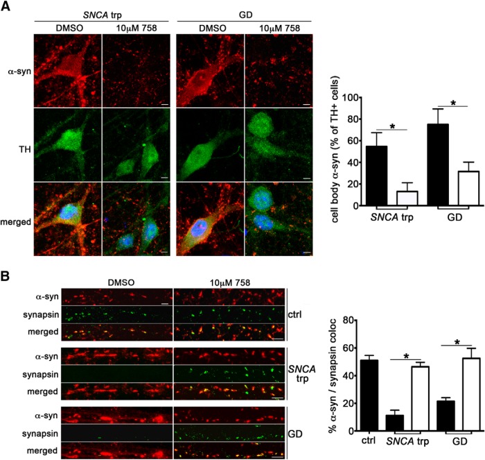
Parkinson's disease (PD) is characterized by the accumulation of α-synuclein (α-syn) within Lewy body inclusions in the nervous system. There are currently no disease-modifying therapies capable of reducing α-syn inclusions in PD. Recent data has indicated that loss-of-function mutations in the GBA1 gene that encodes lysosomal β-glucocerebrosidase (GCase) represent an important risk factor for PD, and can lead to α-syn accumulation. Here we use a small-molecule modulator of GCase to determine whether GCase activation within lysosomes can reduce α-syn levels and ameliorate downstream toxicity. Using induced pluripotent stem cell (iPSC)-derived human midbrain dopamine (DA) neurons from synucleinopathy patients with different PD-linked mutations, we find that a non-inhibitory small molecule modulator of GCase specifically enhanced activity within lysosomal compartments. This resulted in reduction of GCase substrates and clearance of pathological α-syn, regardless of the disease causing mutations. Importantly, the reduction of α-syn was sufficient to reverse downstream cellular pathologies induced by α-syn, including perturbations in hydrolase maturation and lysosomal dysfunction. These results indicate that enhancement of a single lysosomal hydrolase, GCase, can effectively reduce α-syn and provide therapeutic benefit in human midbrain neurons. This suggests that GCase activators may prove beneficial as treatments for PD and related synucleinopathies.

Significance statement: The presence of Lewy body inclusions comprised of fibrillar α-syn within affected regions of PD brain has been firmly documented, however no treatments exist that are capable of clearing Lewy bodies. Here, we used a mechanistic-based approach to examine the effect of GCase activation on α-syn clearance in human midbrain DA models that naturally accumulate α-syn through genetic mutations. Small molecule-mediated activation of GCase was effective at reducing α-syn inclusions in neurons, as well as associated downstream toxicity, demonstrating a therapeutic effect. Our work provides an example of how human iPSC-derived midbrain models could be used for testing potential treatments for neurodegenerative disorders, and identifies GCase as a critical therapeutic convergence point for a wide range of synucleinopathies.

Keywords: Parkinson's disease; glucocerebrosidase; induced pluripotent stem cells; lysosomes; synucleinopathy; α-synuclein.


Figures

Figure 1.
Figure 1.
Small molecule modulator 758 enhances GCase activity within lysosomal compartments and reduces α-syn. Inducible H4 cells that express α-syn under the control of a tetracycline-responsive promoter (tet-off) were treated with 10 μm 758 and harvested 3 d later. A, Lysates from H4 α-syn cells treated with DOX for 48 h to turn off α-syn expression were analyzed by Western blot. GAPDH was used as a loading control. Right, Quantification of α-syn levels with the C20 antibody normalized to GAPDH. Values are expressed as fold-change compared with +DOX (n = 3). B, Lysosomal GCase activity was measured in living H4 cells treated with DOX for 48 h by quantifying the degradation of PFB-FDGlu over 3 h. Activity within lysosomal compartments was achieved by measuring the response to baf A1 (n = 4). C, Treatment of 10 μm 758 for 3 d elevates the lysosomal GCase activity but does not alter non-lysosomal activity. GCase activity in H4 cells was quantified by measuring fluorescence upon the cleavage of PFB-FD-Glu substrate over time. RFU, relative fluorescent units. Parallel cultures were treated with baf A1 to obtain lysosome and non-lysosomal activity. D, Quantification and statistical analysis of total, lysosomal, and non-lysosomal activities from the kinetic data obtained in C. E, Triton-soluble lysates from 758-treated H4 cells were analyzed by Western blot for α-syn using either C20, syn303, or LB509 antibodies. Molecular weight marker is indicated in kilodaltons. Right, Quantification of α-syn using C20 normalized to GAPDH (n = 3). For all quantifications, values are the mean ± SEM. *p < 0.05, ***p < 0.001.
Figure 2.
Figure 2.
758 directly enhances GCase activity within lysosomes and reduces lipid substrates in human midbrain synucleinopathy models. A, 758 was added to whole-cell lysates extracted from day 90 SNCA trp neurons and activity was measured by the cleavage of 4-MU-Gluc (n = 3). B, SNCA trp neurons were incubated for 8 d with 758, washed, and whole-cell lysates were used for activity analysis with 4-MU-Gluc (n = 4). CBE was added in parallel samples to distinguish between GBA1 (lysosomal) and GBA2 (cytosolic)-derived hydrolysis of 4-MU-Gluc. C, Lysosomal GCase activity was measured in living neurons at day 120 using PFB-FD-Glu through quantifying the response to baf A1 (AUC obtained from a kinetic activity assay; n = 4). D, Measurement of GluCer from lysosome-enriched fractions or hexosylsphingosine (HexSphing) from whole-cell lysates obtained from control and SNCA trp d200 neurons treated for 14 d with DMSO or 758. Values were normalized to inorganic phosphate (Pi; n = 4). For all quantifications, values are the mean ± SEM. *p < 0.05.
Figure 3.
Figure 3.
Characterization of PARK9 iPSC and differentiation into midbrain DA neurons. Fibroblasts from a patient harboring PARK9 mutations (L1059R/c.3176T>C; L1085WfsX1088/c.3253delC) were reprogrammed into iPSCs by retroviral expression of OCT4, SOX2, cMYC, and KLF4. A, Immunofluorescence analysis of pluripotency markers of PARK9 iPSCs. B, G-banding karyotype analysis of PARK9 iPSCs demonstrates normal male genomic phenotype. C, Hematoxylin and eosin-stained sections of teratomas demonstrate pluripotency of PARK9 iPSCs through differentiation into mesoderm-, endoderm-, and ectoderm-derived structures. D, Western blot analysis of PARK9 iPSC differentiated into midbrain DA neurons at day 90 shows expression of TH and synaptophysin. Coomassie brilliant blue (CBB) is shown as a loading control (n = 3). Analysis of 3 different samples is shown. E, Immunostaining analysis of FOXA2 (red) and TH (green) of PARK9 midbrain neurons at day 60. Scale bar, 10 μm. Right, Quantification of neurons expressing both TH and FOXA2, compared with previously established lines from healthy controls (Mazzulli et al., 2016). F, Sequential extraction/Western blot analysis of α-syn using antibody C20. Day 90 neuronal lysates from three different healthy controls or two PARK9 clones were separated into Triton-soluble (T-sol) and -insoluble (T-insol) fractions. Loading controls include neural-specific β-iii-Tubulin, GAPDH, or vimentin (vim). G, GCase activity from whole-cell extracts using 4-MU-Gluc, obtained from PARK9 neurons at day 90 (n = 4). For all quantifications, values are the mean ± SEM. *p < 0.05.
Figure 4.
Figure 4.
Reduction of α-syn by 758 in human midbrain synucleinopathy models. SNCA trp (A) or GD (N370S/ c.84dupG; B) patient neurons at day 120 were incubated with 0, 5, or 10 μm 758 for 8 d, extracted in 1% Triton X-100 buffer, and analyzed by Western blot for α-syn (antibodies syn211, C20, or LB509). Bottom, Quantification of α-syn normalized to loading control β-iii-tubulin using syn211 (n = 4). C, Western blot analysis and quantification of α-syn from day 90 neuronal lysates of PARK9 or iPD patients (n = 4). D, Western blot and quantification of α-syn from A53T α-syn midbrain neurons (n = 3). For all quantifications, values are the mean ± SEM. *p < 0.05. Molecular weight marker is indicated in kilodaltons.
Figure 5.
Figure 5.
Reduction of cell body accumulation of α-syn and restoration of synaptic localization by 758. Human midbrain neurons from SNCA trp and GD patients at day 120 were treated with 758 for 8 d, fixed, and analyzed by immunofluorescence. A, Cell body accumulation of α-syn detected with LB509 (red) and dopaminergic neurons were detected by TH staining (green). Nuclei were detected with DAPI (blue). Scale bar, 5 μm. Right, Quantification of TH-positive cells that contained α-syn in the cell body (n = 6). B, Analysis of synaptic α-syn (LB509, red) in neurites by colocalization with a synaptic marker, synapsin (green). Scale bar, 5 μm. Right, Quantification of α-syn puncta that colocalized with synapsin (percentage of total puncta calculated per field-of-view, n = 6). For each quantification, values are the mean ± SEM. *p < 0.05.
Figure 6.
Figure 6.
Reduction of pathological α-syn aggregates in human synucleinopathy midbrain models by 758. A, Immunofluorescence analysis of α-syn using antibody LB509 (red) in SNCA trp and GD neurons treated with 10 μm 758 for 8 d. Amyloidogenic structures were detected with Thio S (green), and nuclei (DAPI) are shown in blue. 758 (10 μm) reduced Thio S staining and restored the normal punctated pattern of α-syn in SNCA trp and GD neurons at day 120. Scale bar, 10 μm. Bottom, Quantification of cells and neurites containing α-syn/Thio S accumulations (n = 4, *p < 0.05). Both type I (diffuse cytoplasmic inclusions) and type II (punctated juxtanuclear) inclusions were quantified. B, Pathological analysis of α-syn was done as described in A in A53T patient neurons at day 90. Scale bars, 5 μm. For all quantifications, values are the mean ± SEM. *p < 0.05.
Figure 7.
Figure 7.
758-mediated reduction of α-syn reduces downstream cellular pathology associated with α-syn accumulation. H4 neuroglioma cells were treated with 5 μm 758 for 5 d and analyzed for changes in protein maturation and trafficking. A, H4 cells were fixed and immunostained for GCase (8E4 antibody, green) and LAMP2 (red) to detect colocalization with lysosomes. Right, Quantification of colocalization (n = 6). B, ER accumulation of GCase by α-syn documented by immunostaining for ER resident, PDI (red), is reversed by DOX or 758 treatment. Right, Quantification of colocalization (n = 6). Scale bars, 10 μm. Nuclei were detected with DAPI (blue). C, Analysis of protein maturation by Endo H treatment followed by Western blot for GCase (8E4) or nicastrin. GAPDH was used as a loading control. Molecular weight marker is indicated in kilodaltons. Right, Quantification of Endo H resistance of GCase or nicastrin. Values are the mean ± SEM. ***p < 0.001, **p < 0.01, *p < 0.05.
Figure 8.
Figure 8.
758 improves protein maturation and lysosomal function in human synucleinopathy midbrain models. AC, Analysis of GCase maturation by Western blot. SNCA trp, GD, PARK9 mutant or iPD neurons at day 120 were treated with 0, 5, and 10 μm 758 for 8 d. GAPDH was used as a loading control. Quantification of post ER–ER ratio is shown below (n = 3, *p < 0.05). D, E, Analysis of Hex B maturation by Western blot of SNCA trp or GD midbrain neurons treated with 10 μm 758 for 8 d. Quantification of mature–immature ratio is shown below (n = 3, *p < 0.05). Molecular weight marker is indicated in kilodaltons. F, Measurement of live-cell lysosomal activity of hex and β-Gal in day 120 SNCA trp and GD neurons (n = 4, *p < 0.05). G, Measurement of proteolysis rate by quantification of acid-soluble amino acids (AA's) excreted in the media over time in living SNCA trp neurons treated with 10 μm 758 (n = 4, *p < 0.05). H, Neurite quantification by neurofilament immunofluorescence intensity in SNCA trp neurons treated with 10 μm 758 for 14 d, normalized to total cell volume (n = 9, *p < 0.05). For all quantifications, values are the mean ± SEM.

Similar articles

Cited by

References

    1. Aflaki E, Stubblefield BK, Maniwang E, Lopez G, Moaven N, Goldin E, Marugan J, Patnaik S, Dutra A, Southall N, Zheng W, Tayebi N, Sidransky E. Macrophage models of Gaucher disease for evaluating disease pathogenesis and candidate drugs. Sci Transl Med. 2014;6:240ra73. doi: 10.1126/scitranslmed.3008659. - DOI - PMC - PubMed
    1. Alcalay RN, Levy OA, Waters CC, Fahn S, Ford B, Kuo SH, Mazzoni P, Pauciulo MW, Nichols WC, Gan-Or Z, Rouleau GA, Chung WK, Wolf P, Oliva P, Keutzer J, Marder K, Zhang X. Glucocerebrosidase activity in Parkinson's disease with and without GBA mutations. Brain. 2015;138:2648–2658. doi: 10.1093/brain/awv179. - DOI - PMC - PubMed
    1. Baba M, Nakajo S, Tu PH, Tomita T, Nakaya K, Lee VM, Trojanowski JQ, Iwatsubo T. Aggregation of alpha-synuclein in Lewy bodies of sporadic Parkinson's disease and dementia with Lewy bodies. Am J Pathol. 1998;152:879–884. - PMC - PubMed
    1. Bae EJ, Yang NY, Lee C, Lee HJ, Kim S, Sardi SP, Lee SJ. Loss of glucocerebrosidase 1 activity causes lysosomal dysfunction and alpha-synuclein aggregation. Exp Mol Med. 2015;47:e153. doi: 10.1038/emm.2014.128. - DOI - PMC - PubMed
    1. Bergmann JE, Grabowski GA. Posttranslational processing of human lysosomal acid beta-glucosidase: a continuum of defects in Gaucher disease type 1 and type 2 fibroblasts. Am J Hum Genet. 1989;44:741–750. - PMC - PubMed

MeSH terms