. 2016 Jul 20;36(29):7740-9.
doi: 10.1523/JNEUROSCI.0554-16.2016.

The Severity of Infection Determines the Localization of Damage and Extent of Sensorineural Hearing Loss in Experimental Pneumococcal Meningitis

Affiliations

The Severity of Infection Determines the Localization of Damage and Extent of Sensorineural Hearing Loss in Experimental Pneumococcal Meningitis

Michael Perny et al. J Neurosci. .

Abstract

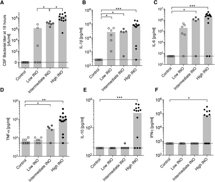
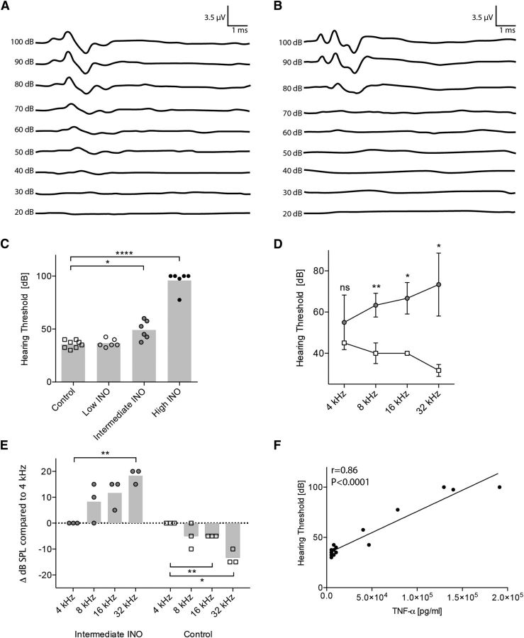
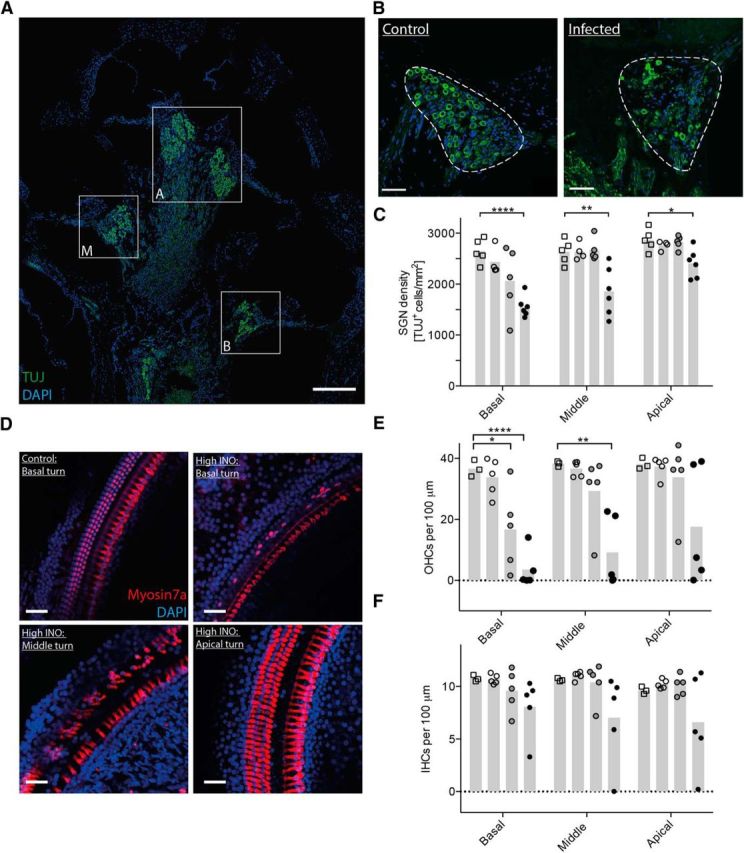
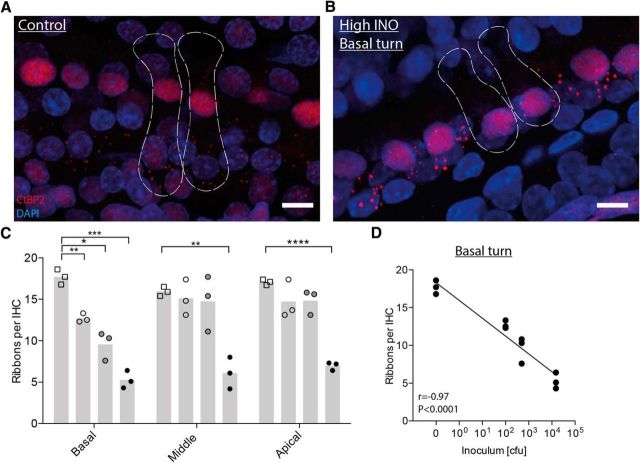
Hearing loss is an important sequela of pneumococcal meningitis (PM), occurring in up to 30% of survivors. The role of the severity of infection on hearing function and pathomorphological consequences in the cochlea secondary to PM have not been investigated to date. Using a well-established model of PM, we systematically investigated the functional hearing outcome and the long-term fate of neurosensory cells in the cochlea, i.e., hair cells and spiral ganglion neurons (SGNs), with a focus on their tonotopic distribution. Intracisternal infection of infant rats with increasing inocula of Streptococcus pneumoniae resulted in a dose-dependent increase in CSF levels of interleukin-1β, interleukin-6, tumor necrosis factor α, interleukin-10, and interferon-γ in acute disease. The severity of long-term hearing loss at 3 weeks after infection, measured by auditory brainstem response recordings, correlated to the initial inoculum dose and to the levels of proinflammatory cytokines determined in the acute phase of PM. Quantitative cochlear histomorphology revealed a significant loss of SGNs and outer hair cells that strongly correlated to the level of infection, with the most severe damage occurring in the basal part of the cochlea. Inner hair cells (IHCs) were not significantly affected throughout the entire cochlea. However, surviving IHCs lost synaptic connectivity to remaining SGNs in all cochlear regions. These findings provide evidence that the inoculum concentration, i.e., severity of infection, is the major determinant of long-term morphological cell pathologies in the cochlea and functional hearing loss.

Significance statement: Hearing loss is a neurofunctional deficit occurring in up to 30% of patients surviving pneumococcal meningitis (PM). Here, we analyze the correlation between the severity of infection and the inflammatory response in the CSF, the tonotopic distribution of neurosensory pathologies in the cochlea, and the long-term hearing function in a rat model of pneumococcal meningitis. Our study identifies the severity of infection as the key determinant of long-term hearing loss, underlining the importance of the prompt institution of antibiotic therapy in patients suffering from PM. Furthermore, our findings reveal in detail the spatial loss of cochlear neurosensory cells, providing new insights into the pathogenesis of meningitis-associated hearing loss that reveal new starting points for the development of otoprotective therapies.

Keywords: animal model; hair cells; pneumococcal meningitis; sensorineural hearing loss; spiral ganglion neurons; streptococcus pneumonia.


Figures

Figure 1.
Figure 1.
Dose-dependent bacterial growth and cytokine profile in the CSF 18 h after infection with S. pneumoniae. A, Dose-dependent growth of S. pneumoniae in the CSF determined at 18 h after infection. B–F, The levels of IL-1β (B), IL-6 (C), TNF-α (D), IL-10 (E), and IFN-γ (F) were measured in the CSF 18 h after infecting the animals intracisternally with three different loads of S. pneumoniae (n = 5–13 animals per group). The means are indicated in the bar graphs. The dashed lines indicate the detection limit for each cytokine. An unpaired t test with Welch's correction was used for single comparisons. *p < 0.05; **p < 0.01; ***p < 0.001.
Figure 2.
Figure 2.
Increased hearing thresholds 3 weeks after infection with S. pneumoniae. A, B, The representative figures show the hearing threshold of a control animal (A) of ∼40 dB and an infected animal (B) with a hearing threshold around 80 dB. C, Increasing inoculum concentrations elevated hearing thresholds in a dose-dependent way. ABR recordings were performed in n = 6–8 animals per group. The means are indicated by the bar graph. An unpaired t test with Welch's correction was used for single comparisons. D, Frequency-dependent hearing thresholds (controls, empty squares; intermediate INO, gray circles; n = 3 animals per group). The mean ± SD is shown. An unpaired t test with Welch's correction was used for single comparisons. E, Frequency-dependent threshold shifts. Data are normalized for each animal individually (threshold at 4 kHz set to 0). A one-sample t test was used for single comparisons. F, Spearman's correlation between TNF-α levels in the CSF during the acute phase and the hearing thresholds in the late phase of disease (3 weeks after infection). *p < 0.05; **p < 0.01; ****p < 0.0001.
Figure 3.
Figure 3.
Immunohistological analysis of spiral ganglion neurons and hair cells 3 weeks after infection. A, Midmodiolar cross section showing the basal (B), middle (M), and apical (A) turns of a control rat immunostained for neurons (TUJ, green) and cell nuclei (DAPI, blue). B, Representative immunostainings of the Rosenthal's canal showing the spiral ganglion (dashed line) in the basal turn of a control animal and an infected animal. C, Neuronal density quantification in the basal, middle, and apical turns in control and infected animals (controls, empty squares; low INO, empty circles; intermediate INO, gray circles; high INO, black circles; n = 4–6 animals per group). The mean is indicated in the bar graph. An unpaired t test with Welch's correction was used for single comparisons. D, Representative confocal images of the Organ of Corti immunostained for hair cells (Myosin7a, red) and cell nuclei (DAPI, blue). All four pictures were made from the same animal. E, F, Quantification of OHCs (E) and IHCs (F) in control and infected animals (controls, empty squares; low INO, empty circles; intermediate INO, gray circles; high INO, black circles; n = 3–5 animals per group). The means are indicated in the bar graphs. An unpaired t test with Welch's correction was used for single comparisons. Scale bars: A, 200 μm; B, D, 50 μm. *p < 0.05; **p < 0.01; ****p < 0.0001.
Figure 4.
Figure 4.
Confocal microscopy analysis of IHC presynaptic ribbons 3 weeks after infection. A, B, Representative confocal images (maximal projection from a focal series) of IHC synapses from the basal turn of a control (A) and an infected (high INO; B) cochlea 3 weeks after infection, immunolabeled for presynaptic ribbons (CtBP2, red) and nuclei (DAPI, blue). The dashed lines show the approximate outlines of two IHCs, determined by immunolabeling for hair cells (Myosin7a, yellow; data not shown). C, Quantification of presynaptic ribbons per IHC for the three cochlear regions in control and infected animals (controls, empty squares; low INO, empty circles; intermediate INO, gray circles; high INO, black circles; n = 3 per group). The means are indicated in the bar graphs. An unpaired t test with Welch's correction was used for single comparisons. D, Spearman's correlation between the number of ribbons per IHC and the initial inoculum load in the basal turn. Scale bars: 10 μm. *p < 0.05; **p < 0.01; ***p < 0.001; ****p < 0.0001.

Similar articles

Cited by

References

    1. Aminpour S, Tinling SP, Brodie HA. Role of tumor necrosis factor-alpha in sensorineural hearing loss after bacterial meningitis. Otol Neurotol. 2005;26:602–609. doi: 10.1097/01.mao.0000178121.28365.0d. - DOI - PubMed
    1. Beurg M, Hafidi A, Skinner L, Cowan G, Hondarrague Y, Mitchell TJ, Dulon D. The mechanism of pneumolysin-induced cochlear hair cell death in the rat. J Physiol. 2005;568:211–227. doi: 10.1113/jphysiol.2005.092478. - DOI - PMC - PubMed
    1. Brand Y, Senn P, Kompis M, Dillier N, Allum JH. Cochlear implantation in children and adults in Switzerland. Swiss Med Wkly. 2014;144:w13909. - PubMed
    1. Brandt CT, Cayé-Thomasen P, Lund SP, Worsøe L, Ostergaard C, Frimodt-Møller N, Espersen F, Thomsen J, Lundgren JD. Hearing loss and cochlear damage in experimental pneumococcal meningitis, with special reference to the role of neutrophil granulocytes. Neurobiol Dis. 2006;23:300–311. doi: 10.1016/j.nbd.2006.03.006. - DOI - PubMed
    1. Brown DL, Desai KK, Vakili BA, Nouneh C, Lee HM, Golub LM. Clinical and biochemical results of the metalloproteinase inhibition with subantimicrobial doses of doxycycline to prevent acute coronary syndromes (MIDAS) pilot trial. Arterioscler Thromb Vasc Biol. 2004;24:733–738. doi: 10.1161/01.ATV.0000121571.78696.dc. - DOI - PubMed

MeSH terms