. 2016 Jul 20;36(29):7768-78.
doi: 10.1523/JNEUROSCI.0154-16.2016.

Ethanol-Induced Motor Impairment Mediated by Inhibition of α7 Nicotinic Receptors

Affiliations

Ethanol-Induced Motor Impairment Mediated by Inhibition of α7 Nicotinic Receptors

John McDaid et al. J Neurosci. .

Abstract

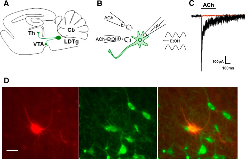
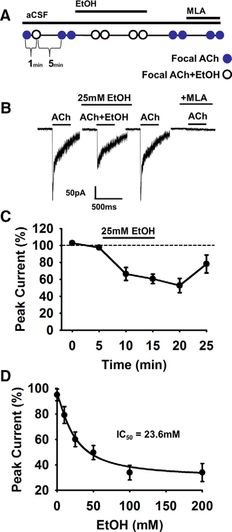
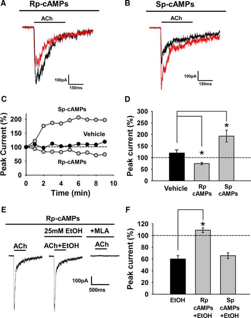
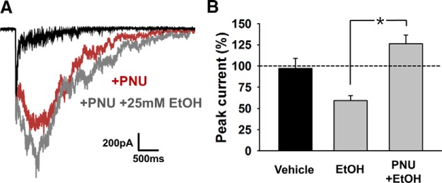
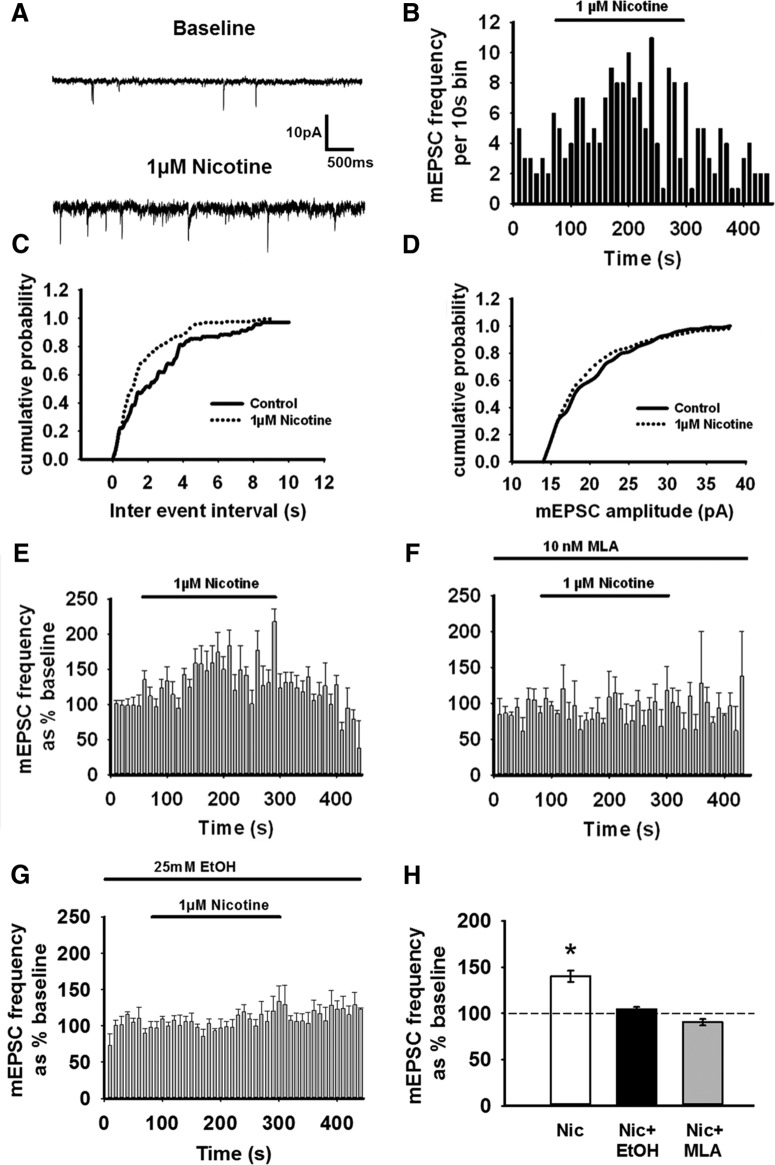
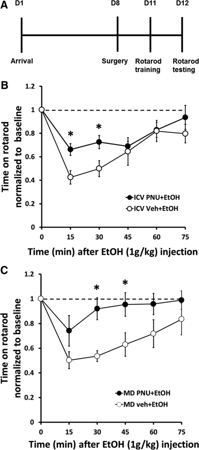
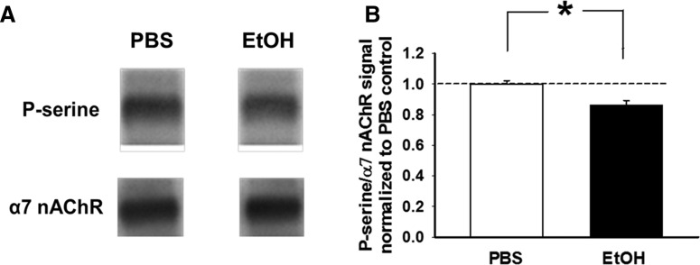
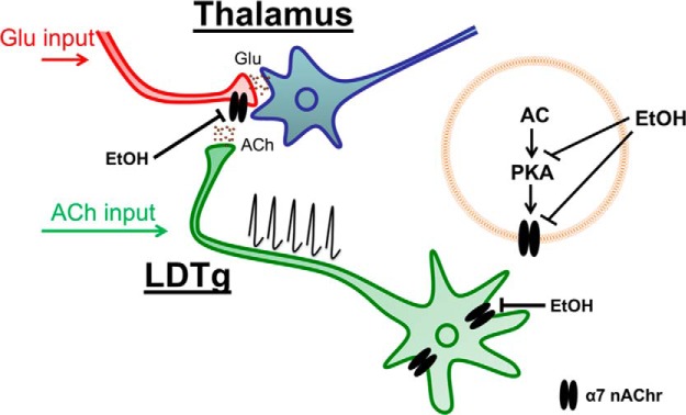
Nicotine and ethanol (EtOH) are among the most widely co-abused substances, and nicotinic acetylcholine receptors (nAChRs) contribute to the behavioral effects of both drugs. Along with their role in addiction, nAChRs also contribute to motor control circuitry. The α7 nAChR subtype is highly expressed in the laterodorsal tegmental nucleus (LDTg), a brainstem cholinergic center that contributes to motor performance through its projections to thalamic motor relay centers, including the mediodorsal thalamus. We demonstrate that EtOH concentrations just above the legal limits for intoxication in humans can inhibit α7 nAChRs in LDTg neurons from rats. This EtOH-induced inhibition is mediated by a decrease in cAMP/PKA signaling. The α7 nAChR-positive allosteric modulator PNU120596 [N-(5-chloro-2,4-dimethoxyphenyl)-N'-(5-methyl-3-isoxazolyl)-urea], which interferes with receptor desensitization, completely eliminated EtOH modulation of these receptors. These data suggest that EtOH inhibits α7 responses through a PKA-dependent enhancement of receptor desensitization. EtOH also inhibited the effects of nicotine at presynaptic α7 nAChRs on glutamate terminals in the mediodorsal thalamus. In vivo administration of PNU120596 either into the cerebral ventricles or directly into the mediodorsal thalamus attenuated EtOH-induced motor impairment. Thus, α7 nAChRs are likely important mediators of the motor impairing effects of moderate EtOH consumption.

Significance statement: The motor-impairing effects of ethanol contribute to intoxication-related injury and death. Here we explore the cellular and neural circuit mechanisms underlying ethanol-induced motor impairment. Physiologically relevant concentrations of ethanol inhibit activity of a nicotinic receptor subtype that is expressed in brain areas associated with motor control. That receptor inhibition is mediated by decreased receptor phosphorylation, suggesting an indirect modulation of cell signaling pathways to achieve the physiological effects.

Keywords: aversion; ethanol; motor impairment; nicotine; protein kinase A; rotarod.


Figures

Figure 1.
Figure 1.
Cholinergic neurons in the LDTg express α7 nAChRs. A, Parasagittal view of the rat brain, illustrating LDTg cholinergic neurons that project to the thalamus and VTA. B, Experimental design consisting of two puffer pipettes, one containing ACh and the other a mixture of ACh and EtOH; focal application of ACh or ACh plus EtOH was performed in conjunction with bath application of EtOH. C, Sample trace of an inward current resulting from focal ACh application (1 mm, 300 ms, Vm = −70 mV, bar denotes application). This current was completely blocked by bath application of the selective α7 nAChR antagonist MLA (10 nm, red trace). D, The recorded cell was loaded with biocytin (0.1%, in red, left) and stained positive for ChAT (in green, middle); the composite image of the recorded cell shows costaining for biocytin and ChAT (in orange, right). Ten of 11 biocytin-labeled neurons, which expressed α7 nAChR current, were also positive for ChAT (scale bar, 25 μm).
Figure 2.
Figure 2.
EtOH dose dependently inhibits α7 nAChRs in LDTg neurons. A, Diagram of experimental protocol: focal application of ACh (300 ms) was followed by focal application of a combination of ACh plus EtOH. After 5 min, ACh was again focally applied to ensure a consistent response. EtOH was then bath applied for 10 min, and the responses to focal ACh plus EtOH were tested at 5 min intervals. This was followed by a washout period and then by bath application of the selective α7 nAChR antagonist MLA (10 nm) to confirm the nAChR subtype. During EtOH bath application, the response to ACh was tested using a pipette with ACh plus EtOH, to ensure that the EtOH concentration remained constant. Repeatability and consistency of the responses were ensured by using the same pressure line for both puffer pipettes and by pulling both pipettes from the same piece of glass to yield similar resistance. Baseline ACh current was the average of the last two currents obtained before bath application of EtOH. EtOH effects were the average of the last two ACh plus EtOH currents before washout. B, Representative α7 nAChR currents recorded from an LDTg neuron during focal application of ACh (1 mm) before, during, and after bath application of 25 mm EtOH. Note the reversal of the EtOH effect on washout. The ACh-evoked current was completely blocked by MLA (10 nm). C, Time course of the effects of bath application of 25 mm EtOH. D, Concentration–effect relationship for EtOH inhibition of peak α7 nAChR currents in LDTg neurons. The EtOH IC50 was 23.6 mm, with a Hill coefficient of −1.6. Vm = −70 mV for all recordings. n = 4, 7, 11, 15, 5, and 3 for 0, 10, 25, 50, 100, and 200 mm EtOH, respectively. ANOVA (F(5,44) = 7.7, p < 0.001), post hoc test with all concentrations compared with aCSF vehicle.
Figure 3.
Figure 3.
EtOH inhibition of α7 nAChR currents in LDTg neurons occurs via inhibition of the PKA pathway. A, Sample ACh (1 mm; 300 ms) responses to the first (black) and fifth (red) ACh applications administered at 1 min intervals with the PKA inhibitor Rp-cAMPs (100 μm) in the recording pipette. B, Sample ACh responses to the first (black) and fifth (red) ACh applications administered at 1 min intervals with the PKA activator Sp-cAMPs (1 mm) in the recording pipette. C, Representative time course of α7 currents with standard internal solution (Vehicle), Rp-cAMPs, or Sp-cAMPs in the recording pipette. Data from three different cells are illustrated. Current amplitudes were normalized to the first ACh response at the start of the recording. D, Summary data for peak current amplitudes normalized to baseline, illustrating that α7 currents were reduced by Rp-cAMPs and increased Sp-cAMPs treatment [ANOVA (F(2,20) = 19.4, post hoc tests), p = 0.03 and p = 0.004 for Rp-cAMPs and Sp-cAMPs vs vehicle control, respectively]. n = 7, 8, and 5 for vehicle, Rp-cAMPs, and Sp-cAMPs, respectively. E, Sample traces illustrating EtOH modulation of α7 currents is reduced by including the PKA inhibitor Rp-cAMPs (100 μm) in the recording electrode. The current was completely blocked by bath application of MLA (10 nm). F, Summary data showing the averaged response magnitudes normalized to baseline after 10 min of recording and immediately before EtOH application. Including the PKA inhibitor Rp-cAMPs (100 μm) in the recording electrode resulted in no inhibition of the α7 response by EtOH (25 mm), suggesting that inhibiting PKA activity occluded the EtOH inhibition of α7 (n = 5). Including the PKA activator Sp-cAMPs (1 mm) augmented the baseline current (see D), but application of EtOH inhibited α7 current amplitudes by a similar percentage to that seen with EtOH treatment alone (n = 5). n = 11, 5, and 5 for EtOH control, Rp-cAMPs, and Sp-cAMPs, respectively; ANOVA (F(2,20) = 18.9, post hoc tests), *p < 0.001 and p = 0.45 for Rp-cAMPs plus EtOH and Sp-cAMPs plus EtOH versus 25 mm EtOH, respectively.
Figure 4.
Figure 4.
Inhibiting α7 nAChR desensitization with PNU120596 blocks the inhibitory effects of EtOH on these receptors. A, Representative traces showing that PNU120596, a positive allosteric modulator of the α7 receptor, prolongs receptor activation by interfering with desensitization. Subsequent application of EtOH (25 mm) did not inhibit α7 currents. B, Summary data showing that PNU120596 significantly reduced the inhibitory effect of EtOH on the peak α7 currents. Data are normalized to baseline peak ACh-induced α7 responses. (EtOH alone vs EtOH with PNU, *p < 0.05).
Figure 5.
Figure 5.
EtOH attenuates nicotine-induced increases in glutamatergic transmission to MD thalamic neurons. A, Representative traces showing that bath application of nicotine (1 μm) increases the frequency, but not the amplitude, of miniature EPSCs recorded from neurons in the MD thalamus. B, Frequency histogram of mEPSCs from a single thalamic neuron recording. C, Cumulative mEPSC frequency distribution for the cell illustrated in A and B. Nicotine significantly increased the frequency of mEPSCs (p < 0.05 by K–S test). D, Cumulative mEPSC amplitude distribution for the cell illustrated in A and B. Nicotine did not affect the amplitude of mEPSCs (p > 0.05 by K–S test). E, Averaged mEPSC frequency histogram for all cells tested, normalized to baseline. Nicotine increased the frequency of mEPSCs (1 μm; p < 0.05). F, Averaged mEPSC frequency histogram for all cells tested, normalized to baseline, showing the response to nicotine in the presence of the selective α7 nAChR antagonist MLA (10 nm). MLA pretreatment blocked the nicotine-induced increase in mEPSC frequency. G, Averaged mEPSC frequency histogram for all cells tested, normalized to baseline, showing the response to nicotine in the presence of EtOH (25 mm). EtOH pretreatment blocked the nicotine-induced increase in mEPSC frequency. H, Summary data showing that both MLA (10 nm) and EtOH (25 mm) blocked the nicotine-induced increase in mEPSC frequency in MD thalamic neurons. Data are normalized to baseline mEPSC frequency before bath application of nicotine (Vm = −70 mV; *p < 0.05 relative to baseline).
Figure 6.
Figure 6.
EtOH-induced motor impairment is attenuated by intracerebroventricular or intra-MD thalamic injection of PNU120596. A, Diagram of the timeline for the behavioral studies. B, Animals previously trained on the accelerating rotarod were first tested for baseline performance with a set of four trials. The times to fall from the rod were averaged within and then between animals in each group to yield group mean performance values. The rats then received intracerebroventricular injections of either vehicle or PNU120596 (20 μl, 100 μm), followed 15 min later by an intraperitoneal injection of EtOH (1 g/kg) and five sets of four trials on the rotarod. Both vehicle- and PNU120596-treated rats showed marked motor impairment after EtOH administration, but PNU120596-treated animals showed less impairment compared with vehicle (*p < 0.05). The rate of performance recovery after EtOH treatment was similar between these groups. PNU120596 treatment in the absence of EtOH had no effect on motor performance (data not shown). C, Another group of animals was treated exactly the same as in B, but they received intra-MD thalamic injections of either vehicle or PNU120596 (1 μl, 100 μm). After EtOH injection(1 g/kg) both vehicle and PNU120596-treated rats showed marked motor impairment after administration of EtOH. However, PNU120596-treated animals showed significantly less impairment of rotarod performance compared with vehicle-treated rats (*p < 0.05). The rate of performance recovery after EtOH treatment was similar between these groups. PNU120596 treatment in the absence of EtOH had no effect on motor performance (data not shown).
Figure 7.
Figure 7.
EtOH decreases the phosphorylation of α7 nAChRs in the LDTg. A, Representative immunoblots show immunoreactive bands for phosphoserine (P-serine) residues and α7 nAChR subunits. LDTg tissue samples were collected 15 min after rats were pretreated with either EtOH (1.0 g/kg, i.p.) or PBS (intraperitoneally), homogenized, and subjected to immunoprecipitation with anti-α7 antibodies. The phosphorylation state of LDTg α7 nAChRs was calculated as the ratio of P-serine/α7 nAChR signal normalized to control (PBS). B, Bar graph illustrates that EtOH decreases the phosphorylation state of LDTg α7 nAChRs in vivo (*p < 0.01).
Figure 8.
Figure 8.
Summary diagram of the proposed circuitry. Cholinergic brainstem projections from LDTg to limbic areas, basal ganglia, and the thalamus contribute to many behaviors, including arousal, reward, and motor control. Our data support the idea that EtOH inhibits α7 nAChRs by modulating the PKA pathway. EtOH inhibition of presynaptic α7 nAChRs in the MD thalamus reduces excitatory drive by interfering with cholinergic modulation of excitatory synapses. We propose that this circuitry contributes to EtOH-induced motor impairment.

Similar articles

Cited by

References

    1. Adalsteinsson E, Sullivan EV, Mayer D, Pfefferbaum A. In vivo quantification of ethanol kinetics in rat brain. Neuropsychopharmacology. 2006;31:2683–2691. doi: 10.1038/sj.npp.1301023. - DOI - PubMed
    1. Aistrup GL, Marszalec W, Narahashi T. Ethanol modulation of nicotinic acetylcholine receptor currents in cultured cortical neurons. Mol Pharmacol. 1999;55:39–49. - PubMed
    1. Alderson HL, Latimer MP, Winn P. Involvement of the laterodorsal tegmental nucleus in the locomotor response to repeated nicotine administration. Neurosci Lett. 2005;380:335–339. doi: 10.1016/j.neulet.2005.01.067. - DOI - PubMed
    1. Al-Rejaie S, Dar MS. Antagonism of ethanol ataxia by intracerebellar nicotine: possible modulation by mouse cerebellar nitric oxide and cGMP. Brain Res Bull. 2006;69:187–196. doi: 10.1016/j.brainresbull.2005.12.002. - DOI - PubMed
    1. Azam L, Winzer-Serhan U, Leslie FM. Co-expression of alpha7 and beta2 nicotinic acetylcholine receptor subunit mRNAs within rat brain cholinergic neurons. Neuroscience. 2003;119:965–977. - PubMed

Publication types

MeSH terms