. 2016 Sep 2;291(36):19018-30.
doi: 10.1074/jbc.M116.746289. Epub 2016 Jul 21.

EspR-dependent ESAT-6 Protein Secretion of Mycobacterium tuberculosis Requires the Presence of Virulence Regulator PhoP

Affiliations

EspR-dependent ESAT-6 Protein Secretion of Mycobacterium tuberculosis Requires the Presence of Virulence Regulator PhoP

Vijjamarri Anil Kumar et al. J Biol Chem. .

Abstract

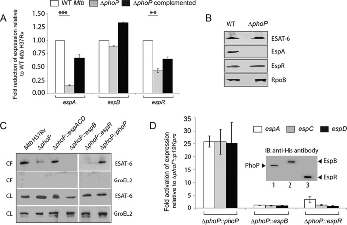
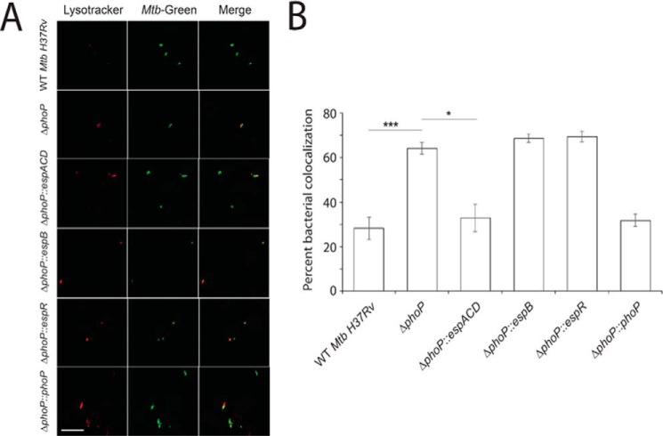
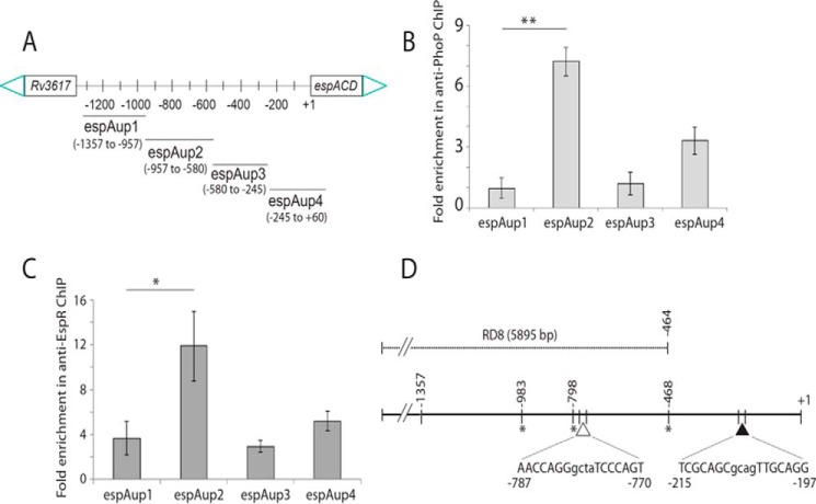
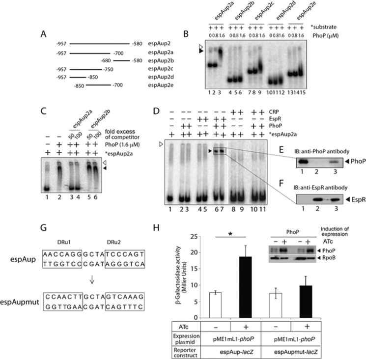
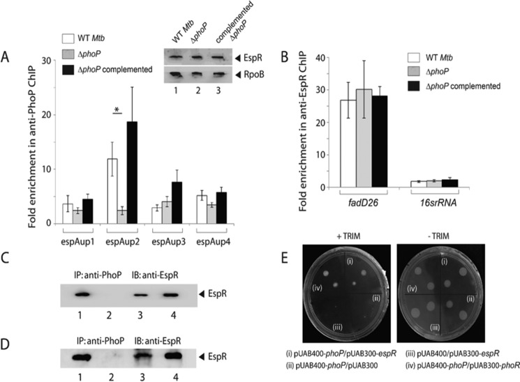
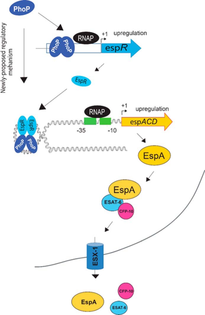
Attenuation of Mycobacterium bovis BCG strain is related to the loss of the RD1-encoded ESX-1 secretion system. The ESX-1 system secretes virulence factor ESAT-6 that plays a critical role in modulation of the host immune system, which is essential for establishment of a productive infection. Previous studies suggest that among the reasons for attenuation of Mycobacterium tuberculosis H37Ra is a mutation in the phoP gene that interferes with the ESX-1 secretion system and inhibits secretion of ESAT-6. Here, we identify a totally different and distinct regulatory mechanism involving PhoP and transcription regulator EspR on transcriptional control of the espACD operon, which is required for ESX-1-dependent ESAT-6 secretion. Although both of these regulators are capable of influencing espACD expression, we show that activation of espACD requires direct recruitment of both PhoP and EspR at the espACD promoter. The most fundamental insights are derived from the inhibition of EspR binding at the espACD regulatory region of the phoP mutant strain because of PhoP-EspR protein-protein interactions. Based on these results, a model is proposed suggesting how PhoP and EspR protein-protein interactions contribute to activation of espACD expression and, in turn, control ESAT-6 secretion, an essential pathogenic determinant of M. tuberculosis Together, these results have significant implications on the mechanism of virulence regulation of M. tuberculosis.

Keywords: DNA-protein interaction; Mycobacterium tuberculosis; gene regulation; microbial pathogenesis; protein secretion.


Figures

FIGURE 1.
FIGURE 1.
PhoP-mediated espACD activation restores ESX-1-dependent ESAT-6 secretion of M. tuberculosis. A, quantitative RT-PCR to examine expression levels of espA, espB, and espR in ΔphoP and ΔphoP-complemented strains relative to WT M. tuberculosis H37Rv. The results represent average values with standard deviations derived from at least three independent RNA preparations (*, p < 0.05; **, p < 0.01; ***, p < 0.001). B, immunoblot analyses of 20 μg of cell extracts of WT and ΔphoP using appropriate antibodies. RpoB was used as a loading control. C, immunoblot analyses of 20 μg of culture filtrates (CF) or cell lysates (CL) of indicated M. tuberculosis strains. αGroEL2 was used as a control to verify cytolysis of cells. Note that complementation of ΔphoP M. tuberculosis with phoP, espB, espR, and espACD was carried out using p19Kpro as the expression vector and compared with ΔphoP M. tuberculosis carrying p19Kpro as the empty vector control (see “Experimental Procedures”). D, expression of espA, espC, and espD in indicated mutant M. tuberculosisH37Rv strains relative to ΔphoP-p19kpro (empty vector control) as measured by RT-PCR analysis. Inset shows ectopic expression of proteins as detected by immunoblotting with anti-His antibody. IB, immunoblot.
FIGURE 2.
FIGURE 2.
PhoP-dependent espACD activation is required for inhibition of phagosome maturation by M. tuberculosis H37Rv. A, macrophages were infected with WT or indicated mutant strains of M. tuberculosis H37Rv for 3 h. The cells were stained with 300 nm LysoTracker for 3 h and then fixed in 4% paraformaldehyde. M. tuberculosis H37Rv strains were labeled with fluorescent phenolic auramine. The confocal images represent the merge of two fluorescence signals (LysoTracker, red; M. tuberculosis, green) and are shown at right. Scale bar, 10 μm. B, percent bacterial co-localization for indicated M. tuberculosisH37Rv strains. The results show the average values with standard error of mean (S.E.) of at least three independent experiments (*, p < 0.05; ***, p < 0.001).
FIGURE 3.
FIGURE 3.
Recruitment of PhoP and EspR at the espACD promoter. A, scheme showing organization of the espACD promoter divided into four fragments (espAup1–espAup4) to probe PhoP and EspR binding. Note that the promoter fragments espAup1–espAup4 span from −957 to −1357, −580 to −957, −245 to −580, and +60 to −245, respectively, relative to the start site of the ORF. To examine PhoP (B) and EspR (C) recruitment within the espACD promoter, ChIP-qPCR was carried out as described under the “Experimental Procedures.” Each data point represents mean of duplicate qPCR measurements using at least three independent M. tuberculosis cultures. Asterisk indicates a statistically significant difference in the ChIP enrichment level compared between espAup1 and espAup2 under the conditions examined (*, p < 0.05; **, p < 0.01). Note that the difference in the signal enrichment between PhoP and EspR antibodies may be attributable to affinity differences between proteins and/or respective antibodies. D, location of relevant protein-binding sites within espACD upstream regulatory region. Note that the EspR-binding sites, indicated by asterisks, are known to be centered around −468, −798, and −983 (relative to the start site of the ORF) (38), and the only reported PhoP-binding site shown by a filled triangle spans from −215 to −197 (37). However, the PhoP-binding site revealed in this study, shown by an empty triangle, spans from −781 to −770 based on sequence similarity with the consensus PhoP-binding motif (37). Importantly, the region upstream of −464, as shown in figure, constitutes part of RD8, including both the PhoP- and EspR-binding sites relevant to espACD activation.
FIGURE 4.
FIGURE 4.
Probing in vitro binding of PhoP and EspR to the espACD regulatory region. A, to probe the core binding site of PhoP within espAup2, indicated promoter fragments (espAup2a–espAup2e) were amplified and end-labeled as described under “Experimental Procedures.” B, EMSA of radiolabeled espAup2-derived promoter fragments with indicated concentrations of M. tuberculosis PhoP shows that the regulator directly binds to espAup2a comprising −957 to −580 of the regulatory region relative to the ORF start site. C, specificity of binding was examined by EMSA in the presence of 50- and 100-fold excess of indicated specific and nonspecific competitors. D, EMSA of end-labeled espAup2a to examine binding of PhoP (0.4 and 0.8 μm in lanes 2 and 3, respectively), EspR (0.1 and 0.2 μm in lanes 4 and 5, respectively), and both PhoP (0.4 and 0.8 μm) and EspR (0.2 μm each) together in lanes 6 and 7, CRP (0.1 and 0.2 μm in lanes 8 and 9, respectively), and both PhoP (0.4 and 0.8 μm) and CRP (0.2 μm each) together in lanes 10 and 11, respectively, were carried out as described under the “Experimental Procedures.” In all cases the position of the radioactive material was determined by exposure to a phosphor-storage screen, and bands were quantified by a phosphorimager (Fuji). The empty and filled arrowheads indicate the wells (at the origin) and the position of the retarded probe, respectively. E and F, to examine the composition of the retarded complex (shown by filled triangles), the relevant band (shown inside the box) representing the complex was excised; protein components were extracted and resolved by SDS-PAGE, and immunoblotted with appropriate antibodies; lane 1 (E) and lane 2 (F) resolve recombinant PhoP and EspR, respectively, as positive controls, and lane 3 (for both E and F) resolves protein sample eluted from the retarded complex. IB, immunoblot. G, nucleotide sequences of the proposed PhoP-binding direct repeat motif consisting of the upstream and downstream repeat units (DRu1 and DRu2, respectively). To construct the mutant promoter espAupmut, changes in both the repeat units were introduced by changing As to Gs and Cs to Ts and vice versa, and the orientation of the DRu2 sequence was then reversed relative to DRu1 (see “Results” for details). Note that the espAupmut represents espAup fragment carrying changes only at the proposed PhoP-binding site. H, PhoP regulates expression of espAup by specific recognition of the direct repeat motif. WT M. smegmatis harboring espAup-lacZ and espAupmut-lacZ fusions were grown, and β-galactosidase activities with or without inducing M. tuberculosis PhoP expression were measured at 24-h time point as described earlier (54). The results show the average values with standard error of mean (S.E.) of at least two independent experiments (*, p < 0.05). Insets compare expression of PhoP in crude extracts containing equal amounts of total protein by Western blotting using anti-PhoP antibody. As loading control, crude extracts were probed with anti-RpoB antibody (Abcam).
FIGURE 5.
FIGURE 5.
Recruitment of EspR at the espACD promoter requires the presence of PhoP. A, ChIP-qPCR was carried out to assess EspR recruitment to the espACD promoter of WT, ΔphoP, and the complemented mutant. Fold PCR enrichment due to EspR binding was determined against PCR signal from the mock sample generated by an IP experiment without adding antibody. Note that anti-EspR ChIP data of Fig. 3C was included here to enable a direct comparison of results from WT and ΔphoP M. tuberculosis. Each data for ChIP experiments represent the mean of duplicate qPCR measurements using at least three independent M. tuberculosis cultures. Asterisk indicates a statistically significant difference in the ChIP enrichment at espAup2 compared between WT M. tuberculosis and the phoP mutant (*, p < 0.05). Inset shows comparable EspR expression in WT, ΔphoP, and complemented ΔphoP as determined by immunoblotting of crude cell lysates (∼20 μg of total protein) using anti-EspR antibody; RpoB was used as a loading control. B, to examine specificity of in vivo EspR recruitment, ChIP-qPCR experiments utilized fadD26 promoter as a positive control using appropriate primer pair (supplemental Table S2). Specificity of enrichment was also verified with 16S rRNA gene-specific primer (supplemental Table S2) using identical anti-EspR IP samples from the WT and the mutant M. tuberculosis strains. C, to examine PhoP-EspR interaction in vivo, crude cell lysates of WT M. tuberculosis H37Rv were immunoprecipitated with anti-PhoP antibody, and IP samples were visualized by immunoblotting with anti-EspR antibody; lane 1, input sample; lane 2, control with mock IP (without adding antibody), lane 3, anti-PhoP IP of crude lysate, and lane 4, recombinant EspR as a positive control. D, to further examine PhoP-EspR interaction in vitro, crude cell lysates of E. coli BL21(DE3) expressing both PhoP and EspR via pRSF-Duet-1 vector as described under the “Experimental Procedures” was immunoprecipitated with anti-PhoP antibody, and IP samples were visualized by Western blot analysis using anti-EspR antibody. The lane composition of the SDS-PAGE remains identical to that of C. E, M-PFC experiment to study interaction of PhoP and EspR involved co-expression of indicated fusion constructs in M. smegmatis, used as a surrogate host. Growth of transformants on 7H10/kanamycin/hygromicin hygromycin in presence of TRIM confirms in vivo protein-protein association between PhoP and EspR. Co-expression of empty vectors and phoP/phoR pair were included as negative and positive control, respectively. All of these strains grew well in absence of TRIM. IB, immunoblot.
FIGURE 6.
FIGURE 6.
Schematic model showing newly proposed mechanism of activation of espACD expression by simultaneous binding of PhoP and EspR. It should be noted that EspR upon binding to DNA has been suggested to form hairpin-like structures (40). Here, we propose that the additional stability of the higher order complex is perhaps attributable to protein-protein interaction between PhoP and EspR. The proteins remain bound to their target sites away from the transcription start site allowing RNA polymerase to initiate transcription. Notably, EspA and EspC proteins are themselves substrates for the ESX-1 secretion system co-secreting ESA-6 and CFP-10.

References

    1. Bitter W., Houben E. N., Bottai D., Brodin P., Brown E. J., Cox J. S., Derbyshire K., Fortune S. M., Gao L. Y., Liu J., Gey van Pittius N. C., Pym A. S., Rubin E. J., Sherman D. R., Cole S. T., and Brosch R. (2009) Systematic genetic nomenclature for type VII secretion systems. PLoS Pathog. 5, e1000507. - PMC - PubMed
    1. Guinn K. M., Hickey M. J., Mathur S. K., Zakel K. L., Grotzke J. E., Lewinsohn D. M., Smith S., and Sherman D. R. (2004) Individual RD1-region genes are required for export of ESAT-6/CFP-10 and for virulence of Mycobacterium tuberculosis. Mol. Microbiol. 51, 359–370 - PMC - PubMed
    1. Hsu T., Hingley-Wilson S. M., Chen B., Chen M., Dai A. Z., Morin P. M., Marks C. B., Padiyar J., Goulding C., Gingery M., Eisenberg D., Russell R. G., Derrick S. C., Collins F. M., Morris S. L., et al. (2003) The primary mechanism of attenuation of bacillus Calmette-Guerin is a loss of secreted lytic function required for invasion of lung interstitial tissue. Proc. Natl. Acad. Sci. U.S.A. 100, 12420–12425 - PMC - PubMed
    1. Simeone R., Bottai D., and Brosch R. (2009) ESX/type VII secretion systems and their role in host-pathogen interaction. Curr. Opin. Microbiol. 12, 4–10 - PubMed
    1. Stanley S. A., Raghavan S., Hwang W. W., and Cox J. S. (2003) Acute infection and macrophage subversion by Mycobacterium tuberculosis require a specialized secretion system. Proc. Natl. Acad. Sci. U.S.A. 100, 13001–13006 - PMC - PubMed

MeSH terms

LinkOut - more resources