. 2016 Sep 9;291(37):19661-73.
doi: 10.1074/jbc.M116.745588. Epub 2016 Jul 21.

Chemically Programmed Bispecific Antibodies in Diabody Format

Affiliations

Chemically Programmed Bispecific Antibodies in Diabody Format

Even Walseng et al. J Biol Chem. .

Abstract

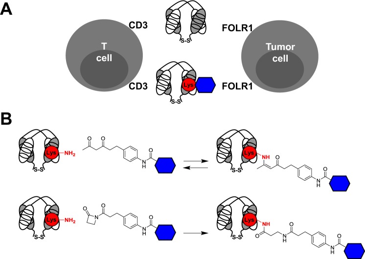
Chemically programmed bispecific antibodies (biAbs) endow target cell-binding small molecules with the ability to recruit and activate effector cells of the immune system. Here we report a platform of chemically programmed biAbs aimed at redirecting cytotoxic T cells to eliminate cancer cells. Two different antibody technologies were merged together to make a novel chemically programmed biAb. This was achieved by combining the humanized anti-hapten monoclonal antibody (mAb) h38C2 with the humanized anti-human CD3 mAb v9 in a clinically investigated diabody format known as Dual-Affinity Re-Targeting (DART). We show that h38C2 × v9 DARTs can readily be equipped with tumor-targeting hapten-derivatized small molecules without causing a systemic response harming healthy tissues. As a proof of concept, we chemically programmed h38C2 × v9 with hapten-folate and demonstrated its selectivity and potency against folate receptor 1 (FOLR1)-expressing ovarian cancer cells in vitro and in vivo Unlike conventional biAbs, chemically programmed biAbs in DART format are highly modular with broad utility in terms of both target and effector cell engagement. Most importantly, they provide tumor-targeting compounds access to the power of cancer immunotherapy.

Keywords: T-cell; antibody engineering; cancer therapy; chemical modification; folate; immunotherapy; ovarian cancer.


Figures

FIGURE 1.
FIGURE 1.
Conventional versus chemically programmed biAbs in DART format. A, biAbs in DART format are comprised of two polypeptides that are linked at their C termini via a disulfide bridge, where each polypeptide contains one of two cognate variable light (white) and heavy chain (gray) domains that form the antigen or hapten binding site. Conventional DARTs described in this study (top) combine a CD3-engaging with a FOLR1-engaging Fv module to bring T cells and tumor cells in close contact and enable the formation of cytolytic synapses. Chemically programmed DARTs described in this study engage the same two antigens; however, FOLR1 binding is mediated by a small molecule (blue hexagon) that is site-specifically and covalently conjugated to the reactive lysine residue (red circle) in the Fv module of humanized anti-β-diketone hapten mAb h38C2. B, mAb h38C2 harbors a reactive Lys residue (red circle) with an unusually low pKa of ∼6.0 at the bottom of its hydrophobic hapten binding site. The nucleophilic ϵ-amino group of this Lys residue can be covalently conjugated to the β-diketone group of the hapten and compounds that incorporate the hapten and a targeting moiety (blue hexagon). This reversible covalent conjugation (top) is stabilized by imine-enamine tautomerism. An irreversible covalent conjugation (bottom) is achieved by replacing the β-diketone group with a β-lactam group.
FIGURE 2.
FIGURE 2.
Chemically programmable DARTs. Two configurations, hv-L (A) and hv-H (B), of h38C2 × v9 DARTs were generated. Each consisted of two polypeptides that are linked at their C termini via a disulfide bridge and that each contain one of two cognate variable light (white) and heavy chain (gray) domains that form the antigen (CD3 for v9) or hapten (β-diketone for h38C2) binding site. The variable domains on each polypeptide are fused with a short polypeptide (G3SG4) that favors diabody over scFv formation. The two polypeptide expression cassettes were cloned under the control of a CMV promoter into mammalian expression vector pCEP4 for transient co-transfection into HEK 293 cells. A C-terminal His6 tag (H6) was included in one of the paired polypeptides to facilitate purification by IMAC. The reactive Lys residue of h38C2 is indicated in red. Note that the hv-L configuration has free light chain N termini, whereas configuration hv-H displays free heavy chain N termini. SP, signal peptide. All amino acid sequences are given in the supplemental information.
FIGURE 3.
FIGURE 3.
Biochemical characterization of chemically programmed and conventional FOLR1 × CD3 DARTs. A, Coomassie Blue-stained SDS-PAGE gel with 2 μg/lane purified representative DARTs (hv-L and fv-L) in reducing (red) and nonreducing (nonred) conditions, showing the expected bands at ∼27.5 and 55 kDa, respectively. A protein marker (in kDa) was run in the center lane. B, size-exclusion chromatography profile of a purified representative DART (hv-L). C, catalytic activity of purified DARTs hv-L (top; open red squares) and hv-H (bottom; open red squares) measured with the fluorogenic retro-aldol substrate methodol. Chemical programming with compound 1 eradicated the catalytic activity of both DARTs (solid red squares). Conventional DART fv-L served as negative control (solid green squares). D, efficacy of the conjugation of compound 1 to DART hv-L as measured by the EZ Biotin Quantitation kit. Conventional DART fv-L following attempted conjugation of compound 1 served as negative control.
FIGURE 4.
FIGURE 4.
Cell surface binding of chemically programmed and conventional FOLR1 × CD3 DARTs. The indicated DARTs were analyzed for binding to human CD3+ T-cell line Jurkat and human FOLR1+ ovarian cancer cell lines IGROV1, OVCAR3, and SKOV3 by flow cytometry at a concentration of 2 μg/ml, using a mouse anti-His6 mAb followed by Alexa Fluor 488-conjugated goat anti-mouse IgG pAbs. The background signal of the secondary reagents alone is shown in pale blue.
FIGURE 5.
FIGURE 5.
Folate derivatives. Trifunctional β-lactam-biotin-folate 1 and β-diketone-biotin-folate 2 contain a folate (a.k.a. pteroyl-glutamate) moiety derivatized at its γ-carboxyl group with a branched linker that incorporates a biotin moiety and an electrophilic hapten functionality enabling covalent conjugation to the nucleophilic ϵ-amino group of the reactive Lys residue in the hapten binding site of mAb h38C2. Bifunctional β-diketone-biotin 3 was used as negative control.
FIGURE 6.
FIGURE 6.
Cell surface binding and crosslinking of chemically programmed and conventional FOLR1 × CD3 DARTs. A, binding of chemically programmed hv-L and hv-H DARTs and conventional fv-L and fv-H DARTs to FOLR1-expressing IGROV1 cells. All DARTs were analyzed by flow cytometry at a concentration of 2 μg/ml (∼40 nm), using a mouse anti-His tag mAb followed by Alexa Fluor 488-conjugated goat anti-mouse IgG pAbs. hv-L and hv-H DARTs were chemically programmed by incubation with 400 nm β-lactam-biotin-folate compound 1 (red) or β-diketone-biotin compound 3 (blue), incubated for 1 h at room temperature, and purified by ultrafiltration. The background signal of the secondary reagents alone is shown in gray. B, crosslinking of CD3+ primary human T cells and FOLR1-expressing OVCAR3 cells in the presence of FOLR1 × CD3 DARTs. hv-L and hv-H DARTs were chemically programmed as noted above. The various DARTs were added at 2 μg/ml to 5 × 10⁴ T cells stained with CellTrace Far Red DDAO-SE. Following incubation for 1 h at room temperature and washing, 5 × 10⁴ OVCAR3 cells stained with CellTracker Blue CMAC were added. The mixtures were then incubated for 1 h at room temperature, gently washed, and fixed with 1% (w/v) paraformaldehyde. The formation of blue/far red cell aggregates was detected by flow cytometry and plotted as percentage of double positive events among all events. The results are representative of three experiments.
FIGURE 7.
FIGURE 7.
Crosslinking of chemically programmed and conventional FOLR1 × CD3 DARTs. A, IGROV1 cells were stained with CellTracker Blue CMAC and T cells or Jurkat cells with CellTrace Far Red DDAO-SE. The indicated DARTs were added at 2 μg/ml to 5 × 10⁴ T cells or Jurkat cells. Following incubation for 1 h at room temperature and washing, 5 × 10⁴ IGROV1 cells were added. The mixtures were then incubated for 1 h at room temperature, gently washed, and fixed with 1% (w/v) paraformaldehyde. The formation of blue/far red cell aggregates was detected by flow cytometry, using a forward scatter/side scatter gate that excluded the smaller non-crosslinked T cells and Jurkat cells from the analysis. The results are representative of three experiments. B, results from all three experiments were plotted as percentage of double positive events among all events following subtraction of the background observed with unprogrammed DART hv-L. Shown are mean values ± S.D.
FIGURE 8.
FIGURE 8.
In vitro activity of chemically programmed and conventional FOLR1 × CD3 DARTs. A, hv-L and hv-H DARTs were chemically programmed with biotin-β-diketone (compound 3) or folate-biotin-β-lactam (compound 1) as above. The cytotoxicity of chemically programmed DARTs hv-L and hv-H and conventional DARTs fv-L and fv-H was tested at 2 μg/ml (black), 0.6 μg/ml (gray), and 0.2 μg/ml (white) with ex vivo expanded primary human T cells (E) and OVCAR3 cells (T; 2.5 × 10⁴/well in a 96-well tissue culture plate) at an E:T ratio of 10:1. In all experiments cytotoxicity was measured with the CytoTox-Glo Cytotoxicity Assay (Promega, Inc.) after 16-h incubation at 37 °C in folate-deficient cell culture medium supplemented with 5% (v/v) human serum. Luminescence was measured in a SpectraMax M5 microplate reader with SoftMax Pro software. Shown are mean values of triplicates ± S.D. B, cytotoxicity of conventional DART fv-L (black), chemically programmed DART hv-L/1 (white), and unprogrammed DART hv-L (gray) was assayed as described in A over a concentration range of 2 ng/ml to 2 μg/ml at half-log intervals with ex vivo expanded primary human T cells and IGROV1 cells at an E:T ratio of 10:1. Luminescence measured after incubation of effector and target cells in the absence of DARTs was subtracted. Shown are mean values of triplicates ± S.D. C, supernatants from B at the 2 μg/ml DART concentration were used to measure interferon-γ release by ELISA. Shown are mean values of triplicates ± S.D.
FIGURE 9.
FIGURE 9.
In vivo activity of chemically programmed and conventional FOLR1 × CD3 DARTs. A, IGROV1/ffluc-cell-engrafted NSG mice in 6 cages were divided into 5 cohorts and treated as described in Fig. 10. Shown are bioluminescence images from day 23 for all 30 animals. B, flow cytometry analysis of ex vivo expanded T cells injected on day 6, using 2 μg/ml mouse anti-CD3, CD4, CD8, or CD28 mAbs followed by Alexa Fluor 488-conjugated goat anti-mouse IgG pAbs.
FIGURE 10.
FIGURE 10.
In vivo activity and safety of chemically programmed and conventional FOLR1 × CD3 DARTs. A total of 30 NSG mice were i.p. injected with 1 × 10⁶ IGROV1/ffluc cells on day 0 and divided into 5 cohorts, each consisting of 6 animals. Following human tumor xenograft establishment and growth, the animals in the 4 treatment cohorts received 1 × 10⁷ ex vivo expanded T cells by i.p. injection and 1 h later 10 μg of the indicated DARTs or PBS alone by i.p. injection on day 6. The animals received a total of 10 daily (day 6 to 15) i.p. injections of DARTs or PBS alone. A, starting on day 2, the animals were imaged every 3–4 days as indicated. The graph shows the mean ± S.D. luminescence signal for the 5 cohorts. Significant differences between cohorts treated with unprogrammed hv-L DART (open red squares) and chemically programmed hv-L/1 DART (solid red squares) were calculated using Microsoft Excel software (two-tailed and heteroscedastic t test; *, p < 0.05; **, p < 0.01). Treatment time points are indicated by black arrows. B, starting on day 0, the animals were weighted on the indicated days. The graph shows the mean ± S.D. weights for the 5 cohorts using the same color code as in A.

Similar articles

Cited by

References

    1. Zhukovsky E. A., Morse R. J., and Maus M. V. (2016) Bispecific antibodies and CARs: generalized immunotherapeutics harnessing T cell redirection. Curr. Opin. Immunol. 40, 24–35 - PMC - PubMed
    1. Kontermann R. E., and Brinkmann U. (2015) Bispecific antibodies. Drug Discov. Today 20, 838–847 - PubMed
    1. Frankel S. R., and Baeuerle P. A. (2013) Targeting T cells to tumor cells using bispecific antibodies. Curr. Opin. Chem. Biol. 17, 385–392 - PubMed
    1. Perez P., Hoffman R. W., Shaw S., Bluestone J. A., and Segal D. M. (1985) Specific targeting of cytotoxic T cells by anti-T3 linked to anti-target cell antibody. Nature 316, 354–356 - PubMed
    1. Rader C. (2014) Chemically programmed antibodies. Trends Biotechnol. 32, 186–197 - PMC - PubMed

Publication types

MeSH terms