. 2016 Sep 9;291(37):19387-400.
doi: 10.1074/jbc.M116.736074. Epub 2016 Jul 20.

Functional Diversity of Cytotoxic tRNase/Immunity Protein Complexes from Burkholderia pseudomallei

Affiliations

Functional Diversity of Cytotoxic tRNase/Immunity Protein Complexes from Burkholderia pseudomallei

Parker M Johnson et al. J Biol Chem. .

Abstract

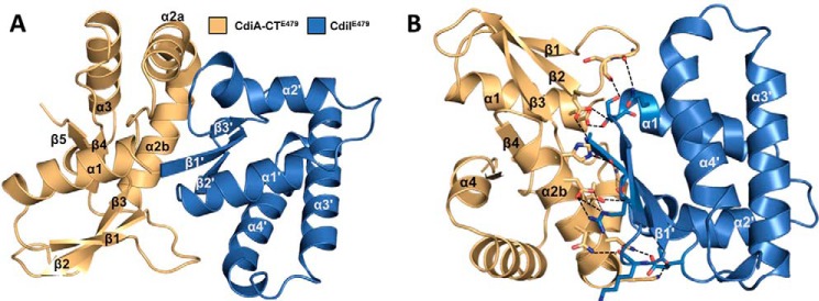
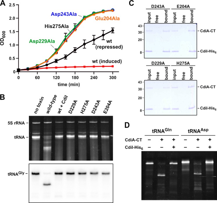
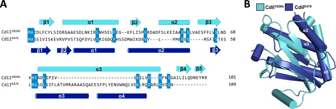
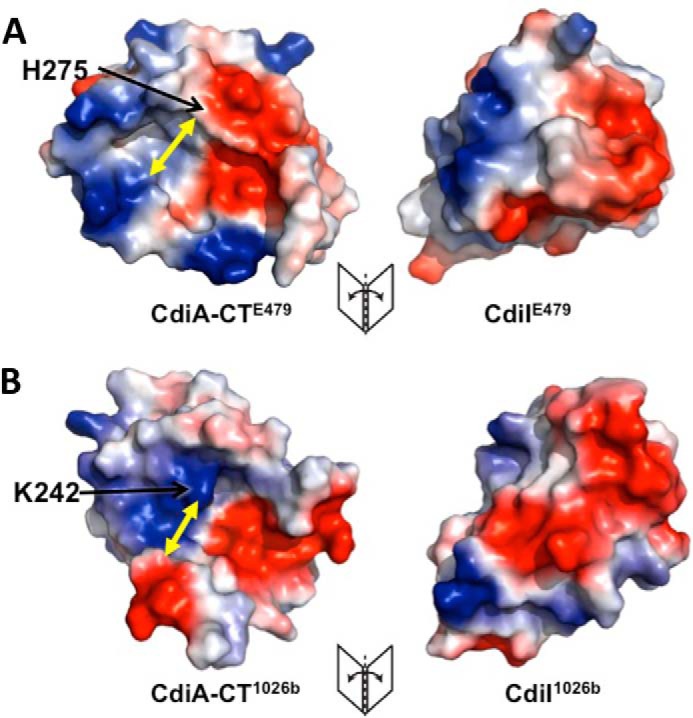
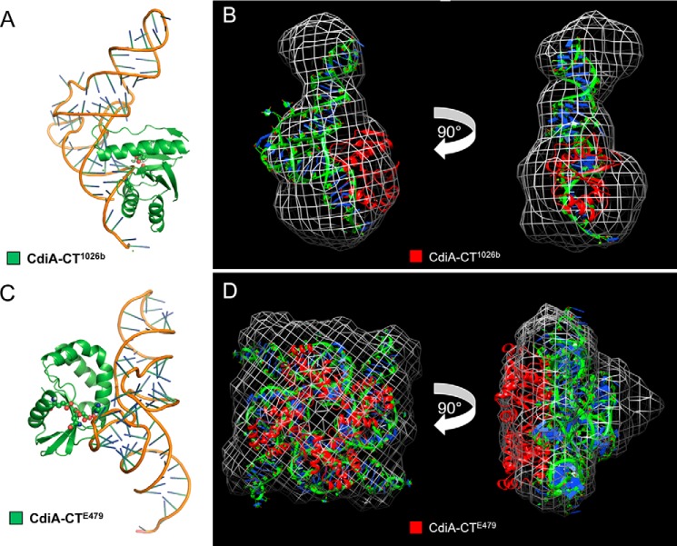
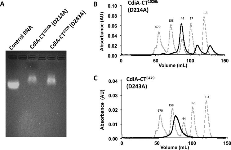
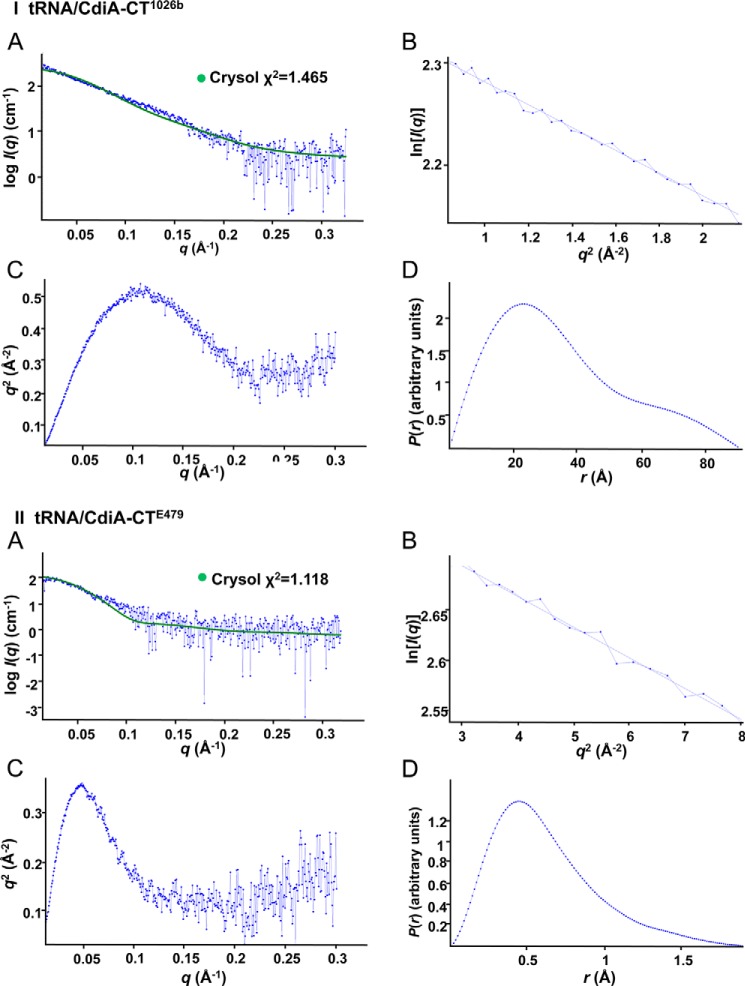
Contact-dependent growth inhibition (CDI) is a widespread mechanism of inter-bacterial competition. CDI(+) bacteria deploy large CdiA effector proteins, which carry variable C-terminal toxin domains (CdiA-CT). CDI(+) cells also produce CdiI immunity proteins that specifically neutralize cognate CdiA-CT toxins to prevent auto-inhibition. Here, we present the crystal structure of the CdiA-CT/CdiI(E479) toxin/immunity protein complex from Burkholderia pseudomallei isolate E479. The CdiA-CT(E479) tRNase domain contains a core α/β-fold that is characteristic of PD(D/E)XK superfamily nucleases. Unexpectedly, the closest structural homolog of CdiA-CT(E479) is another CDI toxin domain from B. pseudomallei 1026b. Although unrelated in sequence, the two B. pseudomallei nuclease domains share similar folds and active-site architectures. By contrast, the CdiI(E479) and CdiI(1026b) immunity proteins share no significant sequence or structural homology. CdiA-CT(E479) and CdiA-CT(1026b) are both tRNases; however, each nuclease cleaves tRNA at a distinct position. We used a molecular docking approach to model each toxin bound to tRNA substrate. The resulting models fit into electron density envelopes generated by small-angle x-ray scattering analysis of catalytically inactive toxin domains bound stably to tRNA. CdiA-CT(E479) is the third CDI toxin found to have structural homology to the PD(D/E)XK superfamily. We propose that CDI systems exploit the inherent sequence variability and active-site plasticity of PD(D/E)XK nucleases to generate toxin diversity. These findings raise the possibility that many other uncharacterized CDI toxins may belong to the PD(D/E)XK superfamily.

Keywords: Burkholderia pseudomallei; crystal structure; protein complex; ribonuclease; small-angle x-ray scattering (SAXS); tRNase; toxin; toxin/immunity complexes.


Figures

FIGURE 1.
FIGURE 1.
Structure of the CdiA-CT/CdiIE479 toxin/immunity protein complex. A, CdiA-CT/CdiIE479 complex is shown as a schematic with secondary structure elements of CdiI indicated with prime symbols. B, CdiA-CT/CdiIE479 complex formation is mediated by electrostatic interactions. Interacting residues are in stick representation with nitrogen and oxygen atoms colored in blue and red (respectively), and bonds are shown as black dashed lines. The view is rotated 180° about the x axis relative to A.
FIGURE 2.
FIGURE 2.
Biolayer interferometry of the CdiA-CT/CdiIE479 binding interaction. Immobilized CdiIE479-His6 was exposed to varying concentrations (1.5–5 μm) of CdiA-CTE479, and the binding interaction and dissociation monitored a wavelength shift (nm). Representative association and dissociation curves are presented with the overall correlation coefficient (R2) shown for the fit.
FIGURE 3.
FIGURE 3.
Sequence and structure alignment of CdiA-CTE479 and CdiA-CT1026b nuclease domains. A, sequence alignment of CdiA-CTE479 and CdiA-CT1026b toxins with proposed active-site residues highlighted in red and conserved residues in blue. Secondary structure elements are colored gold and green for CdiA-CTE479 and CdiA-CT1026b, respectively. B, superimposition of CdiA-CTE479 and CdiA-CT1026b nuclease domains. Secondary structure elements that superimpose are color-coded in gold (CdiA-CTE479) and green (CdiA-CT1026b), and those that do not align rendered in white (CdiA-CTE479) and gray (CdiA-CT1026b). C, active site of CdiA-CTE479 and CdiA-CT1026b nuclease domain. Predicted active-site residues are shown in stick representation (nitrogen and oxygen atoms are colored blue and red, respectively).
FIGURE 4.
FIGURE 4.
CdiA-CTE479 growth inhibition and tRNase activities. A, growth inhibition activity of CdiA-CTE479 variants. The indicated toxins were expressed in E. coli cells from a rhamnose-inducible promoter as described under “Experimental Procedures.” Expression was induced at 0 min, and cell growth was monitored by measuring the optical density at 600 nm (OD600). The curve labeled repressed corresponds to un-induced cells carrying the wild-type CdiA-CTE479 construct. The average ± S.E. from three independent biological replicates is presented. B, in vitro nuclease assays. The indicated CdiA-CTE479 variants were purified and incubated with total E. coli RNA. Reactions were run on denaturing 6% polyacrylamide gels and stained with ethidium bromide. C, mutant CdiA-CTE479 domains bind to CdiIE479 immunity protein. Isolated toxin domains were mixed with purified CdiIE479-His6 and then subjected to Ni2+-affinity chromatography. Lanes labeled input show the protein mixtures loaded onto the column; free lanes show proteins that failed to bind the column, and bound indicates proteins eluted from the column with imidazole. Prior work has shown that CdiA-CTE479 does not bind to Ni2+-NTA-agarose resin (16). D, CdiA-CTE479 cleaves unmodified tRNAs produced by in vitro transcription. E. coli tRNAGln and tRNAAsp transcripts were incubated with purified CdiA-CTE479 and CdiIE479, and reactions were analyzed on denaturing 6% polyacrylamide gels stained with ethidium bromide. Experiments in B–D were repeated twice with essentially identical results. Representative data are shown for each experiment.
FIGURE 5.
FIGURE 5.
Sequence and structure comparison of CdiIE479 and CdiI1026b immunity proteins. A, structure-based sequence alignment of CdiIE479 (blue) and CdiI1026b (cyan) with secondary structure elements indicated above and below the sequence alignment. Conserved residues are highlighted in blue. B, superimposition of CdiIE479 and CdiI1026b structures. Secondary elements that partially or fully superimpose are labeled.
FIGURE 6.
FIGURE 6.
CdiA-CT/CdiIE479 and CdiA-CT/CdiI1026b complexes interact through distinct electrostatic surfaces. A, electrostatic surface map of the CdiA-CT/CdiIE479 complex interface. Negative, positive, and neutral surfaces regions are shown in red, blue, and white, respectively. B, electrostatic surface map of the CdiA-CT/CdiI1026b complex interface. Yellow arrows indicate the minimal width of each active-site pocket.
FIGURE 7.
FIGURE 7.
Computational modeling and SAXS analysis of tRNA/CdiA-CT complexes. A, CdiA-CT1026b (green schematic) binding with its active-site residues adjacent to the backbone of the tRNACys (PDB code 1B23) amino acceptor stem loop with active-site residues shown as spheres (oxygen and nitrogen atoms colored red and blue, respectively). B, SAXS electron density envelope (white mesh) fitted with the docking solution showing the monomeric CdiA-CT1026b toxin (red schematic) bound to tRNACys (green and blue). C, CdiA-CTE479 (green schematic) binding with its active-site residues adjacent to the backbone of the tRNACys T-loop with active-site residues shown as spheres (oxygen and nitrogen atoms colored red and blue, respectively). D, SAXS electron density envelope (white mesh) fitted with the docking solution showing the tetrameric CdiA-CTE479 toxin bound to four molecules of tRNACys (green and blue).
FIGURE 8.
FIGURE 8.
Inactive CdiA-CTE479 and CdiA-CT1026b toxin domains bind to endogenous tRNA. A, agarose gel analysis of catalytically inactive CdiA-CTE479 and CdiA-CT1026b toxins purified under non-denaturing conditions. Control RNA is from yeast (Sigma). B, size-exclusion chromatography of the purified tRNA/CdiA-CT(D214A)1026b complex. C, size-exclusion chromatography of the purified tRNA/CdiA-CT(D243A)E479 complex. Chromatography migration standards are as follows: bovine thyroglobulin (670 kDa), bovine γ-globulin (158 kDa), chicken ovalbumin (44 kDa), horse myoglobin (17 kDa), and vitamin B12 (1.3 kDa). Each experiment was carried out in triplicate with similar results. Representative data are shown for each experiment.
FIGURE 9.
FIGURE 9.
SAXS analyses of tRNA/CdiA-CT complexes. Plots for tRNA/CdiA-CT1026b (I) and tRNA/CdiA-CTE479 (II) SAXS data. A, log I(q) versus q plot with experimental SAXS profile shown in blue and the corresponding structural model fitted data via Crysol (41) shown in green. B, Guinier plots. C, Kratky plots. D, P(r) plots.

References

    1. Aoki S. K., Diner E. J., de Roodenbeke C. T., Burgess B. R., Poole S. J., Braaten B. A., Jones A. M., Webb J. S., Hayes C. S., Cotter P. A., and Low D. A. (2010) A widespread family of polymorphic contact-dependent toxin delivery systems in bacteria. Nature 468, 439–442 - PMC - PubMed
    1. Ruhe Z. C., Low D. A., and Hayes C. S. (2013) Bacterial contact-dependent growth inhibition. Trends Microbiol. 21, 230–237 - PMC - PubMed
    1. Willett J. L., Ruhe Z. C., Goulding C. W., Low D. A., and Hayes C. S. (2015) Contact-dependent growth inhibition (CDI) and CdiB/CdiA two-partner secretion proteins. J. Mol. Biol. 427, 3754–3765 - PMC - PubMed
    1. Makhov A. M., Hannah J. H., Brennan M. J., Trus B. L., Kocsis E., Conway J. F., Wingfield P. T., Simon M. N., and Steven A. C. (1994) Filamentous hemagglutinin of Bordetella pertussis. A bacterial adhesin formed as a 50-nm monomeric rigid rod based on a 19-residue repeat motif rich in β strands and turns. J. Mol. Biol. 241, 110–124 - PubMed
    1. Kajava A. V., Cheng N., Cleaver R., Kessel M., Simon M. N., Willery E., Jacob-Dubuisson F., Locht C., and Steven A. C. (2001) β-Helix model for the filamentous haemagglutinin adhesin of Bordetella pertussis and related bacterial secretory proteins. Mol. Microbiol. 42, 279–292 - PubMed

Publication types

MeSH terms