Case Reports
. 2016:2016:1460480.
doi: 10.1155/2016/1460480. Epub 2016 Apr 30.

Coexistent Congenital Diaphragmatic Hernia with Extrapulmonary Sequestration

Affiliations
Case Reports

Coexistent Congenital Diaphragmatic Hernia with Extrapulmonary Sequestration

Nao Kawamura et al. Can Respir J. 2016.

Abstract

Bronchopulmonary foregut malformations are a heterogeneous but interrelated group of abnormalities that may contain more than one histologic feature. It is helpful to be familiar with the presentation and imaging features of bronchopulmonary foregut malformations presenting as a congenital mass or mass-like lesion, as imaging plays a central role in the evaluation of these lesions since, when symptomatic, clinical features are usually nonspecific. With imaging, the presence of other associated lesions can be determined, facilitating appropriate management to prevent the potential complications. We report a case of coexisting extralobar pulmonary sequestration and ipsilateral diaphragmatic hernia in a term neonate.


Figures

Figure 1
Figure 1
(a) T1-weighted: coronal MRI imaging at 28-week gestation showing left pulmonary bronchopulmonary foregut malformation and herniated bowel in the left hemithorax and displacement of the cardiac structures to the right side. (b) T2-weighted: coronal MRI imaging at 28-week gestation showing left pulmonary bronchopulmonary foregut malformation and herniated bowel in the left hemithorax and displacement of the cardiac structures to the right side.
Figure 2
Figure 2
Chest radiograph showing left diaphragmatic hernia and contralateral shift of the heart and mediastinum.
Figure 3
Figure 3
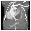
Coronal postcontrast CT image showing bronchopulmonary sequestration in a left paraspinal location. Herniated bowel loops and the superior mesenteric artery are seen in the left hemithorax. A branch from coeliac artery is seen supplying spleen, which is located in the left lower paraspinal region.
Figure 4
Figure 4
Coronal CT image showing feeding artery to sequestrated lung segment arising from the descending aorta and draining vein from the sequestration entering into left atrium.

References

    1. Biyyam D. R., Chapman T., Ferguson M. R., Deutsch G., Dighe M. K. Congenital lung abnor malities: embryologic features, prenatal diagnosis, and postnatal radiologic pathologic correlation. Radiographics. 2010;30(6):1721–1738. doi: 10.1148/rg.306105508. - DOI - PubMed
    1. Taylor G. A., Atalabi O. M., Estroff J. A. Imaging of congenital diaphragmatic hernias. Pediatric Radiology. 2009;39(1):1–16. doi: 10.1007/s00247-008-0917-7. - DOI - PubMed
    1. Felson B. The many faces of pulmonary sequestration. Seminars in Roentgenology. 1972;7(1):3–16. doi: 10.1016/0037-198x(72)90026-0. - DOI - PubMed
    1. Kocaoğlu M., Frush D. P., Uğurel M. S., Somuncu I. Bronchopulmonary foregut malformations presenting as mass lesions in children: spectrum of imaging findings. Diagnostic and Interventional Radiology. 2010;16(2):153–161. doi: 10.4261/1305-3825.dir.2063-08.2. - DOI - PubMed
    1. Felker R. E., Tonkin I. L. D. Imaging of pulmonary sequestration. American Journal of Roentgenology. 1990;154(2):241–249. doi: 10.2214/ajr.154.2.2105007. - DOI - PubMed

Publication types

MeSH terms

LinkOut - more resources