. 2016 Jun 30:10:308.
doi: 10.3389/fnins.2016.00308. eCollection 2016.

Altered Cerebral Blood Flow Covariance Network in Schizophrenia

Affiliations

Altered Cerebral Blood Flow Covariance Network in Schizophrenia

Feng Liu et al. Front Neurosci. .

Abstract

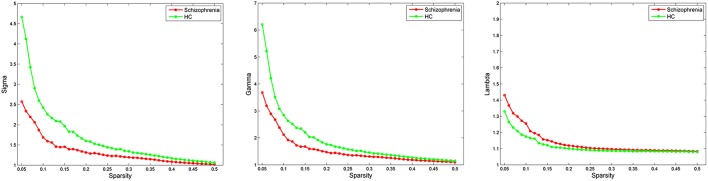
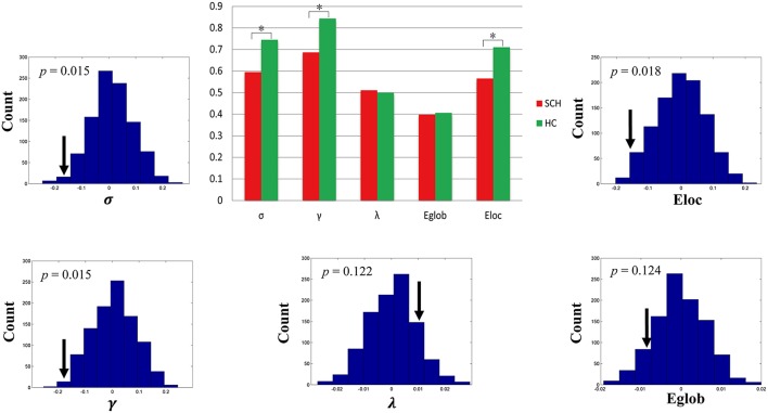
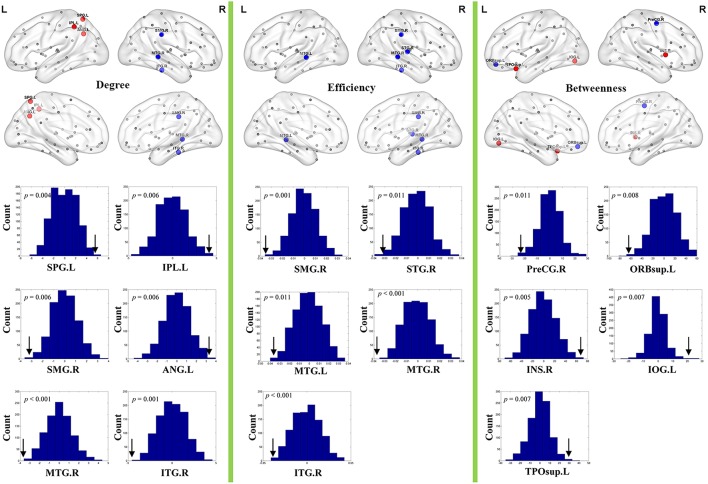
Many studies have shown abnormal cerebral blood flow (CBF) in schizophrenia; however, it remains unclear how topological properties of CBF network are altered in this disorder. Here, arterial spin labeling (ASL) MRI was employed to measure resting-state CBF in 96 schizophrenia patients and 91 healthy controls. CBF covariance network of each group was constructed by calculating across-subject CBF covariance between 90 brain regions. Graph theory was used to compare intergroup differences in global and nodal topological measures of the network. Both schizophrenia patients and healthy controls had small-world topology in CBF covariance networks, implying an optimal balance between functional segregation and integration. Compared with healthy controls, schizophrenia patients showed reduced small-worldness, normalized clustering coefficient and local efficiency of the network, suggesting a shift toward randomized network topology in schizophrenia. Furthermore, schizophrenia patients exhibited altered nodal centrality in the perceptual-, affective-, language-, and spatial-related regions, indicating functional disturbance of these systems in schizophrenia. This study demonstrated for the first time that schizophrenia patients have disrupted topological properties in CBF covariance network, which provides a new perspective (efficiency of blood flow distribution between brain regions) for understanding neural mechanisms of schizophrenia.

Keywords: arterial spin labeling; cerebral blood flow; covariance network; efficiency; schizophrenia; small world.


Figures

Figure 1
Figure 1
Small-world parameters of CBF covariance network as function of sparsity thresholds. Both the schizophrenia and control groups show a small-worldness (σ) >1, normalized clustering coefficient (γ) >1 and normalized characteristic path length (λ) approximately equal to 1, indicating that both groups exhibited a small-world topology. HC, healthy controls.
Figure 2
Figure 2
Global topological differences in CBF covariance network between schizophrenia patients and healthy controls. Each measure is expressed as the integrated area under the curve (AUC). The black stars in bar plots denote statistically significant differences between the two groups (permutation test, p < 0.05). The histogram plots around the bar plots are null distributions of permutation tests of global network measures and the real measures are marked with black arrows. SCH, schizophrenia; HC, healthy controls.
Figure 3
Figure 3
Nodal topological differences in CBF covariance networks between schizophrenia patients and healthy controls (permutation test, p < 1/90). Results are rendered using the BrainNet viewer (Xia et al., 2013). Red and blue spheres represent regions with significantly increased and decreased nodal properties in schizophrenia, respectively. Gray spheres represent regions without significant intergroup difference. The histogram plots below the render plots are the null distributions of permutation tests of significant nodal network measures and the real measures are marked with black arrows. ANG, angular gyrus; INS, insula; IPL, inferior parietal lobule; IOG, inferior occipital gyrus; ITG, inferior temporal gyrus; L, left; MTG, middle temporal gyrus; PreCG, precentral gyrus; R, right; ORBsup, orbital part of superior frontal gyrus; SMG, supramarginal gyrus; SPL, superior parietal lobule; STG, superior temporal gyrus; TPOsup, superior part of temporal pole.

References

    1. Achard S., Salvador R., Whitcher B., Suckling J., Bullmore E. (2006). A resilient, low-frequency, small-world human brain functional network with highly connected association cortical hubs. J. Neurosci. 26, 63–72. 10.1523/JNEUROSCI.3874-05.2006 - DOI - PMC - PubMed
    1. Agarwal S. M., Danivas V., Amaresha A. C., Shivakumar V., Kalmady S. V., Bose A., et al. . (2015). Cognitive mapping deficits in schizophrenia: evidence from clinical correlates of visuospatial transformations. Psychiatry Res. 228, 304–311. 10.1016/j.psychres.2015.05.096 - DOI - PubMed
    1. Alexander-Bloch A., Giedd J. N., Bullmore E. (2013). Imaging structural co-variance between human brain regions. Nat. Rev. Neurosci. 14, 322–336. 10.1038/nrn3465 - DOI - PMC - PubMed
    1. Bassett D. S., Bullmore E. (2006). Small-world brain networks. Neuroscientist 12, 512–523. 10.1177/1073858406293182 - DOI - PubMed
    1. Bassett D. S., Bullmore E., Verchinski B. A., Mattay V. S., Weinberger D. R., Meyer-Lindenberg A. (2008). Hierarchical organization of human cortical networks in health and schizophrenia. J. Neurosci. 28, 9239–9248. 10.1523/JNEUROSCI.1929-08.2008 - DOI - PMC - PubMed

LinkOut - more resources