

Current Applications and Future Perspectives of the Use of 3D Printing in Anatomical Training and Neurosurgery
- PMID: 27445707
- PMCID: PMC4919320
- DOI: 10.3389/fnana.2016.00069
Current Applications and Future Perspectives of the Use of 3D Printing in Anatomical Training and Neurosurgery
Abstract
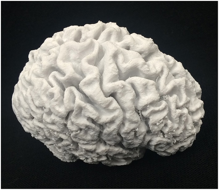
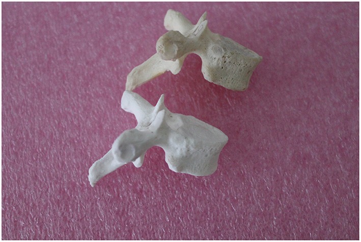
3D printing is a form of rapid prototyping technology, which has led to innovative new applications in biomedicine. It facilitates the production of highly accurate three dimensional objects from substrate materials. The inherent accuracy and other properties of 3D printing have allowed it to have exciting applications in anatomy education and surgery, with the specialty of neurosurgery having benefited particularly well. This article presents the findings of a literature review of the Pubmed and Web of Science databases investigating the applications of 3D printing in anatomy and surgical education, and neurosurgery. A number of applications within these fields were found, with many significantly improving the quality of anatomy and surgical education, and the practice of neurosurgery. They also offered advantages over existing approaches and practices. It is envisaged that the number of useful applications will rise in the coming years, particularly as the costs of this technology decrease and its uptake rises.
Keywords: 3D printing; anatomy; computer aided design (CAD); education; neurosurgery; rapid prototyping; surgery.
Figures
References
-
- Berman B. (2012). 3-D printing: the new industrial revolution. Bus. Horizons 55, 155–162. 10.1016/j.bushor.2011.11.003 - DOI
-
- Chambers S. B., Deehan D. J., Gillinder S., Holland J. P. (2015). Cadaveric surgical training improves surgeon confidence. RCS Bull. 97, E1–E4. 10.1308/147363515x14134529299349 - DOI
LinkOut - more resources
Full Text Sources
Other Literature Sources
Miscellaneous