. 2016 Jun 30:7:273.
doi: 10.3389/fphys.2016.00273. eCollection 2016.

Uptake of Plasmin-PN-1 Complexes in Early Human Atheroma

Affiliations

Uptake of Plasmin-PN-1 Complexes in Early Human Atheroma

Kamel Boukais et al. Front Physiol. .

Abstract

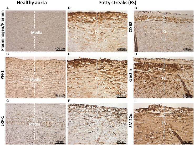
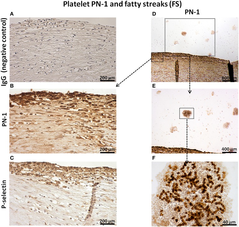
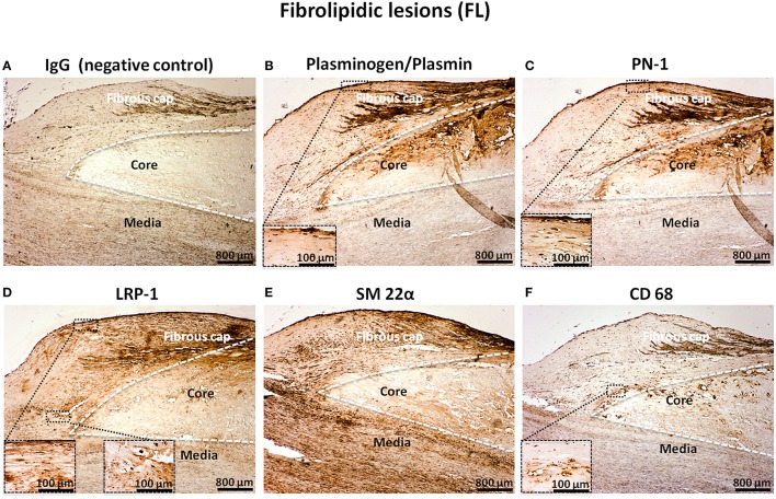
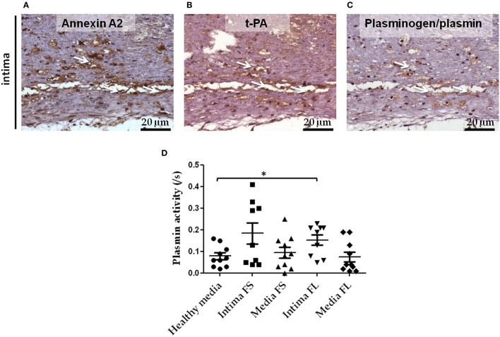
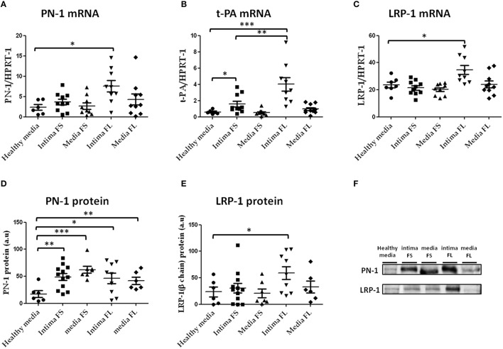
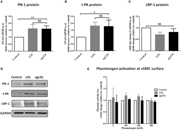
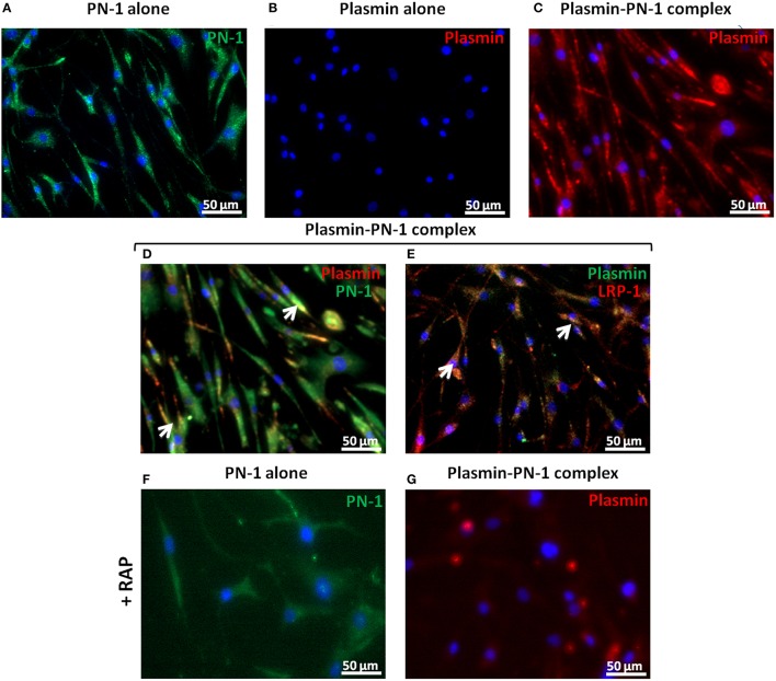
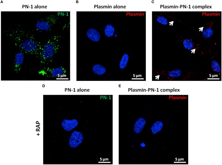
Zymogens are delivered to the arterial wall by radial transmural convection. Plasminogen can be activated within the arterial wall to produce plasmin, which is involved in evolution of the atherosclerotic plaque. Vascular smooth muscle cells (vSMCs) protect the vessels from proteolytic injury due to atherosclerosis development by highly expressing endocytic LDL receptor-related protein-1 (LRP-1), and by producing anti-proteases, such as Protease Nexin-1 (PN-1). PN-1 is able to form covalent complexes with plasmin. We hypothesized that plasmin-PN-1 complexes could be internalized via LRP-1 by vSMCs during the early stages of human atheroma. LRP-1 is also responsible for the capture of aggregated LDL in human atheroma. Plasmin activity and immunohistochemical analyses of early human atheroma showed that the plasminergic system is activated within the arterial wall, where intimal foam cells, including vSMCs and platelets, are the major sites of PN-1 accumulation. Both PN-1 and LRP-1 are overexpressed in early atheroma at both messenger and protein levels. Cell biology studies demonstrated an increased expression of PN-1 and tissue plasminogen activator by vSMCs in response to LDL. Plasmin-PN-1 complexes are internalized via LRP-1 in vSMCs, whereas plasmin alone is not. Tissue PN-1 interacts with plasmin in early human atheroma via two complementary mechanisms: plasmin inhibition and tissue uptake of plasmin-PN-1 complexes via LRP-1 in vSMCs. Despite this potential protective effect, plasminogen activation by vSMCs remains abnormally elevated in the intima in early stages of human atheroma.

Keywords: and atherosclerosis; antiproteases; endocytosis; proteases; vascular smooth muscle cells.


Figures

Figure 1
Figure 1
Cellular localization of plasminogen/plasmin, PN-1, LRP-1, CD68, α actin, and transgelin (SM 22α) in intima of fatty streaks (FS). Histological sections of healthy aortas and serial sections of FS. Immunostaining of plasminogen/plasmin in healthy aortas (A) and FS (D). Immunosignal of PN-1 in healthy aortas (B), FS (E). Immunosignal of LRP-1 in healthy aortas (C) and FS (F). Phagocytosis marker (CD 68) staining (G). Smooth muscle cell marker staining [(α actin, H) and (SM 22α, I)]. The negative control is shown in Figure 2A.
Figure 2
Figure 2
Platelet PN-1 and fatty streaks (FS). Serial sections of FS. IgG (negative control, A). Immunosignal of PN-1 in FS (B,D,E) and in luminal platelet aggregates (D,E,F). Immunosignal of p-selectin (C).
Figure 3
Figure 3
Cellular localization of plasminogen/plasmin, PN-1, LRP-1, SM 22α, and CD68 in fibrolipidic lesions (FL). Serial sections of FL. IgG (negative control, A). Plasminogen/plasmin staining (B). Immunosignal of PN-1 (C). Immunostaining of LRP-1 (D). Smooth muscle cell marker staining (SM 22α, E). Phagocytosis marker (CD 68) staining (F).
Figure 4
Figure 4
Colocalization of Annexin A2, t-PA, and plasminogen in serial sections of FS intima. Immunostaining of Annexin A2 (A), t-PA (B), and plasminogen (C) in FS. White arrows indicate the area in which Annexin A2, t-PA, and plasminogen immunostaining were colocalized. Plasmin activity in conditioned medium of healthy media, intima and media SL and FL (D): healthy media n = 10 vs. intima of FL n = 9; *p < 0.05. The non-parametric Mann-Whitney test was used.
Figure 5
Figure 5
PN-1 (A), tissue plasminogen activator (t-PA; B), and LRP-1(C) mRNA expression and PN-1 (D, F) and LRP-1 (E, F) protein expression in human aortic tissues (healthy aortas, intima and media of FS, intima and media of FL). PN-1 (A): healthy media n = 6 vs. intima of FL n = 9; *p < 0.05, t-PA (B): healthy media n = 6 vs. intima of FL n = 9; ***p < 0.001, intima of FS n = 10 vs. intima of FL n = 9; **p < 0.01, healthy media: n = 6 vs. intima of FS n = 10; *p < 0.05. LRP-1 (C): healthy media n = 7 vs. intima of FL n = 9; *p < 0.05. PN-1 protein expression (D,F): healthy media n = 6 vs. intima of FS n = 12; **p < 0.01, vs. media of FS n = 7; ***p < 0.001, vs. intima of FL n = 9; *p < 0.05, vs. media of FL n = 6; **p < 0.01. LRP-1 protein (E,F): healthy media n = 6 vs. intima of FL n = 9; *p < 0.05. The non-parametric Mann-Whitney test was used. There was no loading control because intima of advanced lesions is acellular and there are no appropriate stable housekeeping proteins.
Figure 6
Figure 6
Effects of native and aggregated (ag) LDL on PN-1 (A,D), t-PA (B,D), LRP-1 (C,D) protein expression and plasminogen activation (E) in vSMCs. The results are expressed relative to control for each experiment. PN-1 protein expression (A,D): LDL 1.62 ± 0.30, agLDL 1.60 ± 0.21; **p < 0.01, n = 5. t-PA protein expression (B,D): LDL 1.82 ± 0.26, agLDL 1.78 ± 0.46; *p < 0.05, n = 5. LRP-1 (C,D) protein expression (n = 5). The plasminogen activation (n = 3 and each experiment done in triplicate) at the surface of vSMCs (E). The paired t-test was used.
Figure 7
Figure 7
Internalization of PN-1, plasmin, and plasmin-PN-1 complexes in vSMCs. PN-1 alone (125 nM, A,F), plasmin alone (25 nM, B), and pre-formed plasmin-PN-1 complexes (C,D,E,G) were added to vSMCs for 2 h at 37°C without (A–E) or with RAP (50 μg/ml, F,G). After cell permeabilization, PN-1 alone (A,F) was detected by Alexa Fluor®488-labeled secondary antibody (green), plasmin alone (B) by Alexa Fluor®555-labeled secondary antibody (red) and nuclei by DAPI staining (blue). Plasmin-PN-1 complexes were revealed with plasmin antibody and detected by Alexa Fluor®555-labeled secondary antibody (C,G, red) or PN-1 and plasmin antibodies and detected with Alexa Fluor®488 (D, green) and 555-labeled secondary antibody (D, red) respectively. Yellow color and white arrows highlight examples of intracellular co-localization of plasmin-PN-1 complexes revealed with PN-1 and plasmin antibodies (D). LRP-1 was detected with Alexa Fluor®555-labeled secondary antibody (E, red), plasmin-PN-1 complexes were revealed with plasmin antibody and detected by Alexa fluor®488-labeled secondary antibody (E, green). Yellow color and white arrows indicate the intracellular colocalization of LRP-1 and plasmin-PN-1 complexes (E).
Figure 8
Figure 8
Internalization of PN-1, plasmin, and plasmin-PN-1 complexes in vSMCs (confocal microscopy). PN-1 alone (125 nM, A,D), plasmin alone (25 nM, B), and pre-formed plasmin-PN-1 complexes (C,E) were added to vSMCs for 2 h at 37°C without (A–C) or with RAP (50 μg/ml, D,E). After cell permeabilization, PN-1 alone (A, D) was detected by Alexa Fluor®488-labeled secondary antibody (green), plasmin alone (B) and plasmin-PN-1 complexes (C,E) by Alexa Fluor®555-labeled secondary antibody (red) and nuclei by DAPI staining (blue). White arrows indicate the intracellular localization of plasmin-PN-1 complexes (C). The images are presented as maximum intensity projection of confocal microscopy.

References

    1. Allahverdian S., Chehroudi A. C., McManus B. M., Abraham T., Francis G. A. (2014). Contribution of intimal smooth muscle cells to cholesterol accumulation and macrophage-like cells in human atherosclerosis. Circulation 129, 1551–1559. 10.1161/CIRCULATIONAHA.113.005015 - DOI - PubMed
    1. Bauriedel G., Hutter R., Welsch U., Bach R., Sievert H., Luderitz B. (1999). Role of smooth muscle cell death in advanced coronary primary lesions: implications for plaque instability. Cardiovasc. Res. 41, 480–488. 10.1016/S0008-6363(98)00318-6 - DOI - PubMed
    1. Borges L. F., Gomez D., Quintana M., Touat Z., Jondeau G., Leclercq A., et al. . (2010). Fibrinolytic activity is associated with presence of cystic medial degeneration in aneurysms of the ascending aorta. Histopathology 57, 917–932. 10.1111/j.1365-2559.2010.03719.x - DOI - PubMed
    1. Boucher P., Gotthardt M. (2004). LRP and PDGF signaling: a pathway to atherosclerosis. Trends Cardiovasc. Med. 14, 55–60. 10.1016/j.tcm.2003.12.001 - DOI - PubMed
    1. Boucher P., Gotthardt M., Li W. P., Anderson R. G., Herz J. (2003). LRP: role in vascular wall integrity and protection from atherosclerosis. Science 300, 329–332. 10.1126/science.1082095 - DOI - PubMed