. 2016 Jun 27:7:76.
doi: 10.3389/fendo.2016.00076. eCollection 2016.

MyomiRs as Markers of Insulin Resistance and Decreased Myogenesis in Skeletal Muscle of Diet-Induced Obese Mice

Affiliations

MyomiRs as Markers of Insulin Resistance and Decreased Myogenesis in Skeletal Muscle of Diet-Induced Obese Mice

Flávia de Toledo Frias et al. Front Endocrinol (Lausanne). .

Abstract

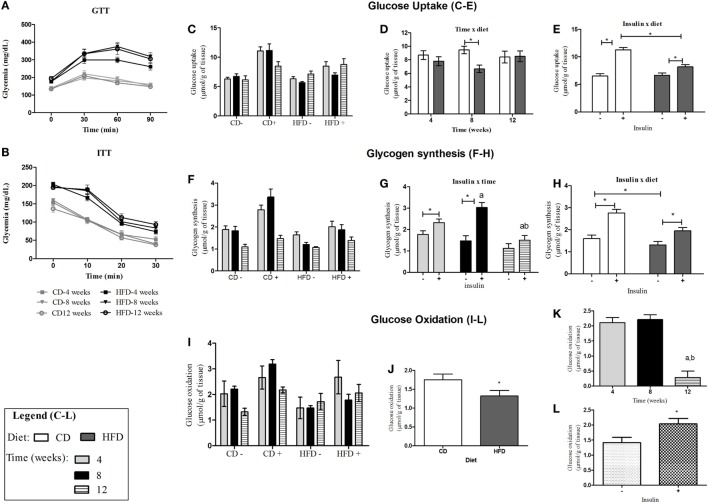
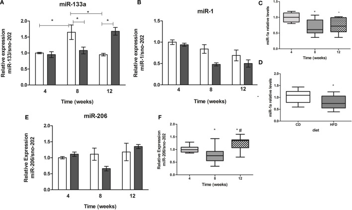
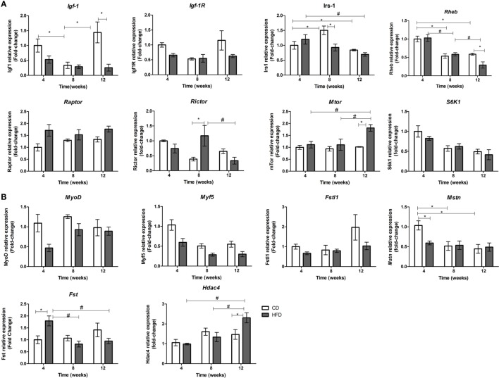
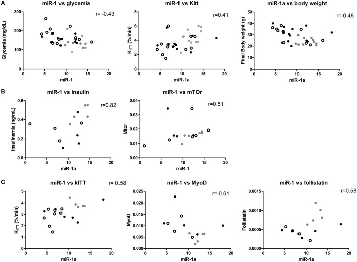
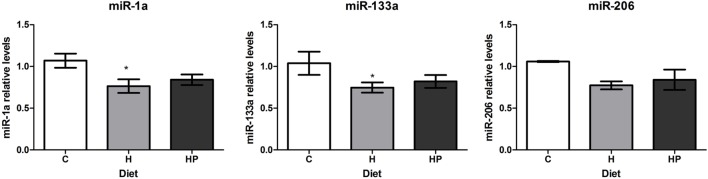
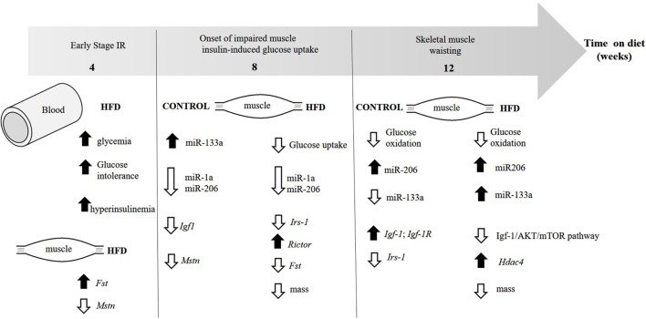
High-fat diet (HFD) feeding causes insulin resistance (IR) in skeletal muscle of mice, which affects skeletal muscle metabolism and function. The involvement of muscle-specific microRNAs in the evolution of skeletal muscle IR during 4, 8, and 12 weeks in HFD-induced obese mice was investigated. After 4 weeks in HFD, mice were obese, hyperglycemic, and hyperinsulinemic; however, their muscles were responsive to insulin stimuli. Expressions of MyomiRs (miR-1, miR-133a, and miR-206) measured in soleus muscles were not different from those found in control mice. After 8 weeks of HFD feeding, glucose uptake was lower in skeletal muscle from obese mice compared to control mice, and we observed a significant decrease in miR-1a in soleus muscle when compared to HFD for 4 weeks. miR-1a expression continued to decay within time. After 12 weeks of HFD, miR-133a expression was upregulated when compared to the control group. Expression of miR-1a was negatively correlated with glycemia and positively correlated with the constant rate of plasma glucose disappearance. Pioglitazone treatment could not reverse decreases of miR-1a levels induced by HFD. Targets of myomiRs involved in insulin-growth factor (IGF)-1 pathway, such as Igf-1, Irs-1, Rheb, and follistatin, were reduced after 12 weeks in HFD and Mtor increased, when compared to the control or HFD for 4 or 8 weeks. These findings suggest for the first time that miR-1 may be a marker of the development of IR in skeletal muscle. Evidence was also presented that impairment in myomiRs expression contributes to decreased myogenesis and skeletal muscle growth reported in diabetes.

Keywords: IGF-1; high-fat diet; insulin resistance; microRNA; myogenesis; myomiRs; skeletal muscle.


Figures

Figure 1
Figure 1
Insulin sensitivity and glucose tolerance in control- or HFD-fed C57BL/6J mice. (A) Glycemic curves obtained from GTT after injection of glucose (2 g/kg/b.w.); (B) glycemic curves obtained from ITT after injection of insulin (0.75 mIU/g b.w.); (C,F,I) glucose metabolism in soleus muscle of control- and HFD-fed animals after stimulation (+) or not (−) with insulin 10 mIU/mL after 4-, 8-, and 12-week feeding period. (C–E): glucose uptake average for each group in the absence/presence of insulin (C), interaction between insulin and diet (D), and interaction between time and diet (E). (F–H): glycogen synthesis average for each group in the absence or presence of insulin (F), interaction between insulin and time (G), and interaction between insulin and diet (H). (I–L): glucose oxidation average for each group in the presence and absence of insulin (I) and effect of diet (J), time (K), and insulin (L) separately on glucose oxidation. a,b and *p < 0.05 as indicated by three-way ANOVA followed by all pairwise multiple comparison test Holm–Sidak for interaction between two factors or effect of just one factor. (a,b) compared to 4 or 8 weeks, respectively. CD, control-fed mice; HFD, high-fat diet-fed mice.
Figure 2
Figure 2
Time-course of muscle-specific microRNAs miR-133a (A), miR-1a (B), and miR-206 (E) expression in soleus muscle of high-fat diet (HFD) and control diet (CD)-fed mice. Total RNA was extracted from soleus muscle of HFD- and CD-fed mice, and 10 ng was used for stem-loop reverse transcription. RT products were used for TaqMan real-time PCR. SnoRNA-202 was used for normalization. *p < 0.05 as indicated by two-way ANOVA followed by Bonferroni posttest; (C,D) effect of diet and time separately on miR-1a expression, respectively, *p < 0.05 as indicated by one-way ANOVA followed by Holm–Sidak method; (F) effect of the time on miR-206 expression, *p < 0.05 as indicated by one-way ANOVA followed by Holm–Sidak method.
Figure 3
Figure 3
Time-course of mRNAs expression involved in IGF-1/PI3K/AKT/MTOR pathway (A) and with myogenesis (B) in soleus muscle of high-fat diet (HFD)- and control diet (CD)-fed mice. Total RNA was extracted from soleus muscle of HFD- and CD-fed mice, and 500 ng was used for reverse transcription. RT products were used for SyBR green real-time PCR. 36B4 gene was used for normalization. *p < 0.05 as indicated by two-way ANOVA followed by all pairwise multiple comparison procedure: Holm–Sidak method. Only significant values for interactions of time and diet are shown. For separate analysis of time and diet, please see the Section “Results.”
Figure 4
Figure 4
miR-1 is correlated with metabolic parameters in mice. (A) Samples from control and obese mice: significant correlations between miR-1, Kitt, and glycemia are shown; (B) only control mice samples were used for this analysis: insulin plasma levels and mTor mRNA significantly correlated with miR-1a levels; and (C) only obese mice were used in these analyses: significant correlations were found between miR-1a and Kitt, MyoD and Fst mRNA. Spearman’s correlation analysis was used to determine the correlation. Gray dot: 4 weeks, open circle: 8 weeks, and black dot: 12 weeks.
Figure 5
Figure 5
Effect of pioglitazone 35 mg/kg/day on muscle-specific microRNAs expression in soleus muscles of HFD-fed mice. Animals were fed for 8 weeks with high-fat diet and treated with pioglitazone for 2 weeks. Total RNA was extracted from soleus muscles for microRNA expression. *p < 0.05 as indicated by one-way ANOVA followed by Holm–Sidak method.
Figure 6
Figure 6
Evolution of the changes in skeletal muscle and insulin sensitivity in diet-induced obese mice. The figure illustrates the time each parameter first appear.

Similar articles

Cited by

References

    1. Abdul-Ghani MA, Defronzo RA. Pathogenesis of insulin resistance in skeletal muscle. J Biomed Biotechnol (2010) 2010:476279.10.1155/2010/476279 - DOI - PMC - PubMed
    1. Olefsky JM, Nolan JJ. Insulin resistance and non-insulin-dependent diabetes mellitus: cellular and molecular mechanisms. Am J Clin Nutr (1995) 61:980S–6S. - PubMed
    1. DeFronzo RA, Tripathy D. Skeletal muscle insulin resistance is the primary defect in type 2 diabetes. Diabetes Care (2009) 32(Suppl 2):S157–63.10.2337/dc09-S302 - DOI - PMC - PubMed
    1. Umegaki H. Sarcopenia and diabetes: hyperglycemia is a risk factor for age-associated muscle mass and functional reduction. J Diabetes Investig (2015) 6:623–4.10.1111/jdi.12365 - DOI - PMC - PubMed
    1. Zanou N, Gailly P. Skeletal muscle hypertrophy and regeneration: interplay between the myogenic regulatory factors (MRFs) and insulin-like growth factors (IGFs) pathways. Cell Mol Life Sci (2013) 70:4117–30.10.1007/s00018-013-1330-4 - DOI - PMC - PubMed

LinkOut - more resources