Review
. 2016 Jun 29:7:82.
doi: 10.3389/fendo.2016.00082. eCollection 2016.

Adrenocortical Gap Junctions and Their Functions

Affiliations
Review

Adrenocortical Gap Junctions and Their Functions

Cheryl L Bell et al. Front Endocrinol (Lausanne). .

Abstract

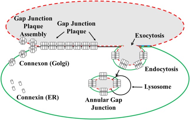
Adrenal cortical steroidogenesis and proliferation are thought to be modulated by gap junction-mediated direct cell-cell communication of regulatory molecules between cells. Such communication is regulated by the number of gap junction channels between contacting cells, the rate at which information flows between these channels, and the rate of channel turnover. Knowledge of the factors regulating gap junction-mediated communication and the turnover process are critical to an understanding of adrenal cortical cell functions, including development, hormonal response to adrenocorticotropin, and neoplastic dedifferentiation. Here, we review what is known about gap junctions in the adrenal gland, with particular attention to their role in adrenocortical cell steroidogenesis and proliferation. Information and insight gained from electrophysiological, molecular biological, and imaging (immunocytochemical, freeze fracture, transmission electron microscopic, and live cell) techniques will be provided.

Keywords: ACTH; connexin; gap junction plaques; gap junction vesicles; steroidogenesis.


Figures

Figure 1
Figure 1
Illustration of the formation and degradation of gap junction plaques and annular gap junctions. Connexin proteins synthesized in the endoplasmic reticulum (ER) oligomerize to form connexon complexes. The connexons are transported to the cell surface and inserted into the plasma membrane where they form hemichannels. These hemichannels can dock with hemichannels of an apposing cell and cluster to form a gap junction plaque, characterized by a 2–4 nm gap between the two cell membranes. Gap junction plaques are removed from the cell surface through endoexocytosis, which results in the formation of an annular gap junction. The annular gap junction is then degraded through lysosomal proteolysis [modified from Ref. (130)].
Figure 2
Figure 2
Characterization of gap junctions in SW-13 adrenocortical tumor cells. The size, location, and structure of gap junction plaques (arrowheads) and annular gap junctions (arrows) have been determined with (A) transmission electron microscopy, (B) freeze-fracture electron microscopy, (C) immunofluorescence, and (D) confocal microscopy. The protoplasmic (P) and extracellular (E) fracture faces are shown in the freeze-fracture replica of the gap junction plaque in (B). Cell borders are defined by cortical actin (green) in (C). Note the lumen of the annular gap junction revealed with confocal microscopy in (D). n, nucleus. Bars: (A) 100 nm, (B) 60 nm, (C) 10 μm, and (D) 0.3 μm. [(A) from Ref. (48), (B) from Ref. (130), (C) from Ref. (131), and (D) from Ref. (132)].
Figure 3
Figure 3
Gap junction (Cx43) localization and dye communication in the intact adrenal gland. Immunohistochemical localization of Cx43 gap junction proteins revealed extensive staining in the zona fasciculata (ZF) and zona reticularis (ZR), while there was limited staining in the zona glomerulosa (ZF) [left panels: (A,C)]. Correspondingly, lucifer yellow dye communication between cells was more abundant in the inner zones of the adrenal cortex (ACTH responsive areas) than in the outer zone [right panels: (B,D)]. Capsule (arrowheads), connective tissue trabecule (arrow), and Cx43 (white puncta). Bars: (A,B) 50 μm and (C,D) 30 μm [modified from Ref. (30)].
Figure 4
Figure 4
Communication in the intact adrenal cortex. Oil Red O staining was used to distinguish the lipid-rich zona fasciculata and zona reticularis from the zona glomerulosa (A,C). Lucifer yellow dye was transferred between the fibroblasts of the connective tissue capsule and trabeculae; however, communication was absent between cells of the zona glomerulosa (B). The cells of the inner two cortical zones, particularly those in the zona fasciculata, exhibited extensive dye communication (D). Note the Lucifer yellow and Oil Red O staining are diffuse and seen throughout the cytoplasm, which somewhat obscures the cell boundaries and nuclei. Bars: (A–D) 30 μm [modified from Ref. (30)].
Figure 5
Figure 5
Cocultured adrenal/granulosa cell pairs. Adrenal cell clusters viewed with optics specific for fluorescein isothiocyanate (A) or rhodamine isothiocyanate (B) were treated with follicle-stimulating hormone (FSH) for 30 min. Abundant protein kinase dissociation was initially observed in granulosa cells (OG) and could be seen, after 5–15 min, in two adrenal cells (Y-1*) that are in contact with the granulosa cell, while two nearby adrenal cells (Y-1) that are not in contact with the granulosa cell failed to dissociate PKA (A). Granulosa cells were identified by pre-labeling them with rhodamine-coated beads (B) [modified from Ref. (59)].
Figure 6
Figure 6
Immunohistochemical demonstration of gap junction (Cx43) distribution in the adrenal gland. Gap junction proteins were abundant in the inner cortex of the mouse adrenal (A). Hypophysectomy led to diminished Cx43 gap junction expression mainly in the zona fasciculata at 33 days post-surgery (B) [modified from Ref. (31)].
Figure 7
Figure 7
Schematic of the role of gap junctions in steroidogenesis and cell proliferation. The binding of ACTH to its receptors on coupled cortical cells stimulates cyclic adenosine monophosphate (cAMP) to activate protein kinase A (PKA), thereby increasing steroidogenesis and decreasing proliferation. Gap junction-mediated movement of cAMP between cells in the population would amplify hormonal responses in contacting cells, thus increasing ACTH-mediated steroidogenesis. An increase in the number of gap junctions, increases ACTH-mediated steroidogenesis while decreasing proliferation. However, if gap junctions are decreased as a result of molecular manipulations or chemical treatments, for example, ACTH stimulation in one cell does not affect steroidogenesis in the second cell, and there would be a loss in coordinated cell function between cells [modified from Ref. (130)].

References

    1. James VHT. The Adrenal Gland Comprehensive Endocrinology. New York, NY: Raven Press, Ltd; (1992).
    1. Decker RS, Donta ST, Larsen WJ, Murray SA. Gap junctions and ACTH sensitivity in Y-1 adrenal tumor cells. J Supramol Struct (1978) 9:497–507.10.1002/jss.400090405 - DOI - PubMed
    1. Meda P, Pepper MS, Traub O, Willecke K, Gros D, Beyer E, et al. Differential expression of gap junction connexins in endocrine and exocrine glands. Endocrinology (1993) 133:2371–8.10.1210/en.133.5.2371 - DOI - PubMed
    1. Munari-Silem Y, Lebrethon MC, Morand I, Rousset B, Saez JM. Gap junction-mediated cell-to-cell communication in bovine and human adrenal cells. A process whereby cells increase their responsiveness to physiological corticotropin concentrations. J Clin Invest (1995) 95:1429–39.10.1172/JCI117813 - DOI - PMC - PubMed
    1. Murray SA, Kumar NM, Gilula NB. Gap junction expression in rat adrenal gland. Intercellular communication through gap junctions. Prog Cell Res (1995) 4:293–6.10.1016/B978-0-444-81929-1.50059-9 - DOI

LinkOut - more resources