

Tertiary Lymphoid Organs in Cancer Tissues
- PMID: 27446075
- PMCID: PMC4916185
- DOI: 10.3389/fimmu.2016.00244
Tertiary Lymphoid Organs in Cancer Tissues
Abstract
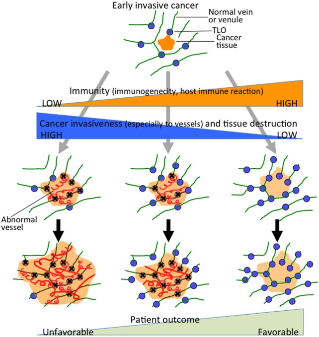
Tertiary lymphoid organs (TLOs) are induced postnatally in non-lymphoid tissues such as those affected by chronic infections, autoimmune diseases, and chronic allograft rejection, and also in cancer tissues. TLOs are thought to provide important lymphocytic functional environments for both cellular and humoral immunity, similar to lymph nodes or Peyer's patches. TLOs have a structure similar to that of lymph nodes or Peyer's patches, including T cell zones, B cell follicles, and high endothelial venules (HEV) without encapsulation. Here, we review recent advances in our knowledge of TLOs in human solid cancers, including their location, structure, methods of evaluation, and clinicopathological impact. We also discuss the formation and/or maintenance of TLOs in cancer tissues in association with the tumor immune microenvironment, cancer invasion, and the tissue structure of the cancer stroma.
Keywords: cancer; tertiary lymphoid organs; tissue structure; tumor immunology; tumor microenvironment.
Figures
Similar articles
-
High Endothelial Venules and Other Blood Vessels: Critical Regulators of Lymphoid Organ Development and Function.Front Immunol. 2017 Feb 3;8:45. doi: 10.3389/fimmu.2017.00045. eCollection 2017. Front Immunol. 2017. PMID: 28217126 Free PMC article. Review.
-
High Endothelial Venules and Lymphatic Vessels in Tertiary Lymphoid Organs: Characteristics, Functions, and Regulation.Front Immunol. 2016 Nov 9;7:491. doi: 10.3389/fimmu.2016.00491. eCollection 2016. Front Immunol. 2016. PMID: 27881983 Free PMC article. Review.
-
Intratumoral tertiary lymphoid organ is a favourable prognosticator in patients with pancreatic cancer.Br J Cancer. 2015 May 26;112(11):1782-90. doi: 10.1038/bjc.2015.145. Epub 2015 May 5. Br J Cancer. 2015. PMID: 25942397 Free PMC article.
-
Role of tertiary lymphoid organs in the regulation of immune responses in the periphery.Cell Mol Life Sci. 2022 Jun 11;79(7):359. doi: 10.1007/s00018-022-04388-x. Cell Mol Life Sci. 2022. PMID: 35689679 Free PMC article. Review.
-
CD4+ T cells of both the naive and the memory phenotype enter rat lymph nodes and Peyer's patches via high endothelial venules: within the tissue their migratory behavior differs.Eur J Immunol. 1997 Dec;27(12):3174-81. doi: 10.1002/eji.1830271214. Eur J Immunol. 1997. PMID: 9464803
Cited by
-
Emerging Roles of Mast Cells in the Regulation of Lymphatic Immuno-Physiology.Front Immunol. 2020 Jun 17;11:1234. doi: 10.3389/fimmu.2020.01234. eCollection 2020. Front Immunol. 2020. PMID: 32625213 Free PMC article. Review.
-
Tertiary lymphoid structures in the primary tumor site of patients with cancer-associated myositis: A case-control study.Front Med (Lausanne). 2023 Jan 4;9:1066858. doi: 10.3389/fmed.2022.1066858. eCollection 2022. Front Med (Lausanne). 2023. PMID: 36687449 Free PMC article.
-
Ectopic high endothelial venules in pancreatic ductal adenocarcinoma: A unique site for targeted delivery.EBioMedicine. 2018 Dec;38:79-88. doi: 10.1016/j.ebiom.2018.11.030. Epub 2018 Nov 27. EBioMedicine. 2018. PMID: 30497977 Free PMC article.
-
Neural reflex control of vascular inflammation.Bioelectron Med. 2020 Jan 31;6:3. doi: 10.1186/s42234-020-0038-7. eCollection 2020. Bioelectron Med. 2020. PMID: 32232111 Free PMC article. Review.
-
Identification of B Cell Subpopulations with Pro- and Anti-Tumorigenic Properties in an Immunocompetent Mouse Model of Head and Neck Squamous Cell Carcinoma.Cells. 2024 Dec 29;14(1):20. doi: 10.3390/cells14010020. Cells. 2024. PMID: 39791721 Free PMC article.
References
Publication types
LinkOut - more resources
Full Text Sources
Other Literature Sources