. 2016 Jun 30:7:253.
doi: 10.3389/fimmu.2016.00253. eCollection 2016.

Partial Protection against Porcine Influenza A Virus by a Hemagglutinin-Expressing Virus Replicon Particle Vaccine in the Absence of Neutralizing Antibodies

Affiliations

Partial Protection against Porcine Influenza A Virus by a Hemagglutinin-Expressing Virus Replicon Particle Vaccine in the Absence of Neutralizing Antibodies

Meret E Ricklin et al. Front Immunol. .

Abstract

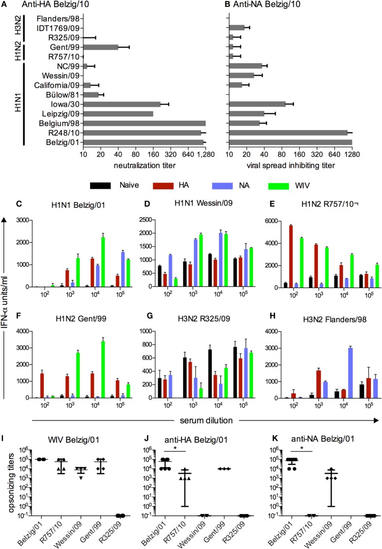
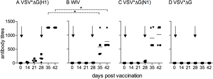
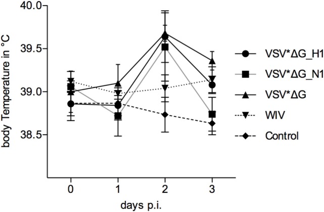
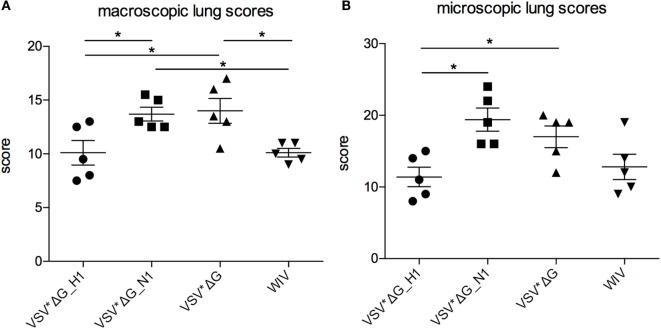
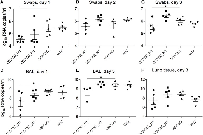
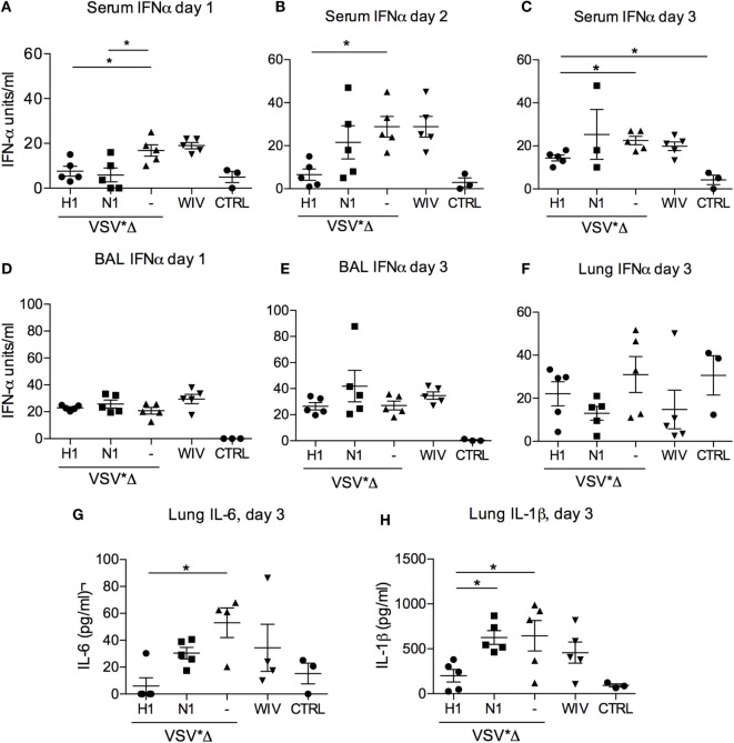
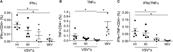
This work was initiated by previous reports demonstrating that mismatched influenza A virus (IAV) vaccines can induce enhanced disease, probably mediated by antibodies. Our aim was, therefore, to investigate if a vaccine inducing opsonizing but not neutralizing antibodies against the hemagglutinin (HA) of a selected heterologous challenge virus would enhance disease or induce protective immune responses in the pig model. To this end, we immunized pigs with either whole inactivated virus (WIV)-vaccine or HA-expressing virus replicon particles (VRP) vaccine based on recombinant vesicular stomatitis virus (VSV). Both types of vaccines induced virus neutralizing and opsonizing antibodies against homologous virus as shown by a highly sensitive plasmacytoid dendritic cell-based opsonization assay. Opsonizing antibodies showed a broader reactivity against heterologous IAV compared with neutralizing antibodies. Pigs immunized with HA-recombinant VRP vaccine were partially protected from infection with a mismatched IAV, which was not neutralized but opsonized by the immune sera. The VRP vaccine reduced lung lesions, lung inflammatory cytokine responses, serum IFN-α responses, and viral loads in the airways. Only the VRP vaccine was able to prime IAV-specific IFNγ/TNFα dual secreting CD4(+) T cells detectable in the peripheral blood. In summary, this work demonstrates that with the virus pair selected, a WIV vaccine inducing opsonizing antibodies against HA which lack neutralizing activity, is neither protective nor does it induce enhanced disease in pigs. In contrast, VRP-expressing HA is efficacious vaccines in swine as they induced both potent antibodies and T-cell immunity resulting in a broader protective value.

Keywords: CD4 T cells; VRP vaccine; hemagglutinin; influenza virus; neutralization; opsonization; pig.


Figures

Figure 1
Figure 1
Functional characterization and cross-reactivity of sera from vaccinated pigs. (A) Neutralizing activity against various IAV of anti-HA sera from pigs that were vaccinated with VSV*ΔG(H1) of Belzig/01. (B) Inhibitory activity of anti-NA sera from pigs vaccinated with VSV*ΔG(N1) of Belzig/01 against virus spread in cell culture. (B). In (A,B), titers were defined as the last serum dilution that inhibited infection or allowed only single cell infection. The sera were collected 3 weeks after the second booster immunization. Mean and SD of six replicates from a representative experiment out of two is shown. (C–H) Different viruses were tested in a pDC-based opsonization assay based for antibody-enhanced pDC activation and IFNα secretion following stimulation with immune complexes. Sera from naive, VSV*ΔG(H1), VSV*ΔG(N1), and WIV vaccinated pigs were serially diluted and tested for their ability to enhance IFNα of pDC stimulated by various IAV. A representative experiment is shown. (I–K) Mean and SD of all pDC-based opsonization assays performed. Opsonizing titers were defined as the highest serum dilution resulting in a significantly higher IFN-α production by enriched pDC as compared with naive serum. Each symbol represents an independent experiment. Asterisks (*) indicate significant differences calculated with the Wilcoxon test (p < 0.05).
Figure 2
Figure 2
Kinetics of antibody responses induced by VSV-based VRP vaccines. Sera from pigs (five animals per vaccine group) that were vaccinated with (A) VSV*ΔG(H1), (B) VSV*ΔG(N1), (C) VSV*ΔG, or (D) WIV were collected at the indicated time points, and tested for neutralization (A,C,D) or inhibition of viral spread (B) against homologous virus (Belzig/01). At day 28, all animals received a booster immunization. Arrows indicate the vaccination time points. Each symbol represents a different animal. Asterisks indicate significant differences between groups at particular days post-vaccination calculated with the Wilcoxon test (p < 0.05).
Figure 3
Figure 3
Body temperature following heterologous challenge with R757/10. VSV*ΔG(H1)-, VSV*ΔG(N1)-, VSV*ΔG-, or WIV-vaccinated animals were challenged with R757/10 (H1N2). In (A), mean values and SDs of body temperatures are shown for each group. In (B), values for individual animals are shown for day 2 p.i.
Figure 4
Figure 4
Lung lesions after heterologous challenge with R757/10. Lung lesions were assessed (A) macroscopically and (B) microscopically from each lobe at 3 days p.i. Scores were determined by a pathologist who was blinded for the treatments and assessed for each lobe. The values were pooled to one value per animal. Asterisks indicate significant differences calculated with the Wilcoxon test (p < 0.05).
Figure 5
Figure 5
Viral RNA loads after heterologous challenge with R757/10. Viral RNA copies per milliliter were assessed by RT-qPCR in swabs on days 1 (A), 2 (B), and 3 (C), in the BAL fluid on day 1 (D) and day 3 (E), and in the lung tissue on day 3 (F). Asterisks (*) indicate significant differences calculated with the Wilcoxon test (p < 0.05).
Figure 6
Figure 6
Innate cytokine responses after heterologous challenge with R757/10. In (A–C) IFNα levels were determined by ELISA in the serum of the infected animals on days 1 (A), 2 (B), and 3 (A) post-infection. (D,E) show the IFNα levels in the BAL on day 1 and 3, respectively. In (F–H) cytokine levels were determined in lung tissue lysates obtained on day 3 post-challenge. “CTRL” stands for samples obtained from non-infected SPF pigs from the same breeding. Each symbol represents a different animal. Asterisks (*) indicate significant differences calculated with the Wilcoxon test (p < 0.05).
Figure 7
Figure 7
Peripheral blood CD4 T-cell responses induced by VSV-based VRP and WIV vaccines. PBMCs were isolated at 3 weeks post-booster vaccination and restimulated with chicken allantoic fluid (CAF) as a mock control or IAV in vitro to determine the percentage of virus-specific IFNγ- (A), TNFα- (B), and dual cytokine (C) producing cells in the CD4 T cell subset. The percentage of virus-specific cytokine producing cells was determined by subtracting the values of the CAF controls from the IAV-stimulated cultures. The gating strategies and CAF/IAV percentages are shown in the Figure S3 in Supplementary Material. Asterisks (*) indicate significant differences calculated with the two-way Mann–Whitney U test (p < 0.05).

References

    1. Zambon MC. Epidemiology and pathogenesis of influenza. J Antimicrob Chemother (1999) 44(Suppl B):3–9. 10.1093/jac/44.suppl_2.3 - DOI - PubMed
    1. Capua I, Alexander DJ. Human health implications of avian influenza viruses and paramyxoviruses. Eur J Clin Microbiol Infect Dis (2004) 23(1):1–6. 10.1007/s10096-003-1059-3 - DOI - PubMed
    1. Yoon SW, Webby RJ, Webster RG. Evolution and ecology of influenza A viruses. Curr Top Microbiol Immunol (2014) 385:359–75. 10.1007/82_2014_396 - DOI - PubMed
    1. Brandenburg B, Koudstaal W, Goudsmit J, Klaren V, Tang C, Bujny MV, et al. Mechanisms of hemagglutinin targeted influenza virus neutralization. PLoS One (2013) 8(12):e80034. 10.1371/journal.pone.0080034 - DOI - PMC - PubMed
    1. Krammer F, Palese P. Influenza virus hemagglutinin stalk-based antibodies and vaccines. Curr Opin Virol (2013) 3(5):521–30. 10.1016/j.coviro.2013.07.007 - DOI - PMC - PubMed