. 2016 Jun 30:7:958.
doi: 10.3389/fpls.2016.00958. eCollection 2016.

Construction of Global Acyl Lipid Metabolic Map by Comparative Genomics and Subcellular Localization Analysis in the Red Alga Cyanidioschyzon merolae

Affiliations

Construction of Global Acyl Lipid Metabolic Map by Comparative Genomics and Subcellular Localization Analysis in the Red Alga Cyanidioschyzon merolae

Natsumi Mori et al. Front Plant Sci. .

Abstract

Pathways of lipid metabolism have been established in land plants, such as Arabidopsis thaliana, but the information on exact pathways is still under study in microalgae. In contrast with Chlamydomonas reinhardtii, which is currently studied extensively, the pathway information in red algae is still in the state in which enzymes and pathways are estimated by analogy with the knowledge in plants. Here we attempt to construct the entire acyl lipid metabolic pathways in a model red alga, Cyanidioschyzon merolae, as an initial basis for future genetic and biochemical studies, by exploiting comparative genomics and localization analysis. First, the data of whole genome clustering by Gclust were used to identify 121 acyl lipid-related enzymes. Then, the localization of 113 of these enzymes was analyzed by GFP-based techniques. We found that most of the predictions on the subcellular localization by existing tools gave erroneous results, probably because these tools had been tuned for plants or green algae. The experimental data in the present study as well as the data reported before in our laboratory will constitute a good training set for tuning these tools. The lipid metabolic map thus constructed show that the lipid metabolic pathways in the red alga are essentially similar to those in A. thaliana, except that the number of enzymes catalyzing individual reactions is quite limited. The absence of fatty acid desaturation to produce oleic and linoleic acids within the plastid, however, highlights the central importance of desaturation and acyl editing in the endoplasmic reticulum, for the synthesis of plastid lipids as well as other cellular lipids. Additionally, some notable characteristics of lipid metabolism in C. merolae were found. For example, phosphatidylcholine is synthesized by the methylation of phosphatidylethanolamine as in yeasts. It is possible that a single 3-ketoacyl-acyl carrier protein synthase is involved in the condensation reactions of fatty acid synthesis in the plastid. We will also discuss on the redundant β-oxidation enzymes, which are characteristic to red algae.

Keywords: Cyanidioschyzon merolae; comparative genomics; lipid metabolism; red alga; subcellular localization.


Figures
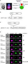
Figure 1
Figure 1
Examples of subcellular localization of GFP-fused proteins. (A) Fluorescence microscopic images showing a C. merolae cell stained with 4′,6-diamidino-2-phenylindole (DAPI; left). The illustration is a summary of cellular structure of C. merolae cell (right). Bar = 2 μm. (B) Structure of a construct expressing a GFP-fused protein. pCEG1 vector contains APCC promoter of C. merolae (PAPCC) (Watanabe et al., 2011), gene for enhanced green fluorescence protein (EGFP) and NOS terminator (NOS). Genomic fragment of N-terminal transit peptide (TP) of lipid metabolic enzymes was inserted into pCEG1 vector and cloned. This construct was transformed into C. merolae cells for transient expression. If necessary, transformed cells were immunostained with anti-GFP antibody. (C) Structure of a construct expressing an HA-tagged protein. pBSHAb-T3′ vector (Ohnuma et al., 2008) contains three repeats of HA tag (3 × HA) and 3′-untranslated region (UTR) of β-tubulin of C. merolae. A full-length sequence of candidate genes was inserted into pBSHAb-T3′ vector containing APCC promoter. This construct was transformed into C. merolae cells, and then subcellular localization of HA-tagged proteins was detected by immunostaining using anti-HA antibody. (D) Fluorescence micrographs of C. merolae cells transiently expressing GFP-fused proteins. These images show that subcellular localization of GFP-fused proteins to plastid (Pt), mitochondrion (Mt), cytosol (Cyt), ER, peroxisome (Per), and dual localization of ER and cytoplasmic membrane (CM). GFP-fused PAP was localized in the periphery of the nucleus. In GFP-fused DGAT, GFP fluorescence was observed as a fog in cytosol. We, therefore, supposed these proteins as ER-localized enzymes. An asterisk indicates an enzyme, whose subcellular localization was examined by immunofluorescence rather than GFP fluorescence. DIC, Nomarski differential interference contrast image; Chlorophyll, autofluorescence from phycobilin and chlorophyll; GFP, GFP fluorescence or immunofluorescence using anti-GFP antibody; Merge, merged images of autofluorescence and green fluorescence from the fusion protein. Bar = 2 μm. Abbreviation of enzyme names is listed in Table 1, Supplementary Table 3.
Figure 2
Figure 2
A map of lipid metabolic pathways in C. merolae based on the results of subcellular localization analysis. This pathway map was created based on the results of subcellular localization analysis (Figure 1D, Supplementary Figures 2–6). Enzyme name is indicated in blue. Locus tag of the genes encoded in the nuclear and the plastid genomes are shown in red and green, respectively. Fatty acids are represented by a combination of the number of carbon atom [X] and the number of double bonds [Y], such as X:Y. The number in the parenthesis indicates position of double bonds. Dashed line indicates flow of transport of substrates. In the plastidic fatty acid synthesis, it is possible that acetyl-CoA:ACP acetyltransferase (ACAT) converts acetyl-CoA to acetyl-ACP in the initial condensation reaction (see Discussion), although putative ACAT was not detected from the genomic data in C. merolae. Some enzymes related to mitochondrial fatty acid synthesis, mitochondrial enoyl-CoA reductase (mtEAR), mitochondrial 3-hydroxyacyl-ACP dehydratase (mtHAD), and mitochondrial 3-ketoacyl-ACP reductase (mtKAR), have not been identified either in plants or in C. merolae. In the synthesis of glycolipids and PG, phosphatidylglycerolphosphate phosphatase was not detected in the genomic data of C. merolae. No PAP candidates were localized in the plastid in C. merolae. Putative FAD4 enzyme is included in this figure, although its localization could not be experimentally determined. In C. merolae, linoleoyl-CoA is synthesized in the ER, and then is transferred to the plastid, but this transport system is largely unknown (dotted lines). Abbreviations: acyl-ACP, acyl-acyl carrier protein; acyl-CoA, acyl-coenzyme A; CDP-DAG, CDP-diacylglycerol; DAG, diacylglycerol; DGDG, digalactosyldiacylglycerol; FA, fatty acid; G3P, glycerol-3-phosphate; LPA, lysophosphatidic acid; LPL, lysophospholipid; MGDG, monogalactosyldiacylglycerol; PA, phosphatidic acid; PC, phosphatidylcholine; PE, phosphatidylethanolamine; PG, phosphatidylglycerol; PGP, phosphatidylglycerolphosphate; PI, phosphatidylinositol; PL, phospholipid; PS, phosphatidylserine; SQDG, sulfoquinovosyldiacylglycerol; TAG, triacylglycerol. Abbreviated names of enzyme and genes are shown according to Table 1, Supplementary Table 3.
Figure 3
Figure 3
β-Oxidation of fatty acids in C. merolae. (A) Schematic domain structure of representative MFPs. We searched for functional domains of these MFPs using the Pfam database (Finn et al., 2014). Gray boxes indicate individual functional domains, namely enoyl-CoA hydratase/isomerase family domain (1), 3-hydroxyacyl-CoA dehydrogenase NAD binding domain (2), 3-hydroxyacyl-CoA dehydrogenase C-terminal domain (3), thiolase N-terminal domain (4), and thiolase C-terminal domain (5). Homo sapience has both peroxisomal MFP (perMFP) and mitochondrial MFP (mtMFP). mtMFP is a complex consisting of α and β subunits (α or β). (B) A pathway map of β-oxidation in C. merolae. This pathway map was created based on the results of subcellular localization analysis (Supplementary Figures 4A,B). For abbreviated names of enzymes and genes, see Table 1.

References

    1. Bates P. D., Stymne S., Ohlrogge J. (2013). Biochemical pathways in seed oil synthesis. Curr. Opin. Plant Biol. 16, 358–364. 10.1016/j.pbi.2013.02.015 - DOI - PubMed
    1. Chapman K. D., Dyer J. M., Mullen R. T. (2012). Biogenesis and functions of lipid droplets in plants: thematic Review Series: Lipid Droplet Synthesis and Metabolism: from Yeast to Man. J. Lipid Res. 53, 215–226. 10.1194/jlr.R021436 - DOI - PMC - PubMed
    1. Clough R. C., Matthis A. L., Barnum S. R., Jaworski J. G. (1992). Purification and characterization of 3-ketoacyl-acyl carrier protein synthase III from spinach. A condensing enzyme utilizing acetyl-coenzyme A to initiate fatty acid synthesis. J. Biol. Chem. 267, 20992–20998. - PubMed
    1. Emanuelsson O., Nielsen H., Brunak S., von Heijne G. (2000). Predicting subcellular localization of proteins based on their N-terminal amino acid sequence. J. Mol. Biol. 300, 1005–1016. 10.1006/jmbi.2000.3903 - DOI - PubMed
    1. Finn R. D., Bateman A., Clements J., Coggill P., Eberhardt R. Y., Eddy S. R., et al. . (2014). Pfam: the protein families database. Nucleic Acids Res. 42, 222–230. 10.1093/nar/gkt1223 - DOI - PMC - PubMed