. 2016 Aug;12(2):1271-1278.
doi: 10.3892/ol.2016.4797. Epub 2016 Jun 28.

SIX1 coordinates with TGFβ signals to induce epithelial-mesenchymal transition in cervical cancer

Affiliations

SIX1 coordinates with TGFβ signals to induce epithelial-mesenchymal transition in cervical cancer

Shu-Hua Sun et al. Oncol Lett. 2016 Aug.

Abstract

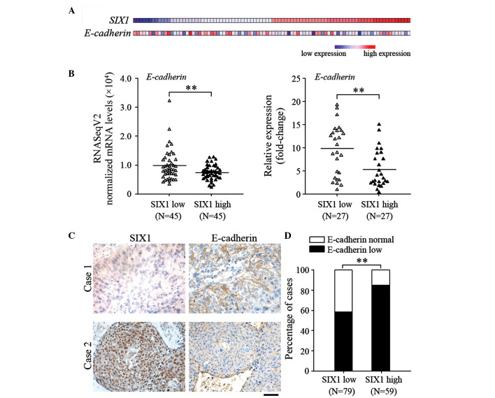
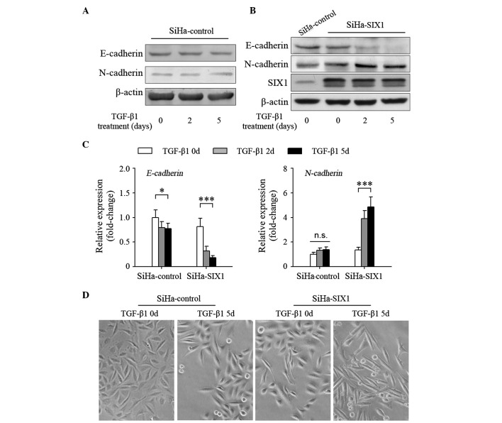
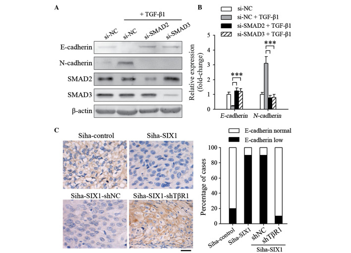
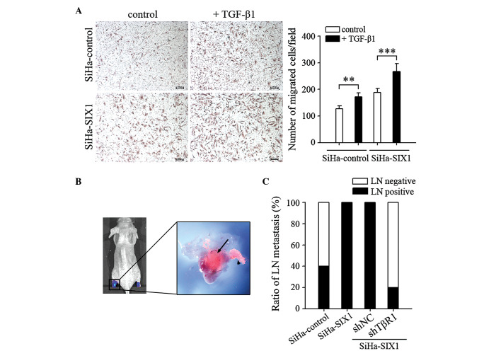
Epithelial-mesenchymal transition (EMT) plays a critical role in promoting tumor invasion and metastasis. However, the key cofactors that modulate the signal transduction to induce EMT have note been fully explored to date. The present study reports that sine oculis homeobox homolog 1 (SIX1) is able to promote EMT of cervical cancer by coordinating with transforming growth factor (TGF)β-SMAD signals. The expression of SIX1 was negatively correlated with the expression of the epithelial marker E-cadherin in two independent groups of cervical cancer specimens. SIX1 could promote the transition of mesenchymal phenotype in the presence of active TGFβ signals in vitro and in vivo. TGFβ-SMAD signals were required for the SIX1-mediated promotion of EMT and metastatic capacity of cervical cancer cells. Together, SIX1 and TGFβ cooperated to induce more remarkable changes in the transition of phenotype than each of them alone, and coordinated to promote cell motility and tumor metastasis in cervical cancer. These results suggest that the coordination of SIX1 and TGFβ signals may be crucial in the EMT program, and that SIX1/TGFβ may be considered a valuable marker for evaluating the metastatic potential of cervical cancer cells, or a therapeutic target in the treatment of cervical cancer.

Keywords: EMT; SIX1; TGFβ; cervical cancer; metastasis.


Figures

Figure 1.
Figure 1.
The expression of SIX1 and E-cadherin is negatively correlated in cervical cancer. (A) The expression profile of SIX1 and E-cadherin in cervical cancer specimens obtained from the TCGA database is represented as a heat map. (B) The TCGA RNA-sequencing data of cervical cancer was divided into two groups by the median level of SIX1 expression. The normalized messenger RNA levels of E-cadherin in the samples are indicated. **False discovery rate <0.05. The expression levels of SIX1 and E-cadherin in human CSCC specimens were detected by reverse transcription-quantitative polymerase chain reaction. (C and D) Immunohistochemical analysis of SIX1 and E-cadherin protein expression in tissue microarrays of human CSCC specimens. (C) Representative images of SIX1 and E-cadherin staining (magnification, ×200; scale bar, 100 µm). (D) Percentage of cases with different intensity of E-cadherin staining. **P<0.01. TCGA, The Cancer Genome Atlas; SIX1, sine oculis homeobox homolog 1; mRNA, messenger RNA; RNASeqV2, RNA-sequencing version 2; CSCC, cervical squamous cell carcinoma.
Figure 2.
Figure 2.
SIX1 enhances TGFβ-induced epithelial-mesenchymal transition in cervical cancer cells. SiHa cells, untransfected or transfected with SIX1-expressing vector, were untreated or treated with TGFβ1 (1 ng/ml) for the indicated times. The protein expression levels of E-cadherin and N-cadherin in (A) SiHa-control and (B) SiHa-SIX1 cells were detected by western blotting. The upper band is phosphorylated SIX and the lower band is unphosphorylated SIX1. Both bands were taken into account for phosphorylation. (C) The messenger RNA expression levels of E-cadherin and N-cadherin were detected by reverse transcription-quantitative polymerase chain reaction. (D) The morphological changes caused by SIX1 or/and TGFβ1 in SiHa cells were observed by microscopy. *P<0.05; ***P<0.001. SIX1, sine oculis homeobox homolog 1; TGF; transforming growth factor; n.s., not significant.
Figure 3.
Figure 3.
TGFβ signals are necessary for SIX1 to induce epithelial-mesenchymal transition in cervical cancer. (A and B) At 24 h post-transfection with the indicated small interfering RNAs, the cells were stimulated with TGFβ1 (1 ng/ml) for 5 days. The expression levels of E-cadherin and N-cadherin were detected by (A) western blotting or (B) reverse transcription-quantitative polymerase chain reaction. (C) SiHa cells were injected subcutaneously into the claw pads of mice to form primary tumors, and the protein expression levels of E-cadherin in the tumors were analyzed by immunohistochemical analysis. Left panel, representative images of SIX1 and E-cadherin staining of primary tumors (magnification, ×400; scale bar, 50 µm). Right panel, percentage of cases (n=10/group) with different intensity of E-cadherin staining. ***P<0.001. TGF; transforming growth factor; SIX1, sine oculis homeobox homolog 1; NC, negative control; sh, small hairpin; si, small interfering; TβR1, TGFβ receptor 1.
Figure 4.
Figure 4.
SIX1 coordinates with TGFβ signals to enhance the metastatic capacity of cervical cancer cells. (A) Migration assay. SiHa cells, untransfected or transfected with SIX1-expressing vector, were untreated or treated with TGFβ1 (1 ng/ml) for 5 days, and their migration ability was assessed (scale bar, 100 µm). (B) In vivo bioluminescence and fluorescence representative images of lymphatic metastasis in mice. Lymph nodes in the popliteal and inguinal regions of the mice were detected by bioluminescence and fluorescence signals, corresponding to luciferase activity and red fluorescent protein expression, respectively. The black arrow indicates tumor cells inside the lymph node, while the arrowhead indicates a tumor-invaded lymphatic vessel. (C) The ratios of lymph node metastasis were calculated (n=10/group). **P<0.01; ***P<0.001. SIX1, sine oculis homeobox homolog 1; TGF; transforming growth factor; LN, lymph node; TβR1, TGFβ receptor 1; NC, negative control; sh, small hairpin.

References

    1. Gao D, Vahdat LT, Wong S, Chang JC, Mittal V. Microenvironmental regulation of epithelial-mesenchymal transitions in cancer. Cancer Res. 2012;72:4883–4889. doi: 10.1158/0008-5472.CAN-12-1223. - DOI - PMC - PubMed
    1. Tsai JH, Yang J. Epithelial-mesenchymal plasticity in carcinoma metastasis. Genes Dev. 2013;27:2192–2206. doi: 10.1101/gad.225334.113. - DOI - PMC - PubMed
    1. Li Y, Wang W, Wang W, Yang R, Wang T, Su T, Weng D, Tao T, Li W, Ma D, Wang S. Correlation of TWIST2 up-regulation and epithelial-mesenchymal transition during tumorigenesis and progression of cervical carcinoma. Gynecol Oncol. 2012;124:112–118. doi: 10.1016/j.ygyno.2011.09.003. - DOI - PubMed
    1. Zhou XM, Zhang H, Han X. Role of epithelial to mesenchymal transition proteins in gynecological cancers: Pathological and therapeutic perspectives. Tumour Biol. 2014;35:9523–9530. doi: 10.1007/s13277-014-2537-1. - DOI - PubMed
    1. Thiery JP, Acloque H, Huang RY, Nieto MA. Epithelial-mesenchymal transitions in development and disease. Cell. 2009;139:871–890. doi: 10.1016/j.cell.2009.11.007. - DOI - PubMed

LinkOut - more resources