

Introduction to ultrasound elastography
- PMID: 27446596
- PMCID: PMC4954857
- DOI: 10.15557/JoU.2016.0013
Introduction to ultrasound elastography
Abstract
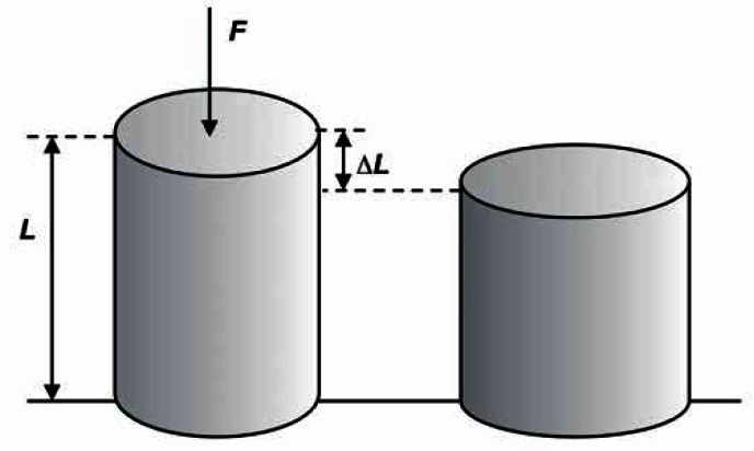
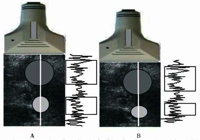
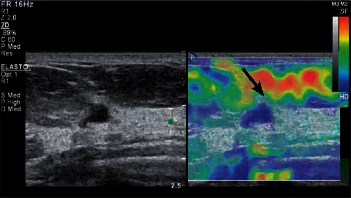
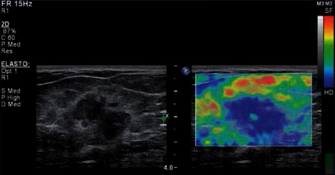
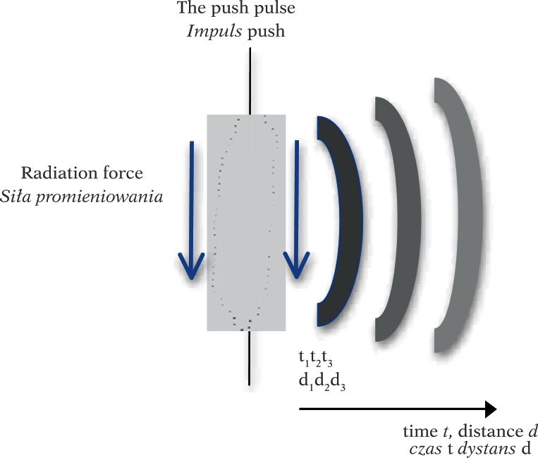
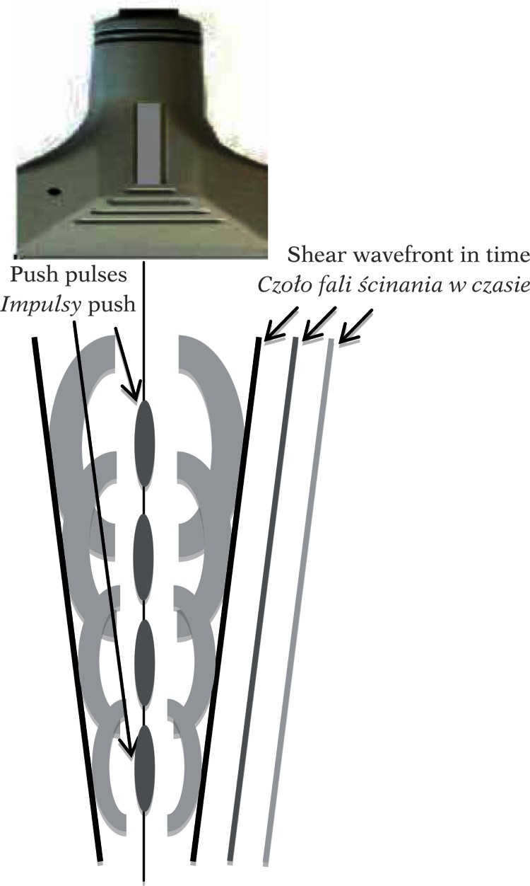
For centuries tissue palpation has been an important diagnostic tool. During palpation, tumors are felt as tissues harder than the surrounding tissues. The significance of palpation is related to the relationship between mechanical properties of different tissue lesions. The assessment of tissue stiffness through palpation is based on the fact that mechanical properties of tissues are changing as a result of various diseases. A higher tissue stiffness translates into a higher elasticity modulus. In the 90's, ultrasonography was extended by the option of examining the stiffness of tissue by estimating the difference in backscattering of ultrasound in compressed and non-compressed tissue. This modality is referred to as the static, compression elastography and is based on tracking the deformation of tissue subjected to the slowly varying compression through the recording of the backscattered echoes. The displacement is estimated using the methods of cross-correlation between consecutive ultrasonic lines of examined tissue, so calculating the degree of similarity of ultrasonic echoes acquired from tissue before and after the compression was applied. The next step in the development of ultrasound palpation was to apply the local remote tissue compression by using the acoustic radiation force generated through the special beam forming of the ultrasonic beam probing the tissue. The acoustic radiation force causes a slight deformation the tissue thereby forming a shear wave propagating in the tissue at different speeds dependent on the stiffness of the tissue. Shear wave elastography, carries great hopes in the field of quantitative imaging of tissue lesions. This article describes the physical basis of both elastographic methods: compression elastography and shear wave elastography.
Od stuleci badanie palpacyjne tkanek stanowi ważne narzędzie diagnostyczne. Nowotwory są zazwyczaj wyczuwane palpacyjnie jako tkanki twardsze od otoczenia. Istotność palpacji związana jest z zależnością wielu zmian tkankowych od ich własności mechanicznych. Palpacyjna ocena twardości lub sztywności tkanek opiera się na fakcie, że wiele chorób powoduje zmiany ich własności mechanicznych. Zwiększona sztywność tkanki oznacza zwiększony moduł sprężystości. W latach 90. ultrasonografia została rozszerzona o opcję badania sztywności tkanek, polegającą na ocenie różnicy w rozproszeniu ultradźwięków na tkance nieuciśniętej i uciśniętej. Jest to statyczna metoda elastografii kompresyjnej. Polega ona na wolnozmiennym ucisku badanego obszaru tkanki i ocenie jej odkształcenia poprzez śledzenie zmian w echach ultradźwiękowych rejestrowanych w kolejnych chwilach uciskania. Wielkość przemieszczenia wyznacza się metodami korelacji wzajemnej pomiędzy następującymi po sobie liniami obrazowymi, a więc obliczając stopień podobieństwa ech ultradźwiękowych przed uciskiem i po ucisku. Kolejnym krokiem w rozwoju palpacji ultradźwiękowej było zastosowanie do lokalnego, zdalnego ucisku tkanki, akustycznej siły promieniowania generowanej przez odpowiednie formownie wiązki ultradźwiękowej sondującej badany narząd. Akustyczna siła promieniowania powoduje niewielkie odkształcenie lub przemieszczenie się tkanki, w wyniku czego powstaje fala ścinania rozchodząca się w tkance z różną prędkością, zależną od sztywności tkanki. Elastografia fali ścinania niesie ogromne nadzieje w zakresie ilościowego obrazowania lokalnych zmian własności tkanki. W artykule opisane zostały podstawy fizyczne obu typów elastografii: kompresyjnej i fali ścinania.
Keywords: dynamic sonoelastography; elastography; static sonoelastography; ultrasonography.
Figures
References
-
- Bamber J, Cosgrove D, Dietrich CF, Fromageau J, Bojunga J, Calliada F, et al. EFSUMB guidelines and recommendations on the clinical use of ultrasound elastography. Part 1: Basic principles and technology. Ultraschall Med. 2013;34:169–184. - PubMed
-
- Ophir J, Céspedes I, Ponnekanti H, Yazdi Y, Li X. Elastography: a quantitative method for imaging the elasticity in biological tissues. Ultrasonic Imaging. 1991;13:111–134. - PubMed
-
- Krouskop TA, Wheeler TM, Kallel F, Garra BS, Hall T. Elastic moduli of breast and prostate tissues under compression. Ultrasonic Imaging. 1998;20:260–274. - PubMed
-
- Itoh A, Ueno E, Tohno E, Kamma H, Takahashi H, Shiina T, et al. Breast disease: clinical application of US elastography for diagnosis. Radiology. 2006;239:341–350. - PubMed
Publication types
LinkOut - more resources
Full Text Sources
Other Literature Sources