. 2016 Jun 28;7(7):2849-60.
doi: 10.1364/BOE.7.002849. eCollection 2016 Jul 1.

Imaging of targeted lipid microbubbles to detect cancer cells using third harmonic generation microscopy

Affiliations

Imaging of targeted lipid microbubbles to detect cancer cells using third harmonic generation microscopy

Kaitlin Harpel et al. Biomed Opt Express. .

Abstract

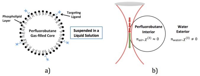
The use of receptor-targeted lipid microbubbles imaged by ultrasound is an innovative method of detecting and localizing disease. However, since ultrasound requires a medium between the transducer and the object being imaged, it is impractical to apply to an exposed surface in a surgical setting where sterile fields need be maintained and ultrasound gel may cause the bubbles to collapse. Multiphoton microscopy (MPM) is an emerging tool for accurate, label-free imaging of tissues and cells with high resolution and contrast. We have recently determined a novel application of MPM to be used for detecting targeted microbubble adherence to the upregulated plectin-receptor on pancreatic tumor cells. Specifically, the third-harmonic generation response can be used to detect bound microbubbles to various cell types presenting MPM as an alternative and useful imaging method. This is an interesting technique that can potentially be translated as a diagnostic tool for the early detection of cancer and inflammatory disorders.

Keywords: (020.4180) Multiphoton processes; (170.1610) Clinical applications; (190.1900) Diagnostic applications of nonlinear optics; (320.7090) Ultrafast lasers.


Figures

Fig. 1
Fig. 1
a) The basic components of the lipid microbubble used in the experiment. The specific targeted ligand (KTLLPTP) used was selective for the plectin-1 receptor. b) Microbubble and focused femtosecond laser beam interaction. THG signal is expected to be generated strongly from the liquid/gas interface.
Fig. 2
Fig. 2
Left: Schematic diagram of our home-built multiphoton microscope. The system has a THG spot size of ~350 nm and an axial resolution of 2μm (with a Nikon 40x objective, NA 1.3) at the excitation wavelength of 1040nm. The system has a THG spot size of ~525 nm and an axial resolution of 3 μm (with a Nikon 40x objective, NA 1.3) at the excitation wavelength of 1560nm. Right: A photograph of the microscope where both excitation laser sources are visible. The 1560nm laser is the the gray box on top and the 1040nm laser source is the black box at the bottom. A Ocean Optics spectrometer (350nm-1100nm detection range) is integrated into the multiphoton microscope for measuring the optical spectrum of the multiphoton excited signals (black box with blue input fiber on the left).
Fig. 3
Fig. 3
Both upper images display lipid microbubbles conjugated with DiI. (a) Image taken by confocal microscopy where bubbles are dispersed and bound to a poly d-lysine cell culture plate with residual DiI washed away. (b) Image taken by multiphoton microscopy (using a 40x Nikon oil objective with a 1.3 NA), specifically THG, where many unbound microbubbles are floating in a solution. (c) Emission spectrum from 1560nm multiphoton microscope displaying emitted THG signal during microbubble imaging compared to the total pump laser. (d) The pump laser spectrum (divided by three) overlaid on the THG spectrum. In a perfect THG process, these curves would be identical. However, it is clear that the THG spectrum is cleaner than the pump laser, and this is likely due to chromatic dispersion, chromatic aberration, and the specific geometry of the focused field.
Fig. 4
Fig. 4
Displays targeted lipid microbubbles conjugated with DiI. (a) The THG signal from the bubbles only (b) The fluorescence signal from the bubbles (c) A composite image of (a), represented in red, and (b), represented in green with the colocalized microbubbles represented with a yellow membrane (d) The measured emission spectrum of the DiI conjugated to the membrane of the lipid microbubbles compared to the thermofisher emission spectrum for DiI.
Fig. 5
Fig. 5
Displays pancreatic cancer cells with the targeted lipid microbubbles bound to the surface of the cells. (a) The THG signal from the bubbles only (b) The fluorescence signal from the bubbles and cells (c) A composite image of (a), represented in red, and (b), represented in green with the colocalized microbubbles represented in yellow (d) An image obtained from confocal microscopy for comparison (e) A spectrum obtained from Thermo Fisher displaying the excitation and emission wavelengths of both calcein and DiI

References

    1. Chen H., Zhen Z., Todd T., Chu P. K., Xie J., “Nanoparticles for Improving Cancer Diagnosis,” Mater. Sci. Eng. R. 74, 35–69 (2013). - PMC - PubMed
    1. König K., “Multiphoton microscopy in life sciences,” J. Microsc. 200(2), 83–104 (2000).10.1046/j.1365-2818.2000.00738.x - DOI - PubMed
    1. Zipfel W. R., Williams R. M., Webb W. W., “Nonlinear magic: Multiphoton microscopy in the biosciences,” Nat. Biotechnol. 21(11), 1369–1377 (2003).10.1038/nbt899 - DOI - PubMed
    1. Helmchen F., Denk W., “Deep tissue two-photon microscopy,” Nat. Methods 2(12), 932–940 (2005).10.1038/nmeth818 - DOI - PubMed
    1. Hoover E. E., Squier J. A., “Advances in multiphoton microscopy technology,” Nat. Photonics 7(2), 93–101 (2013).10.1038/nphoton.2012.361 - DOI - PMC - PubMed