. 2016 May;2(3):328-339.
doi: 10.1016/j.jcmgh.2015.12.008.

Intestinal dysbiosis contributes to the delayed gastrointestinal transit in high-fat diet fed mice

Affiliations

Intestinal dysbiosis contributes to the delayed gastrointestinal transit in high-fat diet fed mice

Mallappa Anitha et al. Cell Mol Gastroenterol Hepatol. 2016 May.

Abstract

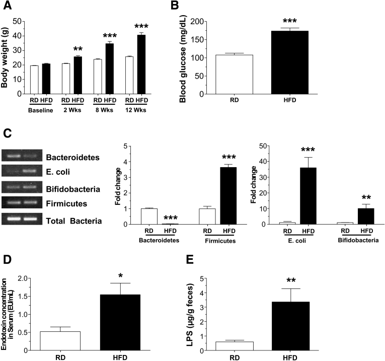
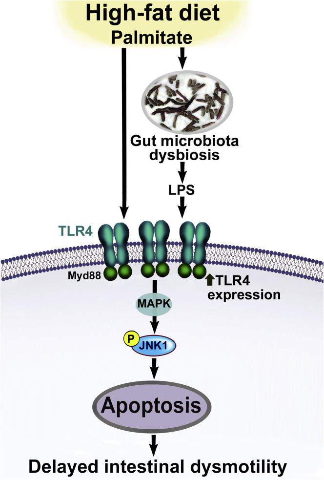
Background & aims: High-fat diet (HFD) feeding is associated with gastrointestinal motility disorders. We recently reported delayed colonic motility in mice fed a HFD mice for 11 weeks. In this study, we investigated the contributing role of gut microbiota in HFD-induced gut dysmotility.

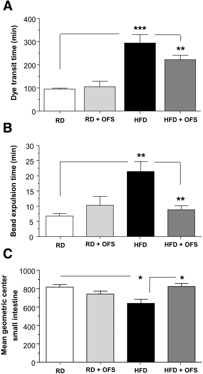
Methods: Male C57BL/6 mice were fed a HFD (60% kcal fat) or a regular/control diet (RD) (18% kcal fat) for 13 weeks. Serum and fecal endotoxin levels were measured, and relative amounts of specific gut bacteria in the feces assessed by real time PCR. Intestinal transit was measured by fluorescent-labeled marker and bead expulsion test. Enteric neurons were assessed by immunostaining. Oligofructose (OFS) supplementation with RD or HFD for 5 weeks was also studied. In vitro studies were performed using primary enteric neurons and an enteric neuronal cell line.

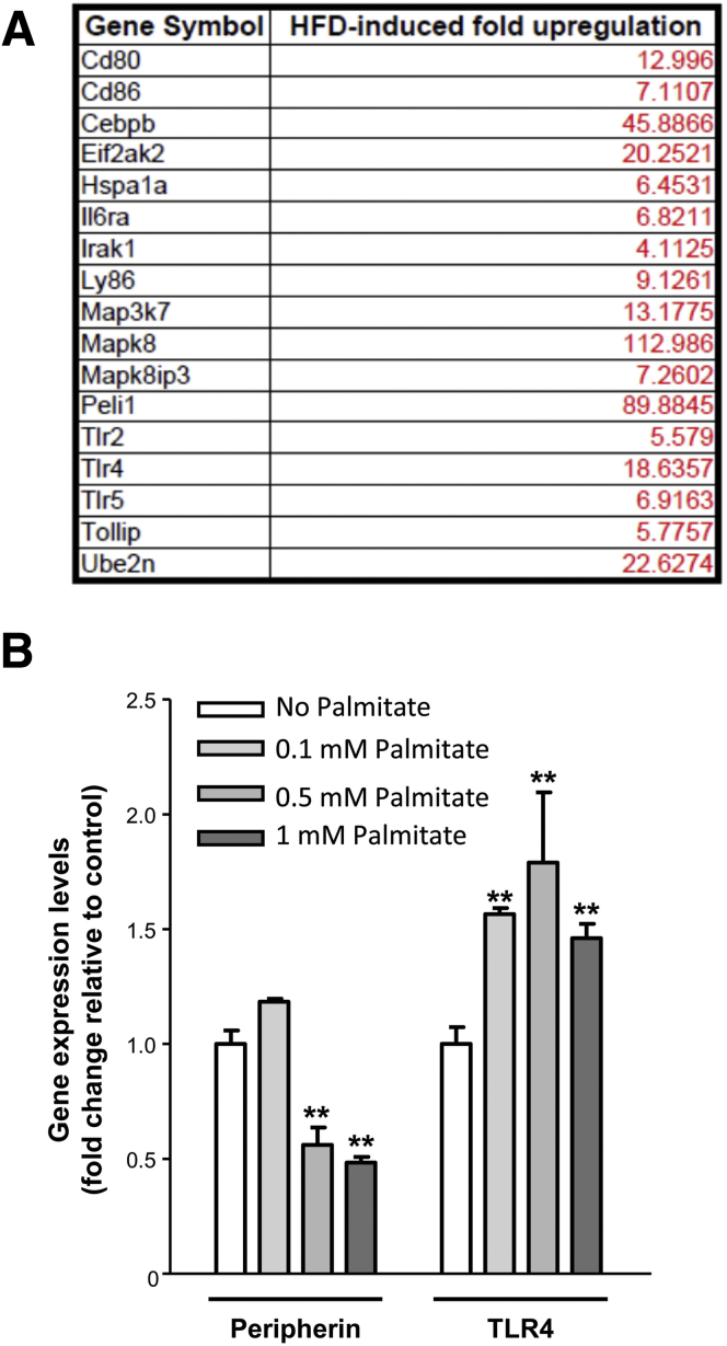
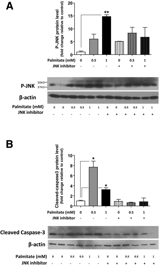
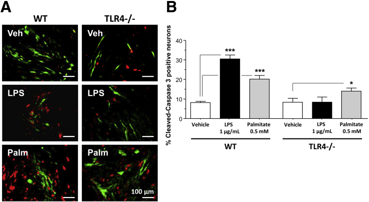
Results: HFD-fed mice had reduced numbers of enteric nitrergic neurons and exhibited delayed gastrointestinal transit compared to RD-fed mice. HFD-fed mice had higher fecal Firmicutes and Escherichia coli and lower Bacteroidetes compared to RD-fed mice. OFS supplementation protected against enteric nitrergic neurons loss in HFD-fed mice, and improved intestinal transit time. OFS supplementation resulted in a reductions in fecal Firmicutes and Escherichia coli and serum endotoxin levels. In vitro, palmitate activation of TLR4 induced enteric neuronal apoptosis in a p-JNK1 dependent pathway. This apoptosis was prevented by a JNK inhibitor and in neurons from TLR4-/- mice.

Conclusions: Together our data suggest that intestinal dysbiosis in HFD fed mice contribute to the delayed intestinal motility by inducing a TLR4-dependant neuronal loss. Manipulation of gut microbiota with OFS improved intestinal motility in HFD mice.

Keywords: LPS; Myenteric neurons; TLR4; colon transit; gut microbiota; palmitate.


Figures

Figure 1
Figure 1
High-fat diet feeding alters gut microbiota and results in endotoxemia in mice. (A) Body weights of mice fed a HFD or RD for up to 12 weeks and (B) fasting blood glucose level after 12 weeks. (C) PCR analyses of gut microbiota in stool from HFD- and RD-fed mice stool showing the relative amount of Bacteroidetes, Firmicutes, E coli, and Bifidobacteria. (D) Serum endotoxin levels and (E) stool LPS levels in RD- and HFD-fed mice. Results are means ± SEM; n = 12 per group. *P < .05, **P < .01, and ***P < .001.
Figure 2
Figure 2
OFS-induced changes in gut microbiota leads to reversal of HF-diet induced enteric neuronal loss. (A) Gut microbiota in stool from mice fed HFD or RD supplemented with or without OFS. (B) Endotoxin levels in serum from mice fed HFD or RD supplemented with or without OFS. (C) Representative photographs of proximal colon whole mount stained for peripherin and nNOS and histograms of neuronal counts. The number of stained neurons was determined per unit area. Scale bars: 50 μm. Results are means ± SEM; n = 6. *P < .05, **P < .01, and ***P < .001.
Figure 3
Figure 3
OFS supplementation leads to reversal of HFD-induced intestinal dysmotility. Assessment of gastrointestinal motility in mice fed a RD or HFD for 13 weeks and an additional 5 weeks with the diet supplemented with or without OFS. (A) Dye transit time after oral gavage with Evans blue dye/methyl cellulose solution, (B) bead expulsion time, and (C) mean geometric center of small intestine in mice fed a RD or HFD for 13 weeks and an additional 5 weeks with the diet supplemented with or without OFS. Results are means ± SEM; n = 6. *P < .05, **P < .01, and ***P < .001.
Figure 4
Figure 4
HFD feeding increases the expression of TLR4 and its downstream target genes in myenteric ganglia. (A) List of highly up-regulated genes in myenteric ganglia isolated by laser capture microdissection from the proximal colon of mice fed RD or HFD for 13 weeks (n = 3). (B) Effect of palmitate on peripherin, and TLR4 gene expression in the IM-PEN cell line as assessed by real-time PCR. Results are means ± SEM; n = 3. **P < .01.
Figure 5
Figure 5
Palmitate induces the phosphorylation of JNK and cleavage of caspase-3 in enteric neurons in vitro. Western blot analysis of (A) JNK phosphorylation and (B) caspase-3 cleavage in IM-PEN neuronal cells cultured in medium supplemented with various concentrations of palmitate in the absence or presence of the JNK inhibitor SP600125. Plotted are means ± SEM; n = 3. *P < .05, **P < .01.
Figure 6
Figure 6
Effect of palmitate on the survival of primary enteric neuronal cells from WT and TLR4-/- mice. (A) Representative photographs of WT or TLR4-/- primary enteric neuronal cells cultured in the presence of vehicle, LPS (1 μg/mL), or palmitate (0.5 mmol/L), and stained for PGP9.5 (green) and cleaved-caspase-3 (red). (B) Plot of cleaved caspase-3–positive neuronal cell counts. Plotted are means ± SEM; n = 6–8. *P < .05, ***P < .001. Palm, palmitate; Veh, vehicle.
Figure 7
Figure 7
A proposed model by which enhanced TLR4 activation leads to myenteric neuronal apoptosis in HFD-fed mice. We propose that a HFD can lead to increased circulating LPS levels, resulting from gut microbiota dysbiosis and activation of TLR4 signaling. In addition, palmitate in HFD can lead to increased TLR4 expression. Together LPS and palmitate lead to enhanced TLR4 signaling, which in turn leads to enhanced mitogen-activated protein kinase (JNK1) signaling and apoptosis of myenteric neurons, and consequent delayed intestinal motility.

Similar articles

Cited by

References

    1. Martin B.C., Barghout V., Cerulli A. Direct medical costs of constipation in the United States. Manag Care Interface. 2006;19:43–49. - PubMed
    1. Bharucha A.E., Dorn S.D., Lembo A. American Gastroenterological Association medical position statement on constipation. Gastroenterology. 2013;144:211–217. - PubMed
    1. Bharucha A.E., Pemberton J.H., Locke G.R., 3rd American Gastroenterological Association technical review on constipation. Gastroenterology. 2013;144:218–238. - PMC - PubMed
    1. Taba Taba Vakili S., Nezami B.G., Shetty A. Association of high dietary saturated fat intake and uncontrolled diabetes with constipation: evidence from the National Health and Nutrition Examination Survey. Neurogastroenterol Motil. 2015;27:1389–1397. - PMC - PubMed
    1. vd Baan-Slootweg O.H., Liem O., Bekkali N. Constipation and colonic transit times in children with morbid obesity. J Pediatr Gastroenterol Nutr. 2011;52:442–445. - PubMed

LinkOut - more resources