. 2016 Jul 7;1(10):e86419.
doi: 10.1172/jci.insight.86419.

Induced regulatory T cells in allograft tolerance via transient mixed chimerism

Affiliations

Induced regulatory T cells in allograft tolerance via transient mixed chimerism

Kiyohiko Hotta et al. JCI Insight. .

Abstract

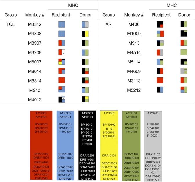
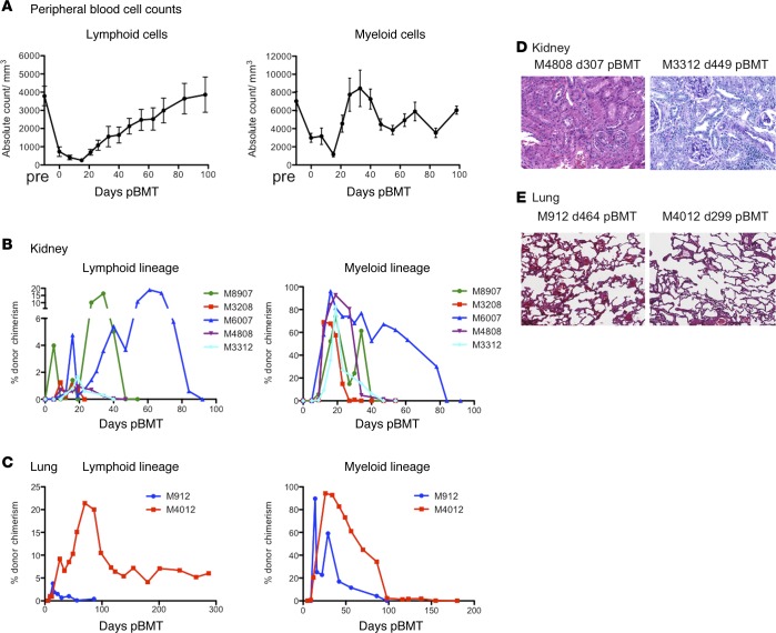
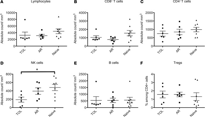
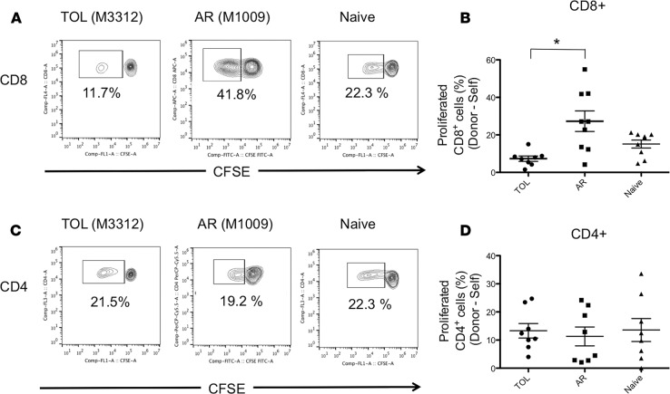
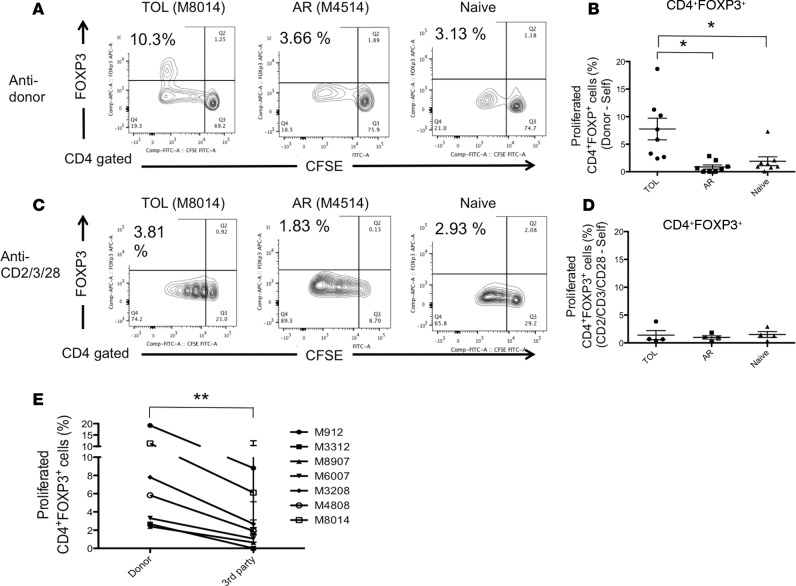
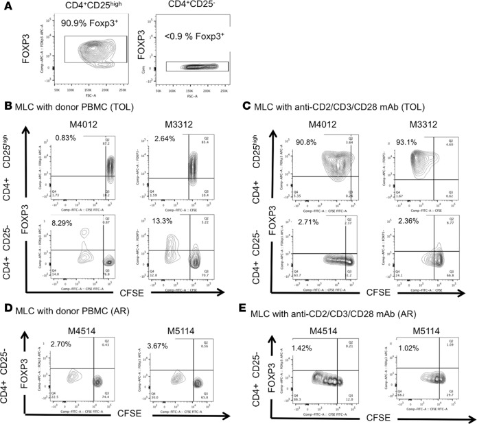
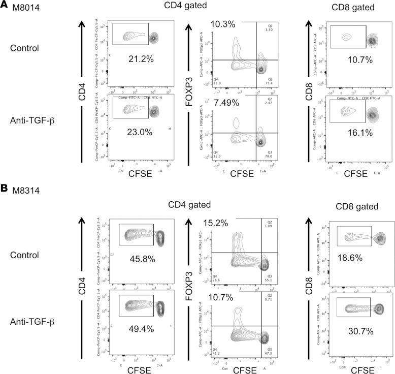
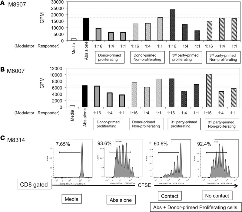
Successful induction of allograft tolerance has been achieved in nonhuman primates (NHPs) and humans via induction of transient hematopoietic chimerism. Since allograft tolerance was achieved in these recipients without durable chimerism, peripheral mechanisms are postulated to play a major role. Here, we report our studies of T cell immunity in NHP recipients that achieved long-term tolerance versus those that rejected the allograft (AR). All kidney, heart, and lung transplant recipients underwent simultaneous or delayed donor bone marrow transplantation (DBMT) following conditioning with a nonmyeloablative regimen. After DBMT, mixed lymphocyte culture with CFSE consistently revealed donor-specific loss of CD8+ T cell responses in tolerant (TOL) recipients, while marked CD4+ T cell proliferation in response to donor antigens was found to persist. Interestingly, a significant proportion of the proliferated CD4+ cells were FOXP3+ in TOL recipients, but not in AR or naive NHPs. In TOL recipients, CD4+FOXP3+ cell proliferation against donor antigens was greater than that observed against third-party antigens. Finally, the expanded Tregs appeared to be induced Tregs (iTregs) that were converted from non-Tregs. These data provide support for the hypothesis that specific induction of iTregs by donor antigens is key to long-term allograft tolerance induced by transient mixed chimerism.


Figures

Figure 1
Figure 1. MHC genotypes of donor-recipient pairs.
The MHC class I (A and B loci) and II (DP, DQ, and DR loci) genes expressed by each recipient monkey and its respective donor are shown and color coded to highlight allelic differences. *Not identified. TOL, tolerant recipient; AR, recipients that developed acute rejection.
Figure 2
Figure 2. Chimerism and histopathology of allografts in the tolerant recipients.
After conditioning, recipients developed transient pancytopenia but recovered by day 30 in the lymphoid cells and by day 20 in the myeloid cells (A); n = 9. Tolerant (TOL) recipients of kidney (B) and lung (C) allografts developed multilineage chimerism in both lymphoid and myeloid lineages. Chimerism in these recipients typically disappeared by day 60 after donor bone marrow transplantation, except for M4012 whose lymphoid chimerism remained detectable (around 5%) until his euthanasia on day 299. Representative biopsies or autopsies of the kidney (D) and the lung allografts (E) in the TOL recipients are shown. No diagnostic abnormality was found in either the kidney or lung allografts. Original magnification, ×100.
Figure 3
Figure 3. Lymphocyte subsets in the peripheral blood in tolerant, rejected, and naive nonhuman primates.
The number of (A) lymphocytes, (B) CD3+CD8+ T cells, (C) CD3+CD4+ T cells, (D) NK cells (CD3CD8+CD16+NKG2a+), (E) B cells (CD3CD20+), and (F) the percentage of Tregs (CD4+CD25+FOXP3+) among peripheral CD4+ T cells. There was no significant difference in the number of lymphocytes, CD8+ T cells, CD4+ T cells, B cells, and Tregs in the peripheral blood among tolerant (TOL), acutely rejected (AR), and naive nonhuman primates (NHPs). The number of NK cells was significantly lower in tolerant NHPs compared to naive NHPs. These assays were performed at 516 ± 179 days in the TOL recipients and 165 ± 42 days in the AR recipients after bone marrow transplantation. Data are presented as the mean ± SEM. *P < 0.05, ANOVA and the Bonferroni multiple-comparison method was used to test for significant differences among 3 groups; n = 6–8 per group.
Figure 4
Figure 4. Loss of anti-donor CD8+ T cell responses despite substantial anti-donor CD4+ T cell responses in tolerant recipients.
The CD3+ cells isolated from tolerant (TOL), acutely rejected (AR), and naive nonhuman primates (NHPs) were labeled with CFSE and were cultured with irradiated self or donor peripheral blood lymphocytes (PBLs) for 5 days. Naive T cells were cultured with irradiated MHC-mismatched PBLs. Cultured cells were then stained for CD4 and CD8. Representative flow cytometric data (A, CD8; C, CD4) and mean of % proliferation relative to the response to the self (B, CD8; D, CD4) are shown. Anti-donor CD8+ T cell hyporesponsiveness was observed in the TOL recipients, which was significantly lower than those observed in the AR recipients (A and B). However, substantial anti-donor CD4+ T cell proliferation was observed in TOL, which was comparable to that observed in the AR and naive NHPs (C and D). Proliferated cells (%) = proliferated cells (%) with donor antigens – proliferated cells (%) with the self. These assays were performed at 520 ± 86 days in the TOL recipients and 175 ± 37 days in the AR recipients after bone marrow transplantation. Data are presented as the mean ± SEM. *P < 0.05, ANOVA and the Bonferroni multiple-comparison method was used to test for significant differences among 3 groups; n = 8 per group.
Figure 5
Figure 5. Allo-specific Treg expansion in tolerant recipients.
CD3+ cells isolated from tolerant (TOL), acutely rejected (AR), and naive nonhuman primates (NHPs) were labeled with CFSE and were cultured with irradiated self, donor cells, or anti-CD2/CD3/CD28 mAbs for 5 days. Naive T cells were cultured with irradiated MHC-mismatched peripheral blood lymphocytes (PBLs). Cultured cells were then stained for CD4 and FOXP3. Representative flow cytometric data (A, anti-donor; C, anti-CD2/CD3/CD28) and mean of % proliferation relative to the response to the self (B, anti-donor; D, anti-CD2/CD3/CD28) are shown. Among these proliferated CD4+ cells after donor antigen stimulation, a significant proportion of proliferating CD4+ cells was FOXP3+ in TOL, while such FOXP3+ cell proliferation was minimal in the AR and naive monkeys (A and B). This Treg expansion appeared alloantigen specific as Treg expansion was not observed by polyclonal stimulation with anti-CD2/CD3/CD28 mAbs (C and D). To examine whether Treg expansion is a donor-specific response, CFSE-labeled recipient PBLs from the TOL recipients (n = 7) were cultured with self, donor, and multiple third-party cells for 5 days, after which cultured cells were stained with FOXP3. Significantly greater FOXP3+ cell proliferation was observed after MLC with the donor PBLs than following stimulation with third-party PBLs (E). Proliferated cells (%) = proliferated cells (%) with donor, third-party antigens or anti-CD2/CD3/CD28 mAbs – proliferated cells (%) with the self. These assays were performed at 520 ± 86 days in TOL recipients and 175 ± 37 days in AR recipients after bone marrow transplantation. Data are presented as the mean ± SEM. *P < 0.05, ANOVA and the Bonferroni multiple-comparison method was used to test for significant differences among 3 groups; n = 8 (anti-donor) and n = 4 (anti-CD2/CD3/CD28 mAbs) per group. **P < 0.001, n = 7. For analysis of the donor-antigen specificity of Treg expansion, we conducted a 2-way ANOVA with factors of animal (experimental unit) and source of cell (donor or third parties).
Figure 6
Figure 6. The expanded Tregs after donor stimulation were derived from non-Treg cells.
The isolated CD4+CD25high cells (Tregs) and CD4+CD25 cells (non-Tregs) from the tolerant (TOL) recipients (M4012: d299 pBMT and M3312: d441 pBMT) were labeled with CFSE and were cultured with irradiated donor peripheral blood lymphocytes (PBLs) or anti-CD2/CD3/CD28 mAbs in the presence of IL-2 (200 IU/ml). More than 90% of sorted CD4+CD25high cells were FOXP3 positive (A, left panel) and less than 0.9% of CD4+CD25 cells were FOXP3 positive (A, right panel). While no significant FOXP3+ cell proliferation was observed in mixed lymphocyte culture (MLC) of CD4+CD25high cells in either recipient (B, upper panels), substantial levels of FOXP3+ cells were detected from MLC of CD4+CD25 cells (B, lower panels). In sharp contrast, although significant FOXP3+ cell proliferation was observed in CD4+CD25high cells after polyclonal stimulation in both recipients (C, upper panels), no conversion from non-Treg to Treg was observed after polyclonal stimulation (C, lower panels). The isolated CD4+CD25 cells (non-Tregs) from the acute rejected recipients (AR) (M4514: d120 pBMT and M5114: d344 pBMT) were labeled with CFSE and were cultured with irradiated donor PBLs or anti-CD2/CD3/CD28 mAbs in the presence of IL-2 (200 IU/ml). Unlike TOL recipients, there was no conversion from non-Treg to Treg observed with both donor (D) and polyclonal stimulation (E). pBMT, post bone marrow transplantation.
Figure 7
Figure 7. TGF-β blockade inhibited Treg expansion and restored anti-donor CD8 responses.
CD3+ cells isolated from 2 tolerant (TOL) recipients, M8014: d212 pBMT (A) and M8314: d252 pBMT (B), were labeled with CFSE and cultured with irradiated donor peripheral blood lymphocytes in the presence of anti–TGF-β (50 mg/ml) or isotype control monoclonal antibody (50 mg/ml) for 5 days. Cultured cells were then stained for CD4, CD8, and FOXP3. Although there was no difference in CD4+ T cell proliferation between anti–TGF-β and isotype control (A and B, left panels), TGF-β blockade inhibited the Treg expansion in the 2 TOL recipients (A and B, middle panels). Furthermore, inhibition of Treg expansion by anti–TGF-β blockade was associated with restoration of anti-donor CD8+ T cell responses in the 2 TOL recipients (A and B, right panels). pBMT, post bone marrow transplantation.
Figure 8
Figure 8. Peripheral blood lymphocytes of the tolerant recipients primed with donor antigens suppressed T cell activation.
Peripheral blood lymphocytes (PBLs) from 3 recipients ,M8907: d821 pBMT (A), M6007: d930 pBMT (B), and M8314: d261 pBMT (C), in the tolerant (TOL) group were labeled with CFSE and cultured with irradiated donor or third-party PBLs. After 5 days of culture, CD4+CSFElow (proliferating) and CD4+CSFEhigh (nonproliferating) cells were sorted and added at a varying ratio into the recipient T cells in the presence of CD3/CD28 bead stimulation. (A and B) A dose-dependent suppression of T cell activation was observed by donor-primed proliferating (CSFElow) cells in both recipients. Although less significant, third-party-primed proliferating cells of a recipient (M8907) also showed a dose-dependent suppression of T cell activation (A). However, such a dose-dependent suppression by the third-party-primed cells was not observed in the other recipient (M6007) (B). Nonproliferating cells primed with either donor or third-party antigens failed to suppress T cell activation. (C) To evaluate whether this suppressive function of the donor-primed CD4+ proliferating cells depends on cell-to-cell contact, isolated CD3+ cells from a TOL recipient (M8314) stained with CFSE were cultured with CD3/CD28 beads. Donor-primed proliferating CD4+ (CSFElow) cells were added directly (contact) or seeded onto Transwell permeable support cell culture inserts (no contact). Since CD4+ cells consisted of both responder CD4+ cells and modulator CD4+ cells in this experiment, T cell activation was evaluated by CD8+ cell proliferation. The ratio of responder to modulator was 1 to 1. Although inhibition of CD8+ cell activation was observed by cell-to-cell contact, such inhibition was not observed in the Transwell system, which suggested that direct cell-to-cell contact is required for suppressive function of donor-primed modulator cells. pBMT, post bone marrow transplantation.

References

    1. Sayegh MH, Carpenter CB. Transplantation 50 years later--progress, challenges, and promises. N Engl J Med. 2004;351(26):2761–2766. doi: 10.1056/NEJMon043418. - DOI - PubMed
    1. Pascual M, Theruvath T, Kawai T, Tolkoff-Rubin N, Cosimi AB. Strategies to improve long-term outcomes after renal transplantation. N Engl J Med. 2002;346(8):580–590. doi: 10.1056/NEJMra011295. - DOI - PubMed
    1. Lamb KE, Lodhi S, Meier-Kriesche HU. Long-term renal allograft survival in the United States: a critical reappraisal. Am J Transplant. 2011;11(3):450–462. doi: 10.1111/j.1600-6143.2010.03283.x. - DOI - PubMed
    1. Kawai T, et al. Mixed allogeneic chimerism and renal allograft tolerance in cynomolgus monkeys. Transplantation. 1995;59(2):256–262. doi: 10.1097/00007890-199501000-00018. - DOI - PubMed
    1. Kawai T, et al. CD154 blockade for induction of mixed chimerism and prolonged renal allograft survival in nonhuman primates. Am J Transplant. 2004;4(9):1391–1398. doi: 10.1111/j.1600-6143.2004.00523.x. - DOI - PubMed