. 2016 Jul 1;57(8):3853-63.
doi: 10.1167/iovs.16-19608.

Cone Photoreceptor Structure in Patients With X-Linked Cone Dysfunction and Red-Green Color Vision Deficiency

Affiliations

Cone Photoreceptor Structure in Patients With X-Linked Cone Dysfunction and Red-Green Color Vision Deficiency

Emily J Patterson et al. Invest Ophthalmol Vis Sci. .

Abstract

Purpose: Mutations in the coding sequence of the L and M opsin genes are often associated with X-linked cone dysfunction (such as Bornholm Eye Disease, BED), though the exact color vision phenotype associated with these disorders is variable. We examined individuals with L/M opsin gene mutations to clarify the link between color vision deficiency and cone dysfunction.

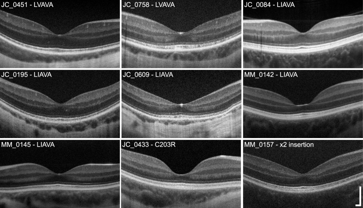
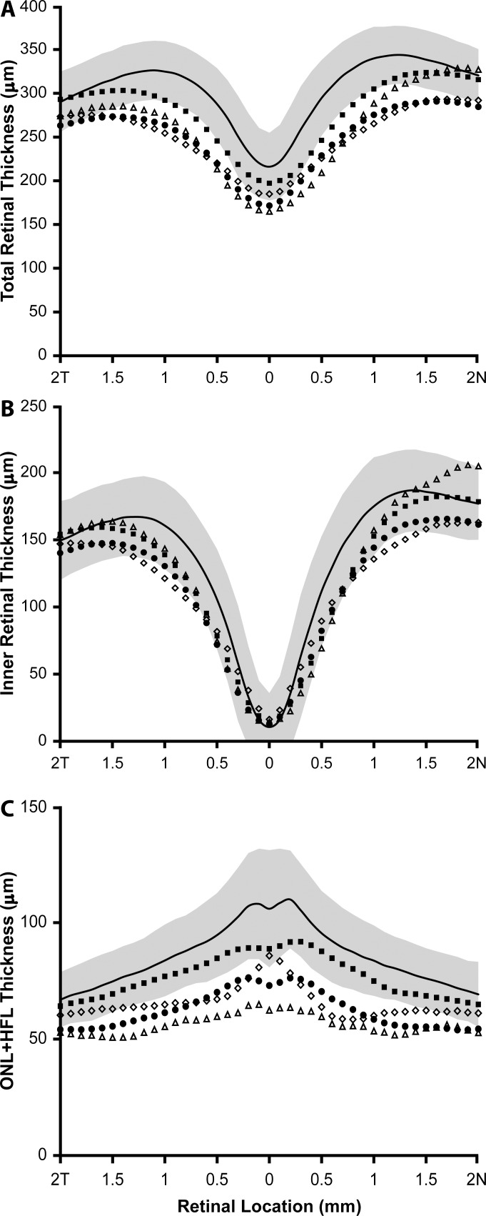
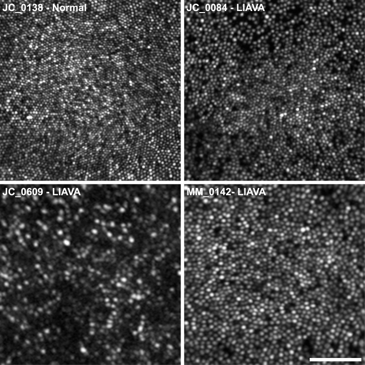
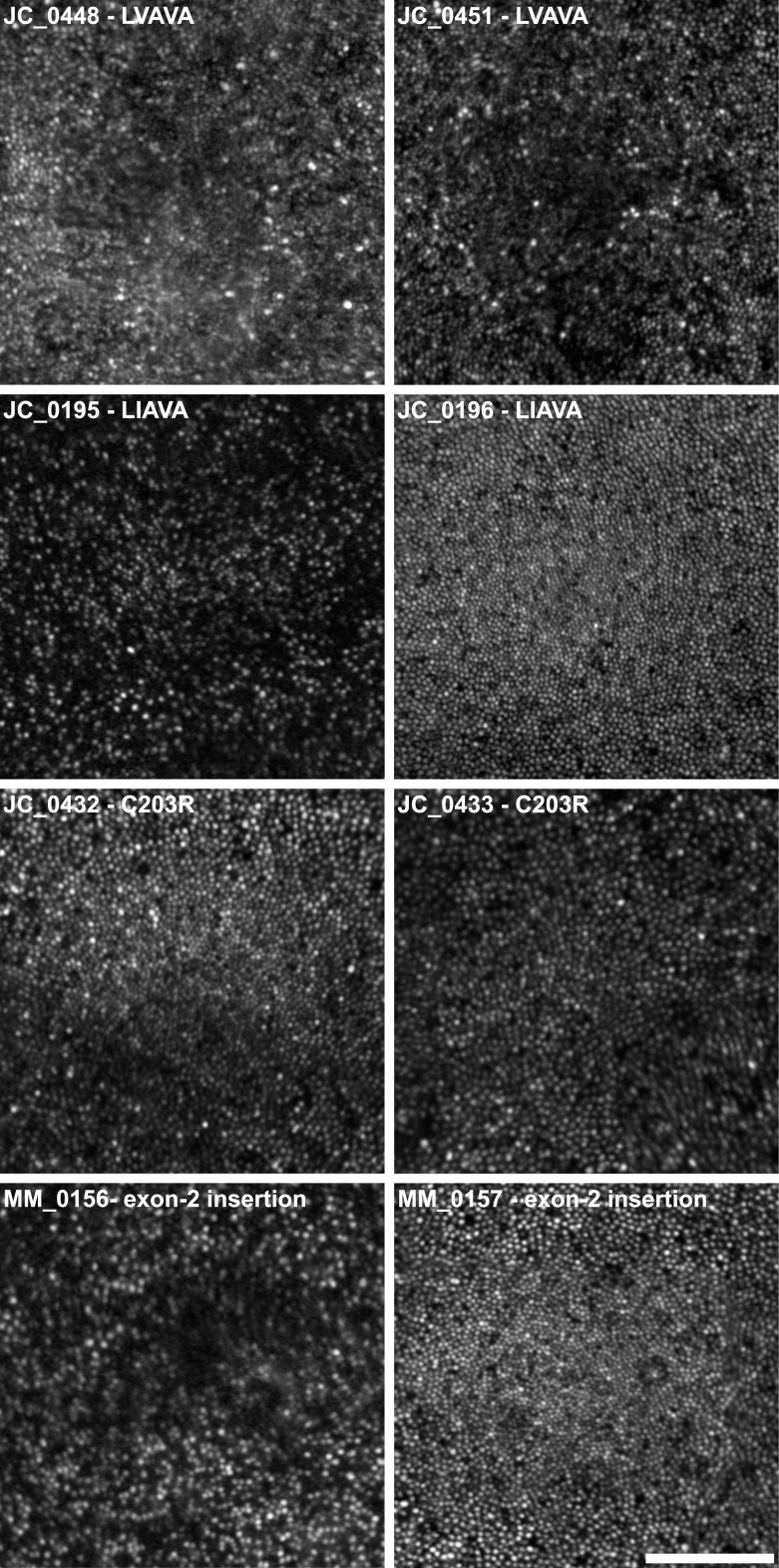
Methods: We recruited 17 males for imaging. The thickness and integrity of the photoreceptor layers were evaluated using spectral-domain optical coherence tomography. Cone density was measured using high-resolution images of the cone mosaic obtained with adaptive optics scanning light ophthalmoscopy. The L/M opsin gene array was characterized in 16 subjects, including at least one subject from each family.

Results: There were six subjects with the LVAVA haplotype encoded by exon 3, seven with LIAVA, two with the Cys203Arg mutation encoded by exon 4, and two with a novel insertion in exon 2. Foveal cone structure and retinal thickness was disrupted to a variable degree, even among related individuals with the same L/M array.

Conclusions: Our findings provide a direct link between disruption of the cone mosaic and L/M opsin variants. We hypothesize that, in addition to large phenotypic differences between different L/M opsin variants, the ratio of expression of first versus downstream genes in the L/M array contributes to phenotypic diversity. While the L/M opsin mutations underlie the cone dysfunction in all of the subjects tested, the color vision defect can be caused either by the same mutation or a gene rearrangement at the same locus.


Figures

Figure 1
Figure 1
Variable disruption in retinal lamination in X-linked cone dysfunction. Shown are high-resolution SD-OCT images (horizontal line scans) through the fovea. One image from each family is displayed, labeled with the subject ID and corresponding genotype. There is clear variability in ONL thickness across subjects, with MM_0142 and JC_0433 having the most pronounced ONL thinning at the fovea. Variation in foveal morphology also is apparent, which has been reported previously. The integrity of the second (IS/OS or EZ) and third (IZ) hyperreflective bands was variable across subjects, ranging from normal to mottled in appearance. Scale bar: 200 μm.
Figure 2
Figure 2
Subjects with X-linked cone dysfunction show reduced outer retinal thickness compared to normals. Plotted are the mean SD-OCT retinal thickness measurements along the horizontal meridian for each group of mutations. (A) Total retinal thickness, (B) inner retinal thickness, (C) ONL + HFL thickness. Solid black lines represent the mean values for 70 normal male controls, with the shaded region representing ± 2 SD from the mean. Filled squares represent the six individuals with the LVAVA haplotype (JC_0447, JC_0448, JC_0451, JC_0683, JC_0758, and JC_10340). Filled circles represent the averaged data from the seven individuals with the LIAVA haplotype (JC_0084, JC_0195, JC_0196, JC_0609, MM_0142, MM_0144, and MM_0145). Open triangles represent the two brothers with Cys203Arg mutations (JC_0432 and JC_0433). Open diamonds represent the two brothers with the novel exon 2 insertion (MM_0156 and MM_0157).
Figure 3
Figure 3
Photoreceptor mosaic disruption is highly variable in X-linked cone dysfunction, even within a given genotype. Foveal AOSLO images are shown for three subjects with the LIAVA haplotype and, for comparison, a previously published normal control with a peak density of 195,030 cones/mm2 (JC_0138). Although many dark spaces are visible in JC_0084's mosaic, there is tight packing at the fovea (peak density = 100,069 cones/mm2). In contrast, MM_0142 has fewer dark spaces but a more loosely packed mosaic (peak density = 53,356 cones/mm2). Subject JC_0609 shows the greatest degree of disruption, with many dark areas, resulting in a much sparser cone mosaic (peak density = 18,927 cones/mm2). Scale bar: 50 μm.
Figure 4
Figure 4
Marked differences in cone mosaic disruption between brothers with X-linked cone dysfunction and OPN1LW/OPN1MW mutations. Foveal AOSLO images are shown from four pairs of brothers. Large variation, not only in the number of cones, but also their apparent size and topography, is evident between JC_0195 and JC_0196, and MM_0156 and MM_0157. For example, MM_0157 shows relatively normal topography, with cone density peaking at the fovea and decreasing with eccentricity (Table 2); MM_0156, on the other hand, has greater disruption at the fovea, with a large area of low reflectivity surrounded by a sparse mosaic of cones. Scale bar: 100 μm.

References

    1. Michaelides M,, Hunt DM,, Moore AT. The cone dysfunction syndromes. Br J Ophthalmol. 2004; 88: 291–297. - PMC - PubMed
    1. Aboshiha J,, Dubis AM,, Carroll J,, Hardcastle AJ,, Michaelides M. The cone dysfunction syndromes. Br J Ophthalmol. 2016; 100: 115–121. - PMC - PubMed
    1. Haim M,, Fledelius HC,, Skarsholm D. X-linked myopia in a Danish family. Acta Ophthalmol. 1988; 66: 450–456. - PubMed
    1. Schwartz M,, Haim M,, Skarsholm D. X-Linked myopia: Bornholm Eye Disease - linkage to DNA markers on the distal part of Xq. Clin Genet. 1990; 38: 281–286. - PubMed
    1. Young TL,, Deeb SS,, Ronan SM,, et al. X-linked high myopia associated with cone dysfunction. Arch Ophthalmol. 2004. ; 122: 897–908. - PubMed

Publication types