. 2016 Jul 22;12(7):e1006195.
doi: 10.1371/journal.pgen.1006195. eCollection 2016 Jul.

Timely Endocytosis of Cytokinetic Enzymes Prevents Premature Spindle Breakage during Mitotic Exit

Affiliations

Timely Endocytosis of Cytokinetic Enzymes Prevents Premature Spindle Breakage during Mitotic Exit

Cheen Fei Chin et al. PLoS Genet. .

Abstract

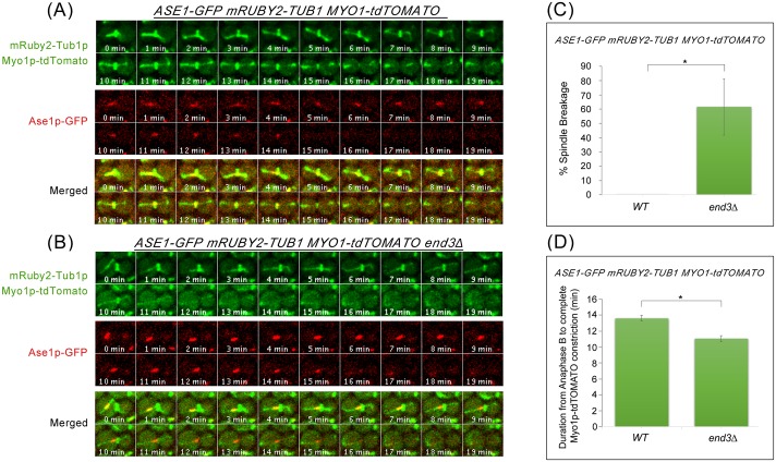
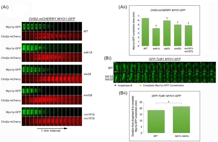
Cytokinesis requires the spatio-temporal coordination of membrane deposition and primary septum (PS) formation at the division site to drive acto-myosin ring (AMR) constriction. It has been demonstrated that AMR constriction invariably occurs only after the mitotic spindle disassembly. It has also been established that Chitin Synthase II (Chs2p) neck localization precedes mitotic spindle disassembly during mitotic exit. As AMR constriction depends upon PS formation, the question arises as to how chitin deposition is regulated so as to prevent premature AMR constriction and mitotic spindle breakage. In this study, we propose that cells regulate the coordination between spindle disassembly and AMR constriction via timely endocytosis of cytokinetic enzymes, Chs2p, Chs3p, and Fks1p. Inhibition of endocytosis leads to over accumulation of cytokinetic enzymes during mitotic exit, which accelerates the constriction of the AMR, and causes spindle breakage that eventually could contribute to monopolar spindle formation in the subsequent round of cell division. Intriguingly, the mitotic spindle breakage observed in endocytosis mutants can be rescued either by deleting or inhibiting the activities of, CHS2, CHS3 and FKS1, which are involved in septum formation. The findings from our study highlight the importance of timely endocytosis of cytokinetic enzymes at the division site in safeguarding mitotic spindle integrity during mitotic exit.


Conflict of interest statement

The authors have declared that no competing interests exist.

Figures

Fig 1
Fig 1. Dynamics of spindle disassembly relative to cytokinetic enzymes’ arrival at the neck and AMR constriction during late mitosis.
(A) GFP-TUB1 MYO1-GFP CHS2-mCHERRY cells were arrested in YPD/Noc at 24°C. After 4 hours, cells were released from metaphase into fresh YPD for 30min. Cells were then mounted in SC/Glu agar pad and examined with time-lapsed microscopy (n = 32). (B) Graph showing Chs2p-mCherry neck fluorescence intensity at the neck relative to mean time of spindle disassembly and mean time of AMR constriction. (C) CHS2-CHERRY CHS3-3mGFP (n = 34), (D) CHS2-CHERRY FKS1-GFP (n = 27), and (E) GFP-TUB1 MYO1-GFP ABP1-mCHERRY cells were treated as described above (A) except FKS1-GFP CHS2-CHERRY cells were mounted on 0.5x YDP agar pad.
Fig 2
Fig 2. Chs2p-GFP accumulates in endocytosis mutants during mitotic exit.
(A) CHS2-GFP ABP1-mCHERRY cells harbouring deletions of key endocytic components were synchronised in metaphase. After release, cells were mounted on SC/Glu agar pad and examined with time-lapsed microscopy. (B) Graph showing Chs2p-GFP mean fluorescence intensity at the neck. Error bars represent SEM.
Fig 3
Fig 3. Chs2p-GFP can be endocytosed when mitotic exit is blocked.
(A) GAL-CHS2 (6S-6A)-GFP SPC42-eqFP cells were arrested in YPR/Noc at 24°C. After 5 hours, 4% GAL was added for 60 min. Cells were then split into YPD/Noc contain DMSO or 32μM Lat B, and images were taken after 100 min. (B) Graph showing percentage of cells with Chs2p-GFP plasma membrane or neck signals. Error bars represent SD. (C) cdc15-2 GAL-CHS2 (6S-6A)-GFP SPC42-eqFP cells were arrested in YPR/Noc at 24°C. After 5 hours, 4% GAL was added for 60 min. Cells were then spun into pre-warmed 37°C YPD/Noc with 32μM LatB for 30 min. Next, cells were released into YPD with 32μM Lat B for 60 min, and then culture was split into YPD with DMSO or 32μM Lat B respectively. Images were taken after 30 min. (D) Graph showing percentage of cells with Chs2p-GFP plasma membrane or neck signals. Error bars represent SD.
Fig 4
Fig 4. Cytokinetic enzymes Chs3p and Fks1p accumulate in endocytosis-defective mutants.
(A) FKS1-GFP ABP1-mCHERRY and (B) CHS3-3mGFP ABP1-mCHERRY cells harbouring deletions of key endocytic components were cultured in YPD at 32°C for 2 hours. Cells were then examined under fluorescence microscope. Error bars represent SD.
Fig 5
Fig 5. Mitotic spindles appear to break during mitotic exit in key endocytosis deletion mutants.
(A) GFP-TUB1 MYO1-GFP cells harbouring deletions of key endocytic components were synchronised in metaphase and then shifted up to 32°C for 30min. After release, cells were mounted on SC/Glu agar pad and examined with time-lapsed microscopy. (B) Graph showing percentage of spindle breakage in endocytosis mutants. Asterisk represents statistical significance as compared to wild-type cells and hash represents no statistical significance as compared to wild-type cells (Student-t test). Error bars represent SD. (C) Table showing the percentage of spindle breakage and p-values for endocytosis mutants.
Fig 6
Fig 6. Uneven distribution of initial location of spindle-halves can be observed in endocytosis mutants undergoing mitotic exit.
GFP-TUB1 MYO1-GFP cells harbouring deletions of key endocytic components were synchronised in metaphase. After 4 hours, 200μM Lat B was added and cells were shifted to 32°C for 30min. Next, cells were released into fresh YPD containing 200μM Lat B for 30 min. Cells were then mounted on SC/Glu agar pad containing 200μM Lat B and subjected to time-lapsed microscopy. The distance of spindle breakdown relative to the bud neck was measured and normalized with cell length as described in Woodruff et al., 2010 (n = 30 for each strain).
Fig 7
Fig 7. end3Δ cells show shorter interval between anaphase B and Myo1p-GFP constriction.
(A) Wild Type and (B) end3Δ cells harbouring mRUBY2-TUB1 MYO1-tdTOMATO ASE1-GFP respectively were synchronised in metaphase and then shifted up to 32°C for 30min. After release, cells were mounted on SC/Glu agar pad and examined with time-lapsed microscopy. (C) Graph showing percentage of spindle breakage in WT and end3Δ respectively (WT = 44, end3Δ = 43). Error bars represent SD. (D) Graph showing duration from anaphase B to Myo1p-tdTOMATO complete constriction (Student t-test, p<0.001; error bars represents SEM).
Fig 8
Fig 8. Premature AMR constriction in endocytosis mutants causes spindle breakage during mitotic exit.
(Ai) CHS2-mCHERRY MYO1-GFP cells harbouring deletions of key endocytic components were synchronised in metaphase and then shifted up to 32°C for 30min. After release, cells were mounted on SC/Glu agar pad and examined with time-lapsed microscopy. (Aii) Graph showing time taken for Myo1p-GFP complete constriction upon Chs2p-mCherry neck signal arrival. Statistical analysis via Mann Whitney-U test. Error bars represent SEM. (Bi) Wild Type and lsb1Δ lsb2Δ cells harbouring GFP-TUB1 MYO1-GFP were arrested in metaphase. After release, cells were mounted on SC/Glu agar pad and subjected to time-lapsed microscopy at 24°C, at 1min intervals. (Bii) Graph showing duration from anaphase B to Myo1p-GFP complete constriction (Student t-test, p<0.001). Error bars represent SEM (n = 60).
Fig 9
Fig 9. Mitotic spindle breakage phenotype in end3Δ cells can be rescued by depleting Chs2p.
(A) Wild Type and end3Δ cells habouring GFP-TUB1 MYO1-GFP ADH1-yeOSTIR1 CHS2-1xMini-AID were synchronised in metaphase. After 4 hours, IAA was added to final concentration of 0.25mM and cells were shifted to 32°C for 30min. Next, cells were released from metaphase into fresh YPD containing 0.25mM IAA for 30min. Cells were then mounted on SC/Glu agar pad containing 0.25mM IAA and subjected to time-lapsed microscopy. (B) Immunoblot showing degradation of Chs2p following treatment with IAA. Samples were collected at metaphase arrest, after addition of IAA and shifting to 32°C for 30min, and before time-lapse microscopy. (C) Graph shows percentage of spindle breakage in WT and end3Δ cells that were not treated and treated with IAA respectively. Error bars represent SD (n>100).
Fig 10
Fig 10. Suppression of cytokinetic enzymes activity via treatment with Caspofungin and Nikkomycin-Z rescues spindle breakage in end3Δ mutant.
(A) GFP-TUB1 MYO1-GFP and GFP-TUB1 MYO1-GFP end3Δ cells were arrested in YPD/Noc at 24°C for 2 hours. Cells were treated with 50ng/ml caspofungin and 1mM nikkomycin-Z respectively for another 2 hours in YPD/Noc. Upon arrested in metaphase, cells were shifted to 32°C for another 30min. Next, cells were released from metaphase into fresh YPD for 30min. Cells were then mounted in SC/Glu agar pad and examined with time-lapsed microscopy. (B) Graph showing the mean percentage of cells with spindle breakage. Error bars represent SD. (C) Table showing the mean percentage of cells with spindle breakage and p-values of statistical analysis (Student-t test).
Fig 11
Fig 11. Suppression of cytokinetic enzymes activity via treatment with Caspofungin and Nikkomycin-Z alters AMR constriction dynamics.
(A) CHS2-GFP MYO1-REDSTAR and CHS2-GFP MYO1-REDSTAR end3Δ cells were treated as in (Fig 10A). (B) Graph showing time taken for complete AMR constriction relative to Chs2p-GFP neck localization. Error bars represent SEM. (C) Table showing the mean time taken for complete Myo1p-GFP constriction relative to Chs2p-GFP neck localization and p-values of statistical analysis (Mann Whitney-U test).
Fig 12
Fig 12. Mitotic spindle breakage in endocytosis mutants contributes to failure in spindle re-establishment in progeny cells.
GFP-TUB1 MYO1-GFP SPC29-RFP cells harbouring deletions of key endocytic components were cultured in YPD for 2 hours at 32°C. Hydroxyurea was added to final concentration of 0.2M. Cells were arrested for 5.5 hours at 32°C and subjected to microscopy analysis. (A) Graph showing percentage of monopolar spindle formation in endocytosis mutants. Error bars represent SD. [(B) and (C)] Images were captured with 9x0.5μm z-planes for GFP-Tub1p Myo1p-GFP and Spc29p- RFP. The images shown were maximum projection of 9 z-planes. (D) Table showing the percentage monopolar spindle formation and p-values for endocytosis mutants (Student-t test).
Fig 13
Fig 13. Monopolar spindles observed in end3Δ cells are not due SPB duplication defect.
(A) Wild Type and (B) end3Δ cells harbouring GFP-TUB1 MYO1-GFP SPC42-eqFP respectively were synchronised in metaphase and then shifted up to 32°C for 30min. After release, cells were mounted on SC/Glu agar pad and examined with time-lapsed microscopy. (C) Graph shows percentage of spindle breakage in WT and end3Δ respectively (WT = 37, end3Δ = 38). Error bars represent SD. (D) WT and end3Δ cells harbouring GFP-TUB1 MYO1-GFP SPC29-RFP respectively were synchronised in metaphase and then shifted up to 32°C for 30min. After release for 60min, cells were spun into YP/Raff with 2% Gal or 2% Glu containing 0.2M HU for 5 hours. Graph showing percentage of monopolar spindles in WT and end3Δ cells (Student t-test, p<0.05). Error bars represent SD.
Fig 14
Fig 14. Proposed model.
Coordination of mitotic spindle disassembly, endocytosis of cytokinetic enzymes endocytosis and cytokinesis during mitotic exit.

Similar articles

Cited by

References

    1. Queralt E, Uhlmann F. Cdk-counteracting phosphatases unlock mitotic exit. Curr Opin Cell Biol. 2008;20(6):661–8. Epub 2008/10/11. 10.1016/j.ceb.2008.09.003 - DOI - PMC - PubMed
    1. Irniger S, Piatti S, Michaelis C, Nasmyth K. Genes involved in sister chromatid separation are needed for B-type cyclin proteolysis in budding yeast. Cell. 1995;81(2):269–78. Epub 1995/04/21. . - PubMed
    1. Rock JM, Lim D, Stach L, Ogrodowicz RW, Keck JM, Jones MH, et al. Activation of the yeast Hippo pathway by phosphorylation-dependent assembly of signaling complexes. Science. 2013;340(6134):871–5. 10.1126/science.1235822 - DOI - PMC - PubMed
    1. Weiss EL. Mitotic exit and separation of mother and daughter cells. Genetics. 2012;192(4):1165–202. Epub 2012/12/06. 10.1534/genetics.112.145516 - DOI - PMC - PubMed
    1. Shaw JA, Mol PC, Bowers B, Silverman SJ, Valdivieso MH, Duran A, et al. The function of chitin synthases 2 and 3 in the Saccharomyces cerevisiae cell cycle. J Cell Biol. 1991;114(1):111–23. Epub 1991/07/01. - PMC - PubMed

Publication types

MeSH terms