. 2016 Aug 9;7(32):52329-52339.
doi: 10.18632/oncotarget.10750.

Up-regulation of fibroblast growth factor 19 and its receptor associates with progression from fatty liver to hepatocellular carcinoma

Affiliations

Up-regulation of fibroblast growth factor 19 and its receptor associates with progression from fatty liver to hepatocellular carcinoma

Yan Li et al. Oncotarget. .

Abstract

Background: Human fibroblast growth factor 19 (FGF19), its receptor (FGFR4) and EpCAM play an important role in cell proliferation, differentiation, motility, and overexpression have been linked to hepatocellular carcinoma (HCC). The aim of this study was to evaluate the FGF19 signals responsible for the progression of HCC arising from fatty liver.

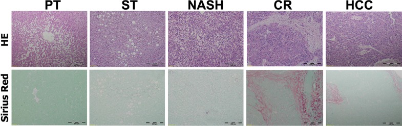
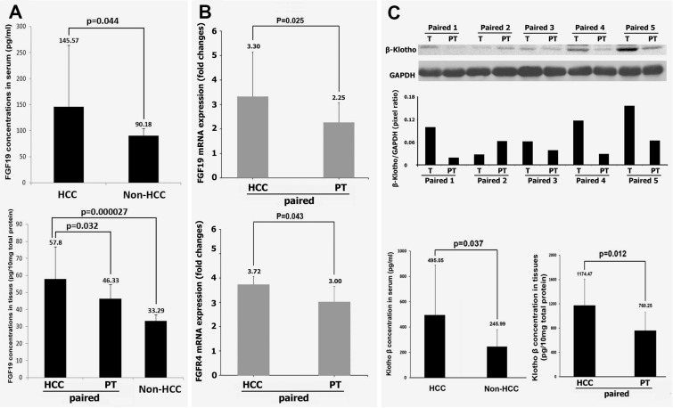
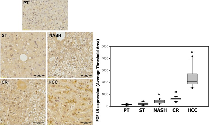
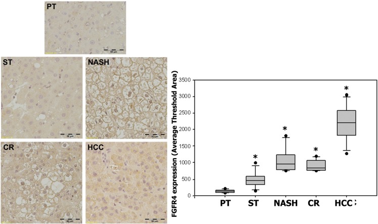
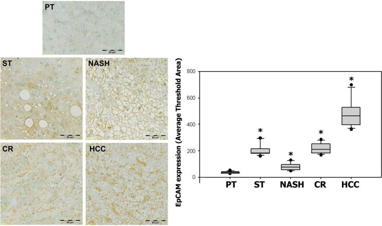
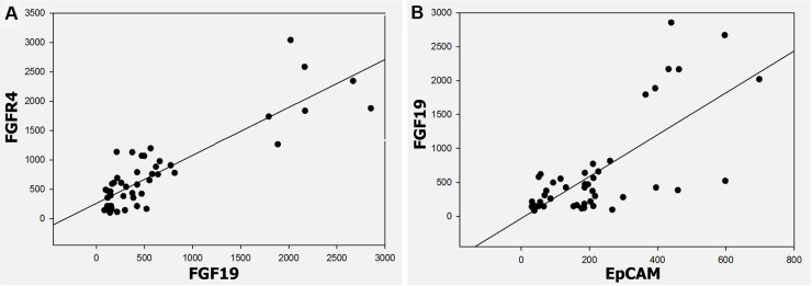
Results: FGF19 level was significantly increased in the HCC patients' serum compared to non-HCC controls. The IHC results demonstrated significant increases of protein expressions of FGF19, FGFR4 and EpCAM in specimens with fatty liver, NASH, cirrhosis, and HCC compared to healthy liver tissue. There was a significant positive correlation between the protein expressions (FGF19, FGFR4, and EpCAM) and histopathologic changes from FL to HCC. Furthermore, FGF19 was positively correlated with FGFR4 and with EpCAM.

Materials and methods: FGF19 protein levels in serum and tissues were determined by ELISA assay. The FGFR4, and EpCAM expression and tissue distribution were further evaluated by immunohistochemical staining in tissue array samples. FGF19, FGFR4 and EpCAM expressions between the different histologic stages of fatty liver steatohepatitis-cirrhosis-HCC carcinogenesis sequence were compared to healthy hepatic tissue.

Conclusions: Overexpression of FGF19/FGFR4 significantly correlated with EpCAM as a marker of hepatic cancer stem cells within the fatty liver-steatosis-cirrhosis-HCC sequence.

Impact: This is the first study to elucidate FGF19/FGFR4 signaling in favor of HCC cells developing as indicated by increased EpCAM within the carcinogenesis sequence from fatty liver to hepatocellular carcinoma. Our study has the potential to yield novel and cost effective screening strategies for HCC patients.

Keywords: FGF19; FGFR4; cancer stem cell; hepatocellular carcinoma.


Conflict of interest statement

All authors have no conflicts of interest.

Figures

Figure 1
Figure 1. Histologic appearances of peritumoral normal and ST-NAHS-cirrhosis-HCC progression
Upper: Representative histologic changes from different stages by Hematoxylin and Eosin (H&E) staining, magnification: 40×. Lower: Representative histologic changes to reveal fibrosis and cirrhosis by Sirius Red staining, magnification: 40×. PT: peritumoral tissues; ST: steatosis with diffuse lipid deposition; NASH: lipid deposition with inflammatory cells infiltration; CR: cirrhosis with regenerative nodule; HCC: hepatocellular carcinoma.
Figure 2
Figure 2. Expressions of FGF19, FGFR4 and of β-Klotho in HCC
(A) the protein levels of FGF19 in serum from 24 HCC patients and 6 non-HCC controls, and in the tissues from 24 paired HCC-peritumoral and 6 non-HCC controls by ELISA analysis. (B) the mRNA levels of FGF19 and FGFR4 from 24 paired HCC-peritumoral tissues. (C) upper: Representative Western blot for β-Klotho protein detection of 5 paired tissues from HCC patients. Lower: the protein levels of β-Klotho in serum from 24 HCC patients and 6 non-HCC controls, and in the tissues from 24 paired HCC-peritumoral tissues by ELISA analysis. T: HCC tumor tissue; PT: peritumoral tissues.
Figure 3
Figure 3. Expression of FGF19 in ST-NAHS-cirrhosis-HCC progression
Representative immunohistochemical staining for FGF19 and the computer quantification of FGF19 expression from different stages. Magnification: 200×, the positive staining shown as brown color. PT: peritumoral tissues; ST: steatosis with diffuse lipid deposition; NASH: lipid deposition with inflammatory cells infiltration; CR: cirrhosis with regenerative nodule; HCC: hepatocellular carcinoma. *p < 0.05 vs PT.
Figure 4
Figure 4. Expression of FGFR4 in ST-NAHS-cirrhosis-HCC progression
Representative immunohistochemical staining for FGFR4 and the computer quantification of FGFR4 expression from different stages. Magnification: 200×, the positive staining shown as brown color. PT: peritumoral tissues; ST: steatosis with diffuse lipid deposition; NASH: lipid deposition with inflammatory cells infiltration; CR: cirrhosis with regenerative nodule; HCC: hepatocellular carcinoma. *p < 0.05 vs PT.
Figure 5
Figure 5. Expression of EpCAM in ST-NAHS-cirrhosis-HCC progression
Representative immunohistochemical staining for EpCAM and the computer quantification of EpCAM expression from different stages. Magnification: 200×, the positive staining shown as brown color. PT: peritumoral tissues; ST: steatosis with diffuse lipid deposition; NASH: lipid deposition with inflammatory cells infiltration; CR: cirrhosis with regenerative nodule; HCC: hepatocellular carcinoma.*p < 0.05 vs PT.
Figure 6
Figure 6. Correlation between FGF19 and FGFR4, FGF19 and EpCAM in ST-NAHS-cirrhosis-HCC progression
(A) Positive correlation of FGF19 and FGFR4 expression in HCC (r = 0.79) (p < 0.001). (B) Positive correlation of FGF19 and EpCAM expression in HCC (r = 0.852) (p < 0.001).

References

    1. El-Serag HB. Hepatocellular carcinoma. N Engl J Med. 2011;365:1118–1127. - PubMed
    1. Jemal A, Bray F, Center MM, Ferlay J, Ward E, Forman D. Global cancer statistics. CA Cancer J Clin. 2011;61:69–90. - PubMed
    1. Li T, Qin LX, Gong X, Zhou J, Sun HC, Wang L, Qiu SJ, Ye QH, Fan J. Clinical characteristics, outcome, and risk factors for early and late intrahepatic recurrence of female patients after curative resection of hepatocellular carcinoma. Surgery. 2014;156:651–660. - PubMed
    1. Onnerhag K, Nilsson PM, Lindgren S. Increased risk of cirrhosis and hepatocellular cancer during long-term follow-up of patients with biopsy-proven NAFLD. Scand J Gastroenterol. 2014;49:1111–8. - PubMed
    1. Schutte K, Schulz C, Poranzke J, Antweiler K, Bornschein J, Bretschneider T, Arend J, Ricke J, Malfertheiner P. Characterization and prognosis of patients with hepatocellular carcinoma (HCC) in the non-cirrhotic liver. BMC gastroenterol. 2014;14:117. - PMC - PubMed

Substances