. 2016 Jul 20;17(7):1170.
doi: 10.3390/ijms17071170.

Structural Analysis of Hand Drawn Bumblebee Bombus terrestris Silk

Affiliations

Structural Analysis of Hand Drawn Bumblebee Bombus terrestris Silk

Andrea L Woodhead et al. Int J Mol Sci. .

Abstract

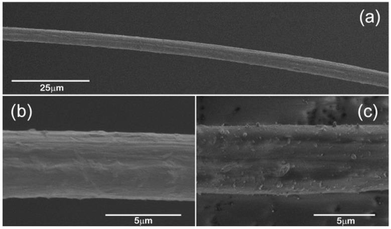
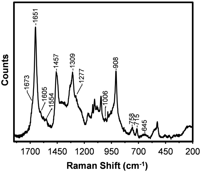
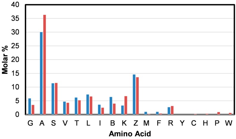
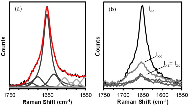
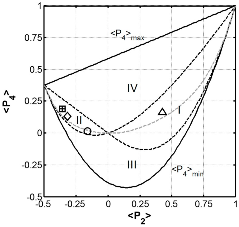
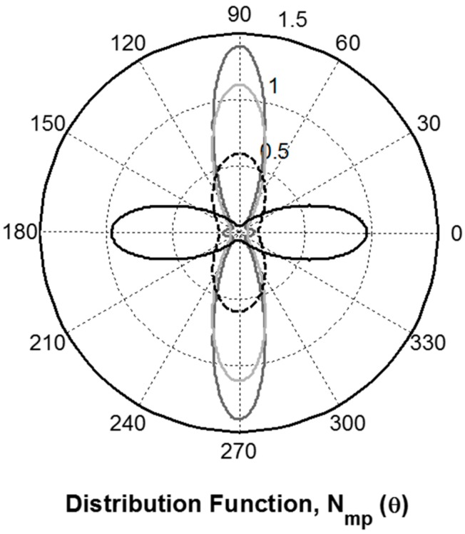
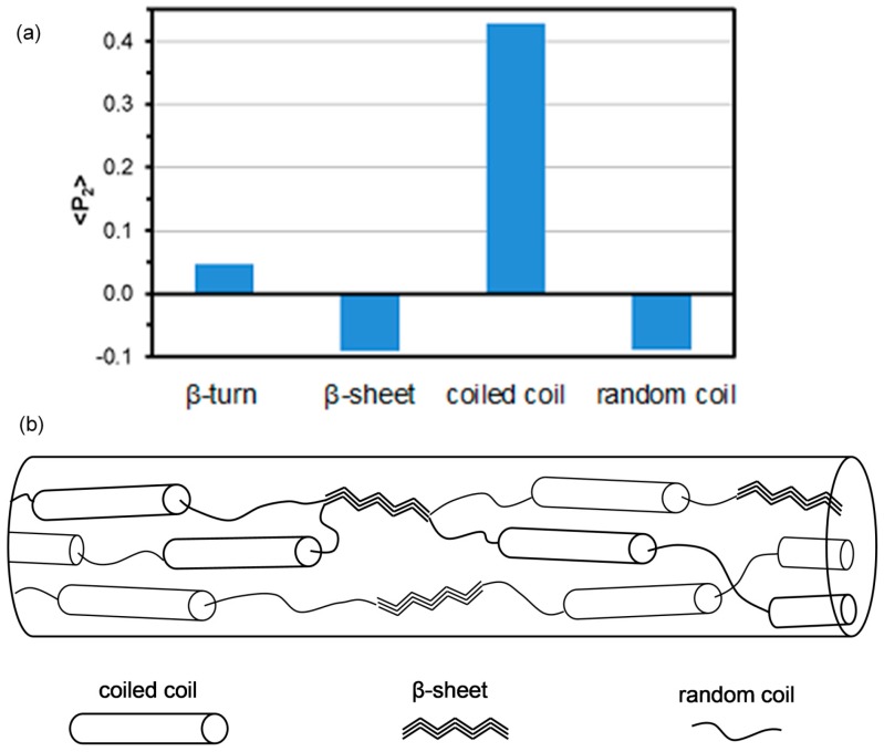
Bombus terrestris, commonly known as the buff-tailed bumblebee, is native to Europe, parts of Africa and Asia. It is commercially bred for use as a pollinator of greenhouse crops. Larvae pupate within a silken cocoon that they construct from proteins produced in modified salivary glands. The amino acid composition and protein structure of hand drawn B. terrestris, silk fibres was investigated through the use of micro-Raman spectroscopy. Spectra were obtained from single fibres drawn from the larvae salivary gland at a rate of 0.14 cm/s. Raman spectroscopy enabled the identification of poly(alanine), poly(alanine-glycine), phenylalanine, tryptophan, and methionine, which is consistent with the results of amino acid analysis. The dominant protein conformation was found to be coiled coil (73%) while the β-sheet content of 10% is, as expected, lower than those reported for hornets and ants. Polarized Raman spectra revealed that the coiled coils were highly aligned along the fibre axis while the β-sheet and random coil components had their peptide carbonyl groups roughly perpendicular to the fibre axis. The protein orientation distribution is compared to those of other natural and recombinant silks. A structural model for the B. terrestris silk fibre is proposed based on these results.

Keywords: Bombus terrestris; Raman spectroscopy; coiled coil; fibre; protein conformation; protein orientation; silk.


Figures

Figure 1
Figure 1
Secondary electron images obtained from (a,b) section of a B. terrestris silk fibre hand drawn at 0.14 cm/s and (c) a fibre hand drawn at 0.10 cm/s.
Figure 2
Figure 2
Raman spectrum obtained from the hand drawn (0.14 cm/s) B. terrestris silk fibre.
Figure 3
Figure 3
The amino acid composition (molar percentage) determined for B. terrestris silk (blue) and determined from amino acid sequences of the four fibrous B. terrestris silk proteins; BterF1: ABW21694, BterF2: ABW21695, BterF3: ABW21696, and BterF4: ABW21697 (red) [26].
Figure 4
Figure 4
The Raman amide I band of the hand drawn B. terrestris fibre. Spectral deconvolution (a); the underlying black trace represents the raw data, the red trace is the sum of the component peaks (dark grey) and components that are not associated with protein conformation and present for fitting purposes only (light grey). Polarized Raman spectra (b).
Figure 5
Figure 5
The graph of the limiting values of ‹P4› as a function of ‹P2›. The (‹P2›, ‹P4›) couple determined for B. terrestris (∆), A. Illawarra (○) fibres are presented along with those of B. mori (+) and S. c. ricini (□) cocoon silks and N. edulis (◊) dragline silk [13,15]. Note that the values determined for B. mori cocoon and S. c. ricini cocoon overlap. The dashed grey line represents the values of ‹P4› when λ4 = 0.
Figure 6
Figure 6
The most probable orientation distribution determined for the hand drawn B. terrestris silk fibre (black trace). A. illawarra fibres (dashed black trace) [13] presented along with those of B. mori and S. c. ricini cocoon (dark grey trace) and N. edulis dragline (light grey trace) silks [16]. The 0° of the polar plot coincides with the fibre direction.
Figure 7
Figure 7
Graph of ‹P2› values obtained from the deconvolution of the polarized hand drawn B. terrestris silk spectra (a); Proposed structure of the B. terrestris silk fibres based on the Raman results (b).

References

    1. Sutherland T.D., Weisman S., Trueman H.E., Sriskantha A., Trueman J.W., Haritos V.S. Conservation of essential design features in coiled coil silks. Mol. Biol. Evol. 2007;24:2424–2432. doi: 10.1093/molbev/msm171. - DOI - PubMed
    1. Atkins E.D.T. A four-strand coiled coil model for some insect fibrous proteins. J. Mol. Biol. 1967;24:139–141. doi: 10.1016/0022-2836(67)90099-X. - DOI
    1. Crewe R.M., Thompson P.R. Oecophylla silk: Functional adaptation in a biopolymer. Naturwissenschaften. 1979;66:57–58. doi: 10.1007/BF00369367. - DOI
    1. Kameda T., Kojima K., Sezutsu H., Zhang Q., Teramoto H., Tamada Y. Hornet (Vespa) Silk composed of coiled-coil proteins. Kobunshi Ronbunshu. 2010;67:641–653. doi: 10.1295/koron.67.641. - DOI
    1. Kameda T., Kojima K., Togawa E., Sezutsu H., Zhang Q., Teramoto H., Tamada Y. Drawing-induced changes in morphology and mechanical properties of hornet silk gel films. Biomacromolecules. 2010;11:1009–1018. doi: 10.1021/bm901472a. - DOI - PubMed

LinkOut - more resources