. 2016 Jul 20;17(7):1174.
doi: 10.3390/ijms17071174.

La Autoantigen Induces Ribosome Binding Protein 1 (RRBP1) Expression through Internal Ribosome Entry Site (IRES)-Mediated Translation during Cellular Stress Condition

Affiliations

La Autoantigen Induces Ribosome Binding Protein 1 (RRBP1) Expression through Internal Ribosome Entry Site (IRES)-Mediated Translation during Cellular Stress Condition

Wenqing Gao et al. Int J Mol Sci. .

Abstract

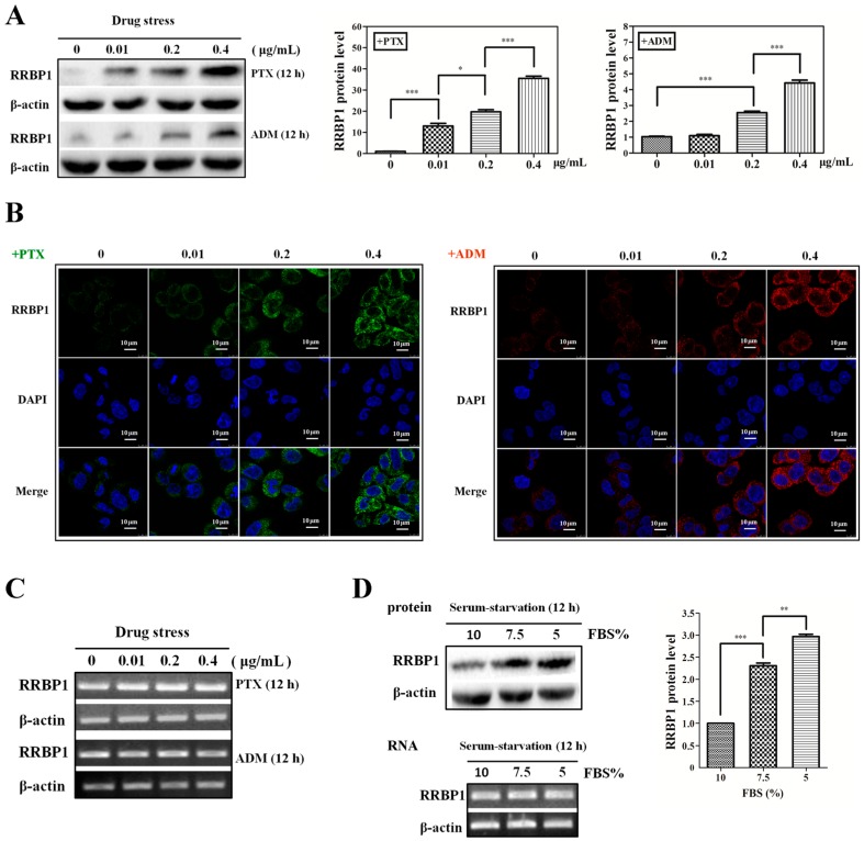
The function of ribosome binding protein 1 (RRBP1) is regulating the transportation and secretion of some intracellular proteins in mammalian cells. Transcription of RRBP1 is induced by various cytokines. However, few studies focused on the process of RRPB1 mRNA translation. The RRBP1 mRNA has a long 5' untranslated region that potentially formed a stable secondary structure. In this study, we show that the 5' UTR of RRBP1 mRNA contains an internal ribosome entry site (IRES). Moreover, the RRBP1 expression is induced by chemotherapeutic drug paclitaxel or adriamycin in human hepatocellular carcinoma cells and accompanied with the increased expression of La autoantigen (La), which binds to RRBP1 IRES element and facilitates translation initiation. Interestingly, we found IRES-mediated RRBP1 translation is also activated during serum-starvation condition which can induce cytoplasmic localization of La. After mapping the entire RRBP1 5' UTR, we determine the core IRES activity is located between nt-237 and -58. Furthermore, two apical GARR loops within the functional RRBP1 IRES elements may be important for La binding. These results strongly suggest an important role for IRES-dependent translation of RRBP1 mRNA in hepatocellular carcinoma cells during cellular stress conditions.

Keywords: La autoantigen (La); adriamycin (ADM); paclitaxel (PTX); ribosome binding protein 1 internal ribosome entry site (RRBP1 IRES); serum-starvation.


Figures

Figure 1
Figure 1
Enhanced protein synthesis contributes to overexpression of ribosome binding protein 1 (RRBP1) during stress conditions. (A) Human hepatocellular carcinoma cells (Bel7402 cells) were treated with paclitaxel (PTX) or adriamycin (ADM) at different doses (0.01, 0.2, 0.4 μg/mL) respectively, for 12 h. The protein expression of RRBP1 was examined via Western blot analysis and normalized to β-actin. The RRBP1/β-actin ratio from treated cells was compared with the ratio from untreated cells, which is set to 1 (*: p < 0.05; ***: p < 0.0005); (B) Immunofluorescence showed dose-dependent cytoplasm expression of RRBP1 in Bel7402 cells treated with PTX or ADM; (C) mRNA expression levels of RRBP1 after treatment with increasing concentrations of PTX or ADM in Bel7402 cells. mRNA levels were determined with reverse transcription-PCR (RT-PCR); and (D) the expression of RRBP1 protein was increased during serum starvation in Bel7402 cells. RT-PCR and Western blotting showed different levels of transcription and translation of RRBP1 in Bel7402 cells growing at serum-starvation and normal conditions (**: p < 0.005; ***: p < 0.0005).
Figure 2
Figure 2
Analysis of internal ribosome entry site (IRES) activity of the RRBP1 5′ UTR sequence. (A) Dual-luciferase reporter vector construction. Positive IRES activity sequence, X-linked inhibitor of apoptosis protein (XIAP) and NF-κB repressing factor (NRF), and detected RRBP1 5′ UTR sequences were inserted between Renilla and firefly luciferase in plasmid pRF with or without promoter simian virus (SV40); (B) both luciferase activities were determined following transfection in human embryonic kidney 293 (HEK293) cells; (C) dual-fluorescent protein reporter vector construction. Positive viral IRES activity sequence, encephalomyocarditis virus (EMCV), and detected RRBP1 5′ UTR sequence were inserted between red and green fluorescent protein genes with or without promoter cytomegalovirus (CMV). The indicated plasmid was then transfected into HEK293 cells and the expression efficiency of the reporter genes were analyzed by fluorescence microscopy; (D) dual-luciferase reporter vector was transfected into Bel7402 and A2780 cell lines and luciferase activities from each cell line was measured, respectively. The ratio of firefly luciferase (FL) to renilla luciferase (RL) represented IRES activity of the inserted 5′ UTR. The expression data are presented as the mean ± SEM (standard error of the mean) of triplicate samples.
Figure 3
Figure 3
The RRBP1 IRES upregulates translation during stress conditions. The construct pR-RRBP1-F shown in Figure 2A was transfected into Bel7402 cells treated with and without increasing concentration (0.01, 0.2 and 0.4 μg/mL) of PTX or ADM, respectively. Meanwhile, the vector was also transfected into Bel7402 cells growing under normal medium and serum-starvation conditions. FL and RL activity were determined and the IRES activity was shown. The expression data are presented as the mean ± SEM of triplicate samples (*: p < 0.05; **: p < 0.005).
Figure 4
Figure 4
La interacted with RRBP1 5′ UTR sequence and enhanced the RRBP1 IRES activity in Bel7402 cells under drug pressure. (A) i: immunoprecipitation analysis of conjugated trans-acting factors (ITAFs) for RRBP1 and XIAP. Samples with (+) or without (−) reverse transcription (r.t.) were exhibited in different lanes; ii: Levels of La bound to RRBP1 bicistronic transcripts were quantified by qPCR and normalized to RRBP1 input levels. The FL/Input ratio from each cell line was compared with the ratio from wild-type Bel7402 cells, which was set to 1 (***: p < 0.0005); (B) Bel7402 cells were treated with PTBP1 siRNA, La siRNA or control, (non-targeting siRNA). Cell lysates were harvested and subjected to Western blot analysis. Bicistronic DNA constructs containing RRBP1 and XIAP IRES elements were transfected into cells after treatment with PTBP1 siRNA, La siRNA, or control siRNA, respectively, and then IRES activities were determined. The expression data are presented as the mean ± SEM of triplicate samples (**: p < 0.005); and (C) Western blot analysis of La expression levels in Bel7402 cells treated with 0.01, 0.2, and 0.4 μg/mL PTX or ADM for 12 h. During serum starvation, Bel7402 cells were growing in medium with 7.5% or 5% FBS for 12 h. Then the cell extracts were tested for both nuclear (Nuc) and cytoplasm (Cyt) expression of La and the total La in the whole cell extracts (WCE). The protein expression for La was normalized to β-actin and the resulting La/β-actin ratio from each treated cells was compared with the ratio from untreated cells, which was set to 1 (*: p < 0.05; ***: p < 0.0005).
Figure 4
Figure 4
La interacted with RRBP1 5′ UTR sequence and enhanced the RRBP1 IRES activity in Bel7402 cells under drug pressure. (A) i: immunoprecipitation analysis of conjugated trans-acting factors (ITAFs) for RRBP1 and XIAP. Samples with (+) or without (−) reverse transcription (r.t.) were exhibited in different lanes; ii: Levels of La bound to RRBP1 bicistronic transcripts were quantified by qPCR and normalized to RRBP1 input levels. The FL/Input ratio from each cell line was compared with the ratio from wild-type Bel7402 cells, which was set to 1 (***: p < 0.0005); (B) Bel7402 cells were treated with PTBP1 siRNA, La siRNA or control, (non-targeting siRNA). Cell lysates were harvested and subjected to Western blot analysis. Bicistronic DNA constructs containing RRBP1 and XIAP IRES elements were transfected into cells after treatment with PTBP1 siRNA, La siRNA, or control siRNA, respectively, and then IRES activities were determined. The expression data are presented as the mean ± SEM of triplicate samples (**: p < 0.005); and (C) Western blot analysis of La expression levels in Bel7402 cells treated with 0.01, 0.2, and 0.4 μg/mL PTX or ADM for 12 h. During serum starvation, Bel7402 cells were growing in medium with 7.5% or 5% FBS for 12 h. Then the cell extracts were tested for both nuclear (Nuc) and cytoplasm (Cyt) expression of La and the total La in the whole cell extracts (WCE). The protein expression for La was normalized to β-actin and the resulting La/β-actin ratio from each treated cells was compared with the ratio from untreated cells, which was set to 1 (*: p < 0.05; ***: p < 0.0005).
Figure 5
Figure 5
Mapping the RRBP1 IRES. (A) Schematic representation of the 5′- and 3’-deleted regions of the RRBP1 5′ UTR and these regions were inserted into pRF between Renilla and firefly luciferase reporter genes; (B) IRES activity of different truncated RRBP1 sequences in HEK293 and Bel7402 cell lines. The expression data are presented as the mean ± SEM of triplicate samples; and (C) schematic representation of the RRBP1 IRES, organized in domain 1–3 (grey line).
Figure 5
Figure 5
Mapping the RRBP1 IRES. (A) Schematic representation of the 5′- and 3’-deleted regions of the RRBP1 5′ UTR and these regions were inserted into pRF between Renilla and firefly luciferase reporter genes; (B) IRES activity of different truncated RRBP1 sequences in HEK293 and Bel7402 cell lines. The expression data are presented as the mean ± SEM of triplicate samples; and (C) schematic representation of the RRBP1 IRES, organized in domain 1–3 (grey line).
Figure 6
Figure 6
Mutational analysis of RRBP1 5′ UTR. (A) Thirteen mutations (with grey or red marker) which were made within the domains 1–3 of RRBP1 5′ UTR were inserted into the vector pRF, respectively. HEK293 and Bel7402 cells were transfected with either wild-type or the mutated versions of RRBP1 5′ UTR. The IRES activity from each cell line was determined and normalized to wild-type IRES (RRBP1, set to 100%). The expression data are presented as the mean ± SEM of triplicate samples (**: p < 0.005; ***: p < 0.0005); (B) the predicted secondary structure of coxsackievirus B3 (CVB3) 5′ UTR was generated by Mfold software indicating the position of the apical GAGA loop; and (C) the predicted secondary structure of RRBP1 IRES (-238 to -58) indicating the potential LA binding position of two apical GARR loops.

Similar articles

Cited by

References

    1. Shatsky I.N., Dmitriev S.E., Terenin I.M., Andreev D.E. Cap- and ires-independent scanning mechanism of translation initiation as an alternative to the concept of cellular iress. Mol. Cells. 2010;30:285–293. doi: 10.1007/s10059-010-0149-1. - DOI - PubMed
    1. Komar A.A., Hatzoglou M. Cellular ires-mediated translation: The war of itafs in pathophysiological states. Cell Cycle. 2011;10:229–240. doi: 10.4161/cc.10.2.14472. - DOI - PMC - PubMed
    1. Oumard A., Hennecke M., Hauser H., Nourbakhsh M. Translation of NRF mRNA is mediated by highly efficient internal ribosome entry. Mol. Cell. Biol. 2000;20:2755–2759. doi: 10.1128/MCB.20.8.2755-2759.2000. - DOI - PMC - PubMed
    1. Arguelles S., Camandola S., Cutler R.G., Ayala A., Mattson M.P. Elongation factor 2 diphthamide is critical for translation of two IRES-dependent protein targets, XIAP and FGF2, under oxidative stress conditions. Free Radic. Biol. Med. 2014;67:131–138. doi: 10.1016/j.freeradbiomed.2013.10.015. - DOI - PMC - PubMed
    1. Subkhankulova T., Mitchell S.A., Willis A.E. Internal ribosome entry segment-mediated initiation of c-myc protein synthesis following genotoxic stress. Biochem. J. 2001;359:183–192. doi: 10.1042/bj3590183. - DOI - PMC - PubMed

MeSH terms

LinkOut - more resources