Review
. 2016 Nov;100(5):441-453.
doi: 10.1002/cpt.435. Epub 2016 Aug 27.

Transporters as Drug Targets in Neurological Diseases

Affiliations
Review

Transporters as Drug Targets in Neurological Diseases

H Qosa et al. Clin Pharmacol Ther. 2016 Nov.

Abstract

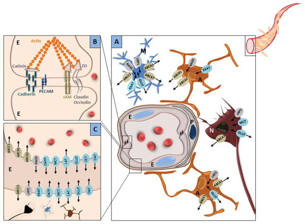
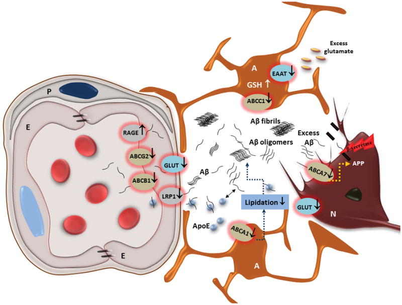
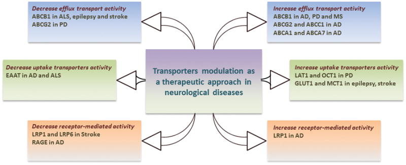
Membrane transport proteins have central physiological function in maintaining cerebral homeostasis. These transporters are expressed in almost all cerebral cells in which they regulate the movement of a wide range of solutes, including endogenous substrates, xenobiotic, and therapeutic drugs. Altered activity/expression of central nervous system (CNS) transporters has been implicated in the onset and progression of multiple neurological diseases. Neurological diseases are heterogeneous diseases that involve complex pathological alterations with only a few treatment options; therefore, there is a great need for the development of novel therapeutic treatments. To that end, transporters have emerged recently to be promising therapeutic targets to halt or slow the course of neurological diseases. The objective of this review is to discuss implications of transporters in neurological diseases and summarize available evidence for targeting transporters as decent therapeutic approach in the treatment of neurological diseases.


Conflict of interest statement

The authors declare no conflicts of interest.

Figures

Figure 1
Figure 1
Localization of transporters at the neurovascular unit. (A) Schematic presentation of neurovascular unit components along with the localization of major transporters in neurons and astrocytes. Neurovascular unit is a modular structure that interconnects neurons (N) and its associated astrocytes (A) and microglia (M) with the cells forming the brain capillaries, the endothelial cells (E) and pericytes (P). (B) The structure of tight junction and its protein elements between two endothelial cells. Endothelial cells of the brain capillaries have several features that distinguish them from those found in the rest of the body. These features include reduced density of caveolae, increased density of mitochondria, and expression of tight and adherence junctions’ proteins, such as occludin, claudins, junction adhesion molecules (JAMs), Zonulae occludin (ZO), VE-cadherin and catenin, that “glue” together the cerebral endothelial cells. (C) Putative localization of the major transporters on brain capillary endothelial. Due to the tightness of the endothelial barrier, paracellular transport is negligible and solutes exchanges can takes place by passive transcellular diffusion, specific transport systems or receptor-mediated transport systems. Wide range of transporters and receptors such as ABCB1, ABCG2, ABCC1, GLUTs, LATs (LAT1 and LAT2), MCTs (MCT1, MCT2, MCT8), OATPs (OATP1A4, OATP2B1, OATP1C1), OCTs (OCT1, OCT2, OCT3) and OAT3, LRP1 and RAGE have been characterized in the endothelium of the brain capillaries. Arrows indicate direction of substrate transport.
Figure 2
Figure 2
Transport proteins in Alzheimer’s disease. Down-regulation of ABCB1, ABCG2 and LRP1 reduce Aβ clearance across the BBB and cause Aβ accumulation in brain parenchyma. Reduced activity of ABCA1 in astrocytes and neurons decreases Aβ clearance via the ApoE clearance pathway, while ABCA7 deficiency accelerates Aβ production, which trigger formation of Aβ oligomers and amyloid plaques. Reduced glutamate uptake by astrocytes through glutamate transporters (EAAT) increases glutamate concentration in synaptic cleft. Increased expression of RAGE increases Aβ entering the brain. Glucose transporters (GLUT) are also reduced which reduce glucose cellular uptake.
Figure 3
Figure 3
Transporters as potential therapeutic targets to halt or slow the course of neurological diseases.

References

    1. Qosa H, Miller DS, Pasinelli P, Trotti D. Regulation of ABC efflux transporters at blood-brain barrier in health and neurological disorders. Brain Res. 2015;1628(Pt B):298–316. - PMC - PubMed
    1. Ye AY, Liu QR, Li CY, Zhao M, Qu H. Human transporter database: comprehensive knowledge and discovery tools in the human transporter genes. PLoS One. 2014;9(2):e88883. - PMC - PubMed
    1. Zhao Z, Nelson AR, Betsholtz C, Zlokovic BV. Establishment and Dysfunction of the Blood-Brain Barrier. Cell. 2015;163(5):1064–78. - PMC - PubMed
    1. Ashraf T, Kao A, Bendayan R. Functional expression of drug transporters in glial cells: potential role on drug delivery to the CNS. Adv Pharmacol. 2014;71:45–111. - PubMed
    1. Zlokovic BV. Neurovascular pathways to neurodegeneration in Alzheimer’s disease and other disorders. Nat Rev Neurosci. 2011;12(12):723–38. - PMC - PubMed

MeSH terms

Substances