. 2016 Oct;15(5):924-39.
doi: 10.1111/acel.12501. Epub 2016 Jul 23.

Proteins that mediate protein aggregation and cytotoxicity distinguish Alzheimer's hippocampus from normal controls

Affiliations

Proteins that mediate protein aggregation and cytotoxicity distinguish Alzheimer's hippocampus from normal controls

Srinivas Ayyadevara et al. Aging Cell. 2016 Oct.

Abstract

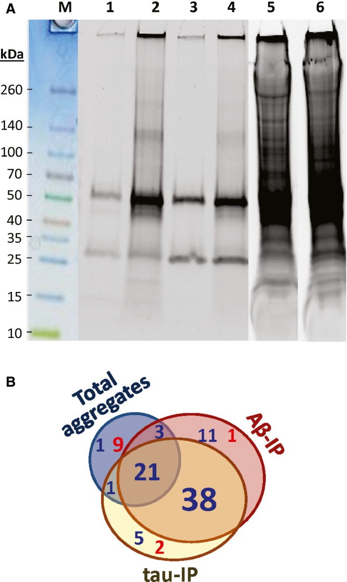
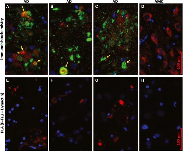
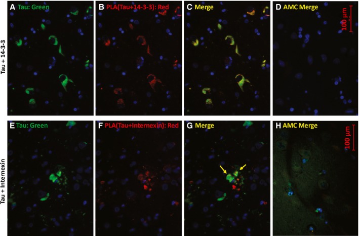
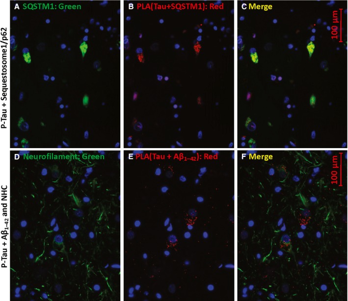
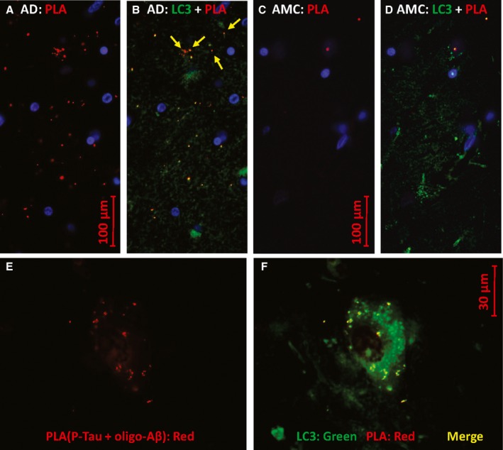
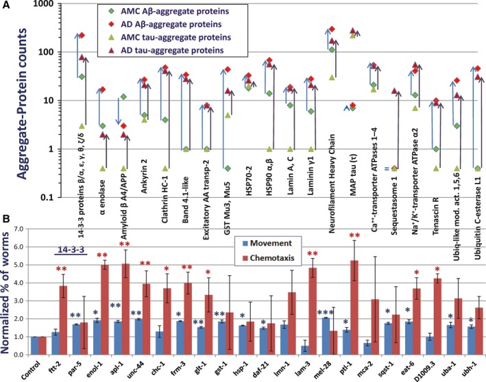
Neurodegenerative diseases are distinguished by characteristic protein aggregates initiated by disease-specific 'seed' proteins; however, roles of other co-aggregated proteins remain largely unexplored. Compact hippocampal aggregates were purified from Alzheimer's and control-subject pools using magnetic-bead immunoaffinity pulldowns. Their components were fractionated by electrophoretic mobility and analyzed by high-resolution proteomics. Although total detergent-insoluble aggregates from Alzheimer's and controls had similar protein content, within the fractions isolated by tau or Aβ1-42 pulldown, the protein constituents of Alzheimer-derived aggregates were more abundant, diverse, and post-translationally modified than those from controls. Tau- and Aβ-containing aggregates were distinguished by multiple components, and yet shared >90% of their protein constituents, implying similar accretion mechanisms. Alzheimer-specific protein enrichment in tau-containing aggregates was corroborated for individuals by three analyses. Five proteins inferred to co-aggregate with tau were confirmed by precise in situ methods, including proximity ligation amplification that requires co-localization within 40 nm. Nematode orthologs of 21 proteins, which showed Alzheimer-specific enrichment in tau-containing aggregates, were assessed for aggregation-promoting roles in C. elegans by RNA-interference 'knockdown'. Fifteen knockdowns (71%) rescued paralysis of worms expressing muscle Aβ, and 12 (57%) rescued chemotaxis disrupted by neuronal Aβ expression. Proteins identified in compact human aggregates, bound by antibody to total tau, were thus shown to play causal roles in aggregation based on nematode models triggered by Aβ1-42 . These observations imply shared mechanisms driving both types of aggregation, and/or aggregate-mediated cross-talk between tau and Aβ. Knowledge of protein components that promote protein accrual in diverse aggregate types implicates common mechanisms and identifies novel targets for drug intervention.

Keywords: Abeta(1-42); Alzheimer (Disease); C. elegans; acetylation (protein); aggregation (protein); beta amyloid; microtubule-associated protein tau; neurodegeneration; neurotoxicity; oxidation (protein); phosphorylation (protein); proteomics.


Figures

Figure 1
Figure 1
Proteins in detergent‐insoluble AD‐derived aggregates are more abundant and diverse than those from controls. (A) Aggregates insoluble in 1% sarcosyl, isolated from hippocampi of normal controls (3 subjects pooled, lanes 1, 3, and 5) or AD (pool of 3, lanes 2, 4, 6). Lanes 1 and 2: pulldown with antibody to Aβ1–42; lanes 3 and 4: pulldown with antibody to tau; lanes 5 and 6: total large insoluble aggregates. (B) Venn diagram of proteins found to be significantly enriched (blue font) or depleted (red) in aggregates from AD relative to controls. More than half of the proteins listed in Table 1 (59/100) were significantly more abundant in both Aβ and tau pulldowns from AD than from controls.
Figure 2
Figure 2
P‐tau coincides with dynactin in situ. Images show hippocampus sections from Alzheimer disease (A–C, E–G) or age‐matched controls (D, H). (A–D) Conventional immunohistochemistry, visualizing proteins with fluor‐tagged secondary antibodies, after incubation with antibodies to P‐tau (Ser202, Thr205; green) and dynactin/p50 (red); nuclei (blue) were stained with DAPI. Light green and yellow (arrows) indicate superposition of red and green fluors. (E–H) Proximity ligation amplification (PLA); red product indicates close proximity (<40 nm separation) of P‐tau (Ser202, Thr205) to dynactin/p50.
Figure 3
Figure 3
Total tau colocalizes with 14‐3‐3 and a subset of internexin in AD hippocampus. Sections were immunostained for total tau (A, E; green), or by proximity ligation amplification (PLA) for total tau in conjunction with 14‐3‐3 proteins (B; red) or with internexin (F; red). C and G are merged images of A+B and E+F, respectively. D and H are the corresponding merged images for AMC hippocampus. Note that in H, the tau (green) exposure was increased ~10‐fold to illustrate the diffuse distribution of normally phosphorylated tau in control tissue.
Figure 4
Figure 4
P‐tau proximity to sequestosome‐1 and Aβ1–42/APP. Sections were immunostained for sequestosome‐1 (SQSTM1, p62; A) or neurofilament heavy chain (NHC, D) and for PLA product (red) showing P‐tau (Thr205) proximal to SQSTM1 (B) or to Aβ1–42 (E). C and D are merged images of A+B and D+E, respectively.
Figure 5
Figure 5
P‐tau co‐localizes with Aβ1–42 oligomers. Antibodies to P‐tau [Thr205] and oligomeric Aβ1–42 (A–D) produce red PLA product if they are within 40 nm. Counterstaining with antibody to LC3B/ATG8, a marker of autophagosomes (B, D, F), appears as green. In E and F, PLA product (red) indicates P‐tau contiguous to Aβ1–42 oligomers. AD hippocampus sections are shown in A, B, E, and F, while C and D show AMC hippocampus.
Figure 6
Figure 6
Knockdown of AD‐enriched aggregate proteins rescues aggregation‐induced traits. (A) Proteins shown are overrepresented (in LCMS spectral counts) in aggregates isolated from AD relative to NC, except microtubule‐associated protein tau, which is hyperphosphorylated but not more abundant in AD (Table 1). Blue arrows connecting diamond symbols show increased abundance of proteins in tau‐IP aggregates; black arrows connecting triangles show increased abundance in Aβ‐IP aggregates. (B) Of 21 protein types more abundant in AD than in NC aggregates, knockdown of 14 (67%) of their nematode orthologs significantly rescued paralysis of a C. elegans strain expressing human Aβ1‐42 in muscle (blue bars), or rescued chemotaxis disrupted by neuronal expression of Aβ1‐42 (red bars). Significance was assessed by 1‐tailed t‐tests (as lower cytotoxicity was predicted) comparing fractions of unparalyzed or chemo‐attracted worms in three independent assays per group (*< 0.05; **< 0.005; ***< 0.0005). Significance by chi‐squared tests within assays was < 5 × 10−5 to 10−16.

Similar articles

Cited by

References

    1. Abdel‐Salam OM (2014) The paths to neurodegeneration in genetic Parkinson's disease. CNS Neurol. Disord. Drug Targets 13, 1485–1512. - PubMed
    1. Aboud O, Parcon PA, DeWall KM, Liu L, Mrak RE, Griffin WS (2015) Aging, Alzheimer's, and APOE genotype influence the expression and neuronal distribution patterns of microtubule motor protein dynactin‐P50. Front Cell Neurosci. 9, 103. - PMC - PubMed
    1. Ahn EH, Kim DW, Shin MJ, Kim HR, Kim SM, Woo SJ, Eom SA, Jo HS, Kim DS, Cho SW, Park J, Eum WS, Choi SY (2014) PEP‐1‐PEA‐15 protects against toxin‐induced neuronal damage in a mouse model of Parkinson's disease. Biochim. Biophys. Acta 1840, 1686–1700. - PubMed
    1. Ayyadevara S, Balasubramaniam M, Gao Y, Yu LR, Alla R, Shmookler Reis RJ (2014) Proteins in aggregates functionally impact multiple neurodegenerative disease models by forming proteasome‐blocking complexes. Aging Cell 14, 35–48. - PMC - PubMed
    1. Ayyadevara S, Mercanti F, Wang XW, Mackintosh SG, Tackett AJ, Prayaga VS, Romeo F, Shmookler Reis RJ, Mehta JL (2016) Age‐ and hypertension‐associated protein aggregates in mouse heart have similar proteomic profiles. Hypertension 67, 1006–1013. - PMC - PubMed

Publication types

LinkOut - more resources