. 2016 Sep 1;197(5):1692-8.
doi: 10.4049/jimmunol.1600940. Epub 2016 Jul 22.

Gut Microbial Membership Modulates CD4 T Cell Reconstitution and Function after Sepsis

Affiliations

Gut Microbial Membership Modulates CD4 T Cell Reconstitution and Function after Sepsis

Javier Cabrera-Perez et al. J Immunol. .

Abstract

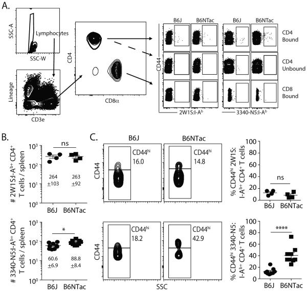
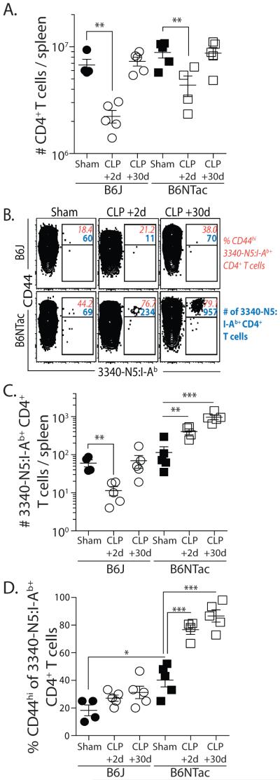
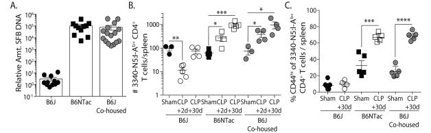
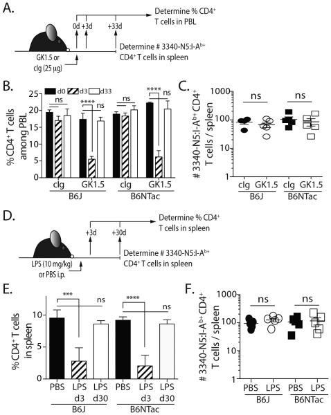
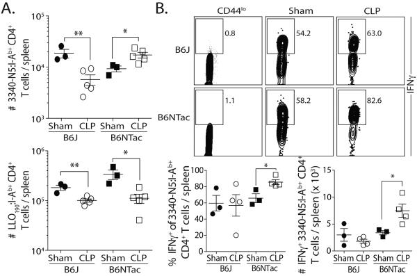
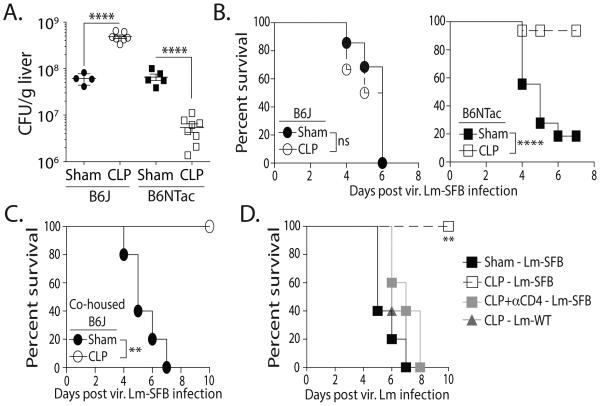
Transient lymphopenia is one hallmark of sepsis, and emergent data indicate the CD4 T cell compartment in sepsis survivors is numerically and functionally altered (when examined at the Ag-specific level) compared with nonseptic control subjects. Previous data from our laboratory demonstrated Ag-independent, lymphopenia-induced homeostatic proliferation to be a contributing mechanism by which CD4 T cells numerically recover in sepsis survivors. However, we reasoned it is also formally possible that some CD4 T cells respond directly to Ag expressed by gut-resident microbes released during polymicrobial sepsis. The effect of gut microbiome leakage on CD4 T cells is currently unknown. In this study, we explored the number and function of endogenous CD4 T cells specific for segmented filamentous bacterium (SFB) after cecal ligation and puncture (CLP)-induced sepsis using mice that either contained or lacked SFB as a normal gut-resident microbe. Interestingly, SFB-specific CD4 T cells underwent Ag-driven proliferation in CLP-treated SFB(+), but not in SFB(-), mice. Moreover, CLP-treated SFB(+) mice showed resistance to secondary lethal infection with recombinant SFB Ag-expressing virulent Listeria (but not wild-type virulent Listeria), suggesting the CLP-induced polymicrobial sepsis primed for a protective response by the SFB-specific CD4 T cells. Thus, our data demonstrate that the numerical recovery and functional responsiveness of Ag-specific CD4 T cells in sepsis survivors is, in part, modulated by the intestinal barrier's health discreetly defined by individual bacterial populations of the host's microbiome.


Figures

Figure 1
Figure 1
Naïve B6J and B6NTac mice differ in number of SFB-specific CD4 T cells and frequency of CD44hi SFB-specific CD4 T cells. (A) Representative flow plots show the gating strategy used in tetramer-enriched cell fractions to detect Ag-specific CD4 T cell populations. Shown are examples used to detect 2W1S:I-Ab- and 3340-N5:I-Ab-specific CD4+ T cells. Gating for p:I-Ab-specific cells was determined using CD8+ T cells as an internal negative control for tetramer binding. (B) Number of 2W1S:I-Ab- and 3340-N5:I-Ab-specific CD4 T cells and (C) frequency of CD44hi 2W1S:I-Ab- and 3340-N5:I-Ab-specific CD4 T cells in the spleens of naïve B6J and B6NTac mice. Statistical significance was determined using group-wise, one-way ANOVA with multiple-testing correction using the Holm-Sidak method, and α = 0.05. Data shown are from 9 mice/group.
Figure 2
Figure 2
Differential response of SFB-specific CD4 T cells in CLP-treated B6J and B6NTac mice. (A) Number of total CD4 T cells in spleens of B6J and B6NTac mice on d 2 and recovery by d 30 after CLP. (B) Representative plots show the number of SFB-specific CD4 T cells and frequency of CD44hi SFB-specific CD4 T cells in sham- and CLP-treated B6J and B6NTac mice. (C-D) Number of SFB-specific CD4 T cells and frequency of CD44hi SFB-specific CD4 T cells in sham- and CLP-treated B6J and B6NTac mice 2 or 30 d after surgery. Statistical significance was determined using group-wise, one-way ANOVA with multiple-testing correction using the Holm-Sidak method, and α = 0.05. Data shown represent 2 independent experiments, with 4-5 mice/group in each experiment.
Figure 3
Figure 3
Horizontal transfer of SFB by co-housing permits the expansion of SFB-specific CD4 T cells in B6J mice after CLP. B6J and B6NTac mice were housed separately or co-housed for 4 weeks. (A) The presence of SFB in fecal pellets in individual mice was determined by PCR prior to sham or CLP surgery. Each symbol represents an individual mouse. (B-C) The number of SFB-specific CD4 T cells and frequency of CD44hi SFB-specific CD4 T cells in the spleens of B6J, B6NTac, and co-housed B6J mice on d 2 and 30 after CLP surgery. Statistical significance was determined using group-wise, one-way ANOVA with multiple-testing correction using the Holm-Sidak method, and α = 0.05. Data shown are the representative from at least 2 independent experiments per population analyzed, with 4-5 mice/group in each experiment.
Figure 4
Figure 4
Transient reduction of CD4 T cells without bacteremia is not sufficient to drive SFB-specific CD4 T cell expansion in B6NTac mice. (A) B6J and B6NTac mice were injected with 25 μg of the anti-CD4 depleting mAb GK1.5 or an isotype control mAb. Mice were bled before mAb injection and on d 3 and 33 after injection. (B) The frequency of CD45.2+CD3+CD4+ T cells in the blood was determined. (C) On d 33, the number of SFB-specific CD4 T cells in the spleens was determined by tetramer enrichment and flow cytometry. (D) B6J and B6NTac mice were injected with 10 mg/kg LPS or PBS. (E) Spleens were collected on d3 and 30 to measure the frequency of CD45.2+CD3+CD4+ T cells. (F) On d 30, the number of SFB-specific CD4 T cells in the spleens was determined by tetramer enrichment and flow cytometry.
Figure 5
Figure 5
SFB colonization changes the function of SFB-specific CD4 T cells in sepsis survivors. (A-B) B6J and B6NTac mice were infected with att. Lm-SFB (107 CFU in 0.1 ml i.v.) 30 d after sham or CLP surgery. (A) Proliferative capacity – The number of LLO190- and SFB-specific CD4 T cells was determined in the spleen 7 d after infection. (B) Functional capacity – 7 d after infection, splenocytes were stimulated for 4 h with PMA/ionomycin. The samples were enriched for SFB-specific CD4 T cells and IFNγ production was determined. Representative flow plots show the frequency of SFB-specific CD4 T cells producing IFNγ. The frequency and number of IFNγ+ SFB-specific CD4 T cells are graphed. Statistical significance in A-B was determined using group-wise, one-way ANOVA analyses followed by multiple-testing correction using the Holm-Sidak method, with α = 0.05. Data shown represent 2 independent experiments, with 3-5 mice/group in each experiment.
Figure 6
Figure 6
SFB colonization influences the protective capacity of SFB-specific CD4 T cells in sepsis survivors to secondary infection. (A-B) B6J and B6NTac mice were infected with vir. Lm-SFB (5 × 10³ CFU in 0.1 ml i.v.) 30 d after sham or CLP surgery. (A) Mice were sacrificed on d 5 post-infection to determine Listeria burden in the liver, while other cohorts (B) were monitored for survival. (C) Additional cohorts of B6J mice co-housed with B6NTac mice for 4 weeks before sham or CLP surgery were similarly infected with vir. Lm-SFB 30 d after surgery. (D) B6NTac mice were infected with vir. wild-type Lm (Lm-WT) or Lm-SFB (5 × 10³ CFU in 0.1 ml i.v.) 30 d after sham or CLP surgery. Some of the CLP-treated mice were depleted of CD4 T cells using the GK1.5 mAb (3 daily injection of 100 μg i.p.) immediately before infection. Survival was monitored. Statistics shown were obtained using Mann-Whitney U test statistics (for B) and one-way ANOVA analyses (for A, C, and D) with multiple-testing correction using the Holm-Sidak method, and α = 0.05. Survival data are cumulative from 3 independent experiments with 5 mice/group in each experiment.

Similar articles

Cited by

References

    1. Shankar-Hari M, Phillips GS, Levy ML, Seymour CW, Liu VX, Deutschman CS, Angus DC, Rubenfeld GD, Singer M, F. Sepsis Definitions Task Developing a New Definition and Assessing New Clinical Criteria for Septic Shock: For the Third International Consensus Definitions for Sepsis and Septic Shock (Sepsis-3) JAMA. 2016;315:775–787. - PMC - PubMed
    1. Vulliamy PE, Perkins ZB, Brohi K, Manson J. Persistent lymphopenia is an independent predictor of mortality in critically ill emergency general surgical patients. Eur. J. Trauma Emerg. Surg. 2015 - PubMed
    1. Hamers L, Kox M, Pickkers P. Sepsis-induced immunoparalysis: mechanisms, markers, and treatment options. Minerva Anestesiol. 2015;81:426–439. - PubMed
    1. Hotchkiss RS, Tinsley KW, Swanson PE, Schmieg RE, Jr., Hui JJ, Chang KC, Osborne DF, Freeman BD, Cobb JP, Buchman TG, Karl IE. Sepsis-induced apoptosis causes progressive profound depletion of B and CD4+ T lymphocytes in humans. J. Immunol. 2001;166:6952–6963. - PubMed
    1. Unsinger J, Kazama H, McDonough JS, Griffith TS, Hotchkiss RS, Ferguson TA. Sepsis-induced apoptosis leads to active suppression of delayed-type hypersensitivity by CD8+ regulatory T cells through a TRAIL-dependent mechanism. J. Immunol. 2010;184:6766–6772. - PMC - PubMed

Publication types

Substances