Case Reports
. 2016 Jul 22:9:358.
doi: 10.1186/s13104-016-2170-8.

Identification and molecular characterization of Corynebacterium xerosis isolated from a sheep cutaneous abscess: first case report in Mexico

Affiliations
Case Reports

Identification and molecular characterization of Corynebacterium xerosis isolated from a sheep cutaneous abscess: first case report in Mexico

Fernando Hernández-León et al. BMC Res Notes. .

Abstract

Background: Corynebacterium xerosis is a commensal organism found in skin and mucous membranes of humans. It is considered an unusual pathogen, and it is rarely found in human and animal clinical samples. Here we describe the isolation of C. xerosis from a 4-months-old Pelifolk lamb located in Tesistán, central western Mexico. This microorganism should be considered for differential diagnosis in cutaneous abscessed lesions in sheep, as it represents a zoonotic risk factor for human infection in sheep farms.

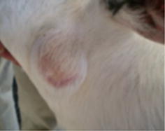
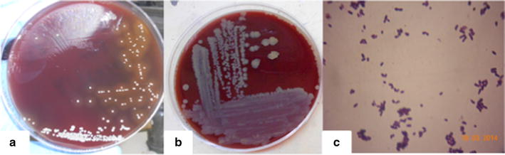
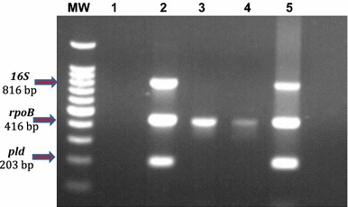
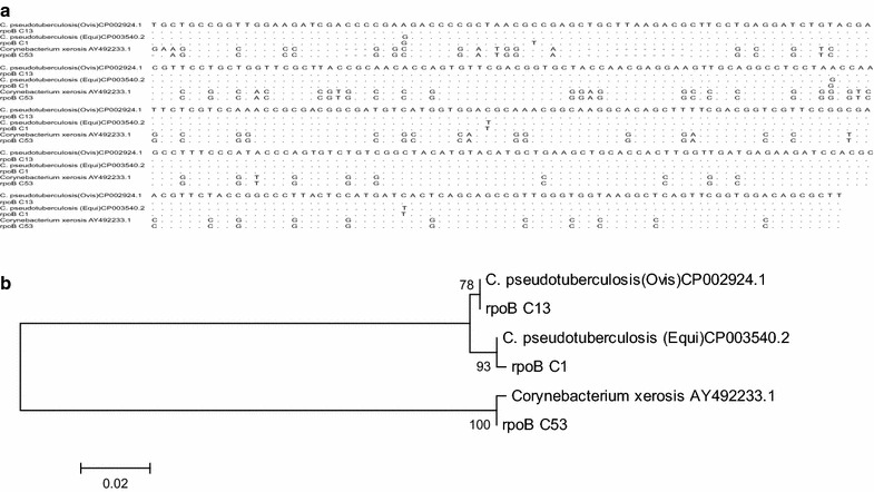
Case presentation: The animal exhibited a hard-consistency, 5 cm diameter abscess, without drainage, in the neck. The presumptive clinical diagnosis was caseous lymphadenitis, caused by Corynebacterium pseudotuberculosis. Samples were obtained by puncture and cultured in 8 % sheep blood agar under microaerophilic conditions. Colonies were non-haemolytic, brown-yellowish and showed microscopic and biochemical features similar to C. pseudotuberculosis, except for the urea test. A multiplex-PCR for the amplification of partial sequences of the pld, rpoB and intergenic fragment from 16S to 23S genes suggested that isolate could be C. xerosis, which was later confirmed by sequencing analysis of the rpoB gene.

Conclusions: This study shows for the first time isolation and molecular characterization of C. xerosis from a clinical sample of an ovine cutaneous abscess in Mexico. This finding highlights the need for differential diagnosis of this pathogen in ovine skin abscesses, as well as epidemiological and control studies of this pathogen in sheep farms.

Keywords: Case report; Corynebacterium pseudotuberculosis; Corynebacterium xerosis; Pld gene; RpoB gene; Sheep; Skin abscess.


Figures

Fig. 1
Fig. 1
Studied abscess. A hard-consistency abscess without drainage was reported in the neck region of a 4-months-old lamb
Fig. 2
Fig. 2
In-vitro Bacteriological culture of Corynebacterium xerosis and Corynebacterium pseudotuberculosis. Bacteria were cultured in 8 % sheep blood agar. a Corynebacterium pseudotuberculosis (ATCC 43924) showed whitish colonies with beta haemolysis. b Corynebacterium xerosis isolate, grew as small yellowish-brown colonies without haemolysis. c Gram-stained smear preparation of Corynebacterium xerosis showing characteristic pleomorphic Gram-positive rods, with club-like ends
Fig. 3
Fig. 3
Multiplex PCR. Amplification of partial sequences of 16S rRNA, rpoB and pld genes. MW lane: molecular weight marker of 1 Kb Plus DNA Ladder™ (Invitrogen). Lane 1: negative control (reaction without template DNA). Lane 2: Corynebacterium pseudotuberculosis biovar equi. Lanes 3–4: Corynebacterium xerosis isolate (10–0.001 ng of DNA, respectively). Lane 5: Corynebacterium pseudotuberculosis biovar ovis
Fig. 4
Fig. 4
Bioinformatic analysis. a Shows the sequence alignment from the sequences used to construct the Phylogenetic tree in (b). Three sequences were downloaded from GenBank: C. pseudotuberculosis biovar ovis (CP002924.1), C. pseudotuberculosis biovar equi (CP003540.2), and Corynebacterium xerosis (AY492233.1). The other three were specimens sent for sequencing (Macrogen, Rockville, MD, USA): rpoB13 (local isolate characterized as C. pseudotuberculosis, biovar ovis), rpoB C1 (C. pseudotuberculosis, biovar equi, reference strain ATCC43924) and rpoB C53 (local isolate, characterized as C. xerosis). b The tree shows the genetic relationship of Corynebacterium pseudotuberculosis and Corynebcaterium xerosis for the rpoB gene. The tree was built from the alignment of partial sequence of the gene. Bootstrap values were obtained generating 1000 random trees, and the strength of each branch is indicated in the respective node

Similar articles

Cited by

References

    1. Vela AI, Gracía E, Fernández A, Domínguez L, Fernández-Garayzábal JF. Isolation of Corynebacterium xerosis from animal clinical specimens. J Clin Microbiol. 2006;2006(44):2242–2243. doi: 10.1128/JCM.02473-05. - DOI - PMC - PubMed
    1. Coyle MB, Lipsky BA. Coryneform bacteria in infectious diseases: clinical and laboratory aspects. Clin Microbiol Rev. 1990;3:227–246. doi: 10.1128/CMR.3.3.227. - DOI - PMC - PubMed
    1. Funke G, Lawson PA, Bernard KA, Collins MD. Most Corynebacterium xerosis strains identified in the routine clinical laboratory correspond to Corynebacterium amycolatum. J Clin Microbiol. 1996;1996(34):1124–1128. - PMC - PubMed
    1. Riegel P, Ruimy R, Christen R, Monteil H. Species identities and antimicrobial susceptibilities of corynebacteria isolated from various clinical sources. Eur J Clin Microbiol Infect Dis. 1996;15:657–662. doi: 10.1007/BF01691153. - DOI - PubMed
    1. Funke G, von Graevenitz A, Clarridge JE, Bernard KA. Clinical microbiology of coryneform bacteria. Clin Microbiol Rev. 1997;10:125–159. - PMC - PubMed

Publication types

MeSH terms

LinkOut - more resources