. 2016 Oct 9;428(20):4228-4241.
doi: 10.1016/j.jmb.2016.07.004. Epub 2016 Jul 21.

Design Principles for SuCESsFul Biosensors: Specific Fluorophore/Analyte Binding and Minimization of Fluorophore/Scaffold Interactions

Affiliations

Design Principles for SuCESsFul Biosensors: Specific Fluorophore/Analyte Binding and Minimization of Fluorophore/Scaffold Interactions

Seymour de Picciotto et al. J Mol Biol. .

Abstract

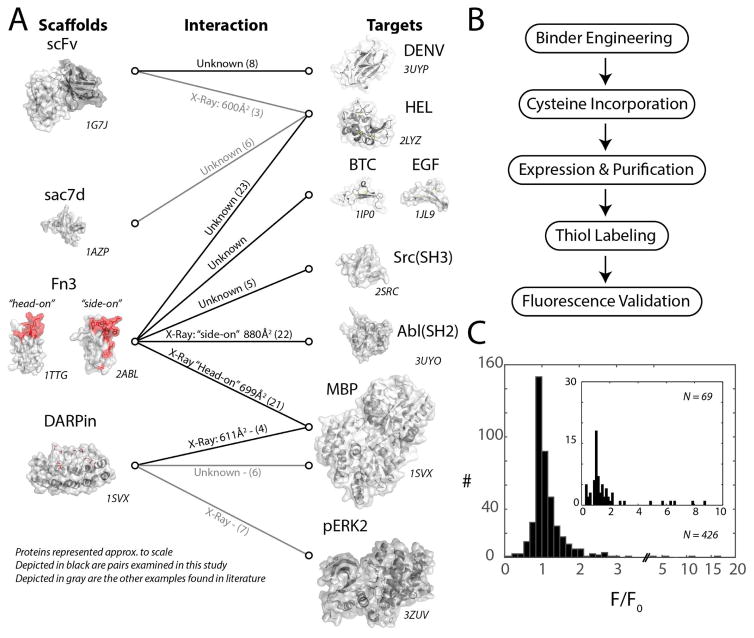
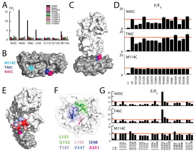
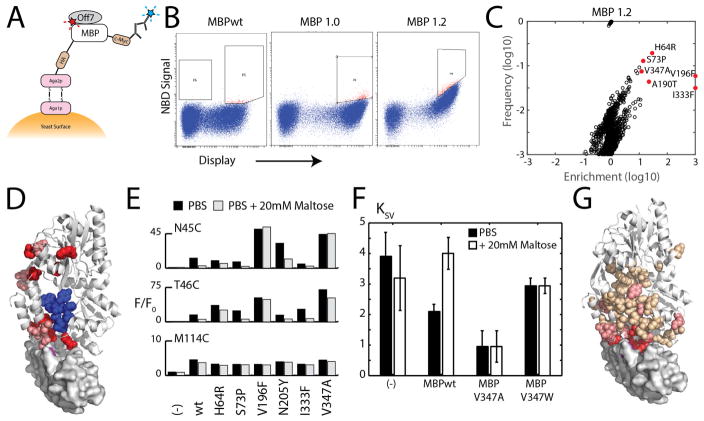
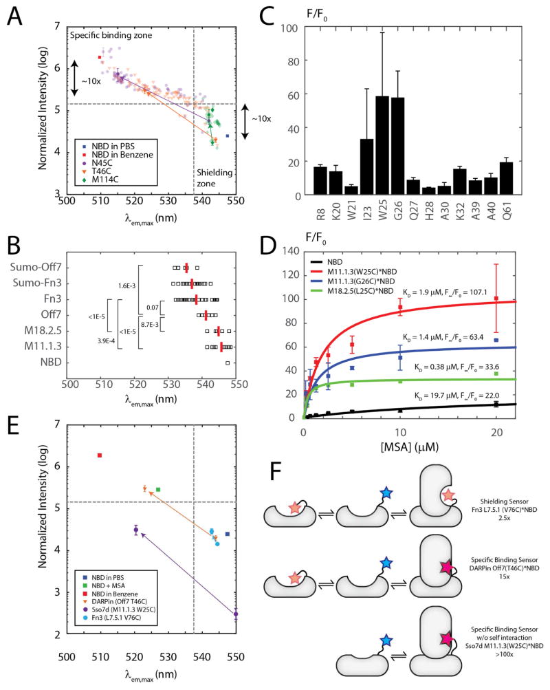
Quantifying protein location and concentration is critical for understanding function in situ. Scaffold conjugated to environment-sensitive fluorophore (SuCESsFul) biosensors, in which a reporting fluorophore is conjugated to a binding scaffold, can, in principle, detect analytes of interest with high temporal and spatial resolution. However, their adoption has been limited due to the extensive empirical screening required for their development. We sought to establish design principles for this class of biosensor by characterizing over 400 biosensors based on various protein analytes, binding proteins, and fluorophores. We found that the brightest readouts are attained when a specific binding pocket for the fluorophore is present on the analyte. Also, interaction of the fluorophore with the binding protein it is conjugated to can raise background fluorescence, considerably limiting sensor dynamic range. Exploiting these two concepts, we designed biosensors that attain a 100-fold increase in fluorescence upon binding to analyte, an order of magnitude improvement over the previously best-reported SuCESsFul biosensor. These design principles should facilitate the development of improved SuCESsFul biosensors.

Keywords: Sso7d scaffold; directed evolution; protein engineering; sensors; solvatochromism.


Conflict of interest statement

The authors declare no competing financial interests.

Figures

Figure 1
Figure 1
Reagentless biosensor workflow and overview. A) Overview of reported reagentless biosensors built from a protein scaffold recognizing another protein. On the left side the various protein scaffolds that have been used and coupled with an environment-sensitive dye are listed. On the right side the target proteins are shown. Dark arrows represent pairs evaluated in this study. The connecting lines indicate the information about the nature of the interaction as well as the reference citation. B) Reagentless biosensor engineering is a five-step process. First, a binder must be engineered with desired specificity and affinity. Second, a chemical moiety for site-specific labeling must be incorporated. This is typically achieved by directed mutagenesis for a cysteine at the desired location. Next, the protein construct is expressed, purified and labeled with a thiol reactive derivative of the fluorescent dye. Finally the construct is evaluated for its fluorescence characteristic in the absence and presence of its target. C) Histogram of the dynamic range of sensors to their target reported as F/F0, the fluorescence intensity ratio between the conditions in presence versus in absence of the target protein. The histogram showed in the inlet is from previously published results.
Figure 2
Figure 2
Mapping the interaction between NBD and MBP in the Off7(N45C)*NBD and Off7(T46C)*NBD sensors. A) Increase in fluorescence signal when adding MBP to Off7 mutants labeled with 10 different fluorophores at 7 positions. B) Top view of the Off7 protein (PBD: 1SVX). C) Side view of the Off7/MBP complex (PDB: 1SVX). D) F/F0 ratios of 3 Off7*NBD sensors with wtMBP and mutants with a single alanine mutation. E) Effects from alanine scanning are plotted against the Off7/MBP structure, red indicates reduced activity, blue increased activity. Deleterious mutations are surrounding a cavity in MBP. F) Residues mutated in the cavity analysis. G) F/F0 ratios for three Off7*NBD sensors with MBP cavity mutants.
Figure 3
Figure 3
Off7(T46C)*NBD Sensor is regulated by MBP conformation. A) Graphical representation of experimental set-up. B) FACS plots showing the improvement of NBD signal produced by MBP mutant displaying yeasts over two selection rounds. C) Enrichment versus frequency plot for the whole gene error prone PCR library. D) Graphical representation of enrichment onto Off7/MBP structure combined from all four libraries. Red and blue indicate positive and negative enrichment respectively. E) F/F0 ratios for the YSD isolated mutants in the absence or presence of maltose. Mutations H64R, V196F and V347A greatly stabilized the activity of the sensor in the presence of maltose. F) Iodide quenching assay, reporting the Stern-Vollmer constant for wild-type MBP, the most strongly activating mutation V347A, and the most deteriorating mutation V347W. G) Weighted combined nitrogen and proton chemical shift perturbations (CSP) are highlighted on the Off7/MBP complex. CSP between 0.025 and 0.05 are shown in beige, between 0.05 and 0.1 in salmon, and CSP > 0.1 in red.
Figure 4
Figure 4
The balance between scaffold rigidity and specific binding pockets. A) Maximum emission wavelength versus signal intensity plotted for the three Off7 sensors in complex with all MBP mutants evaluated in this study. In addition, free NBD in PBS and benzene is depicted. B) Maximum emission wavelength of NBD labeled scaffolds in the absence of antigen. C) F/F0 ratio for M11.1.3*NBD conjugates. D) Titrations of NBD and NBD labeled constructs with MSA. E) Maximum emission wavelength vs intensity for the best Fn3, DARPin, and Sso7d sensors. F) Mechanistic drawing of the three types of reagentless biosensors. The top one describes a sensors that suffers from pre-activation and the fluorophore undergoes shielding upon binding, yielding a small F/F0 ratio. The middle one describes a sensor that may have some pre-activation but that alleviates this drawback by having a strong fluorescence enhancement due to a fluorophore binding pocket. The bottom scenario describes a sensor that possesses both ideal properties: little self-interaction and specific binding pocket interaction on the analyte.

References

    1. Zadran S, et al. Fluorescence resonance energy transfer (FRET)-based biosensors: visualizing cellular dynamics and bioenergetics. Appl Microbiol Biotechnol. 2012;96:895–902. - PubMed
    1. Burr M, Koshland DE. Use of ‘Reporter Groups’ in Structure-Function Studies of Proteins. Proc Natl Acad Sci. 1964;52:1017–1024. - PMC - PubMed
    1. Renard M, et al. Knowledge-based design of reagentless fluorescent biosensors from recombinant antibodies. J Mol Biol. 2002;318:429–42. - PubMed
    1. Brient-Litzler E, Pluckthun A, Bedouelle H. Knowledge-based design of reagentless fluorescent biosensors from a designed ankyrin repeat protein. Protein Eng Des Sel. 2010;23:229–41. - PubMed
    1. Gulyani A, et al. A biosensor generated via high-throughput screening quantifies cell edge Src dynamics. Nat Chem Biol. 2011;7:437–44. - PMC - PubMed

Publication types

LinkOut - more resources