. 2016 Aug 23;7(34):54983-54997.
doi: 10.18632/oncotarget.10713.

NHERF1, a novel GPER associated protein, increases stability and activation of GPER in ER-positive breast cancer

Affiliations

NHERF1, a novel GPER associated protein, increases stability and activation of GPER in ER-positive breast cancer

Ran Meng et al. Oncotarget. .

Abstract

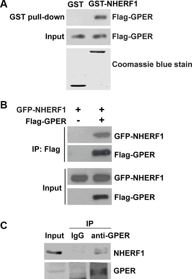
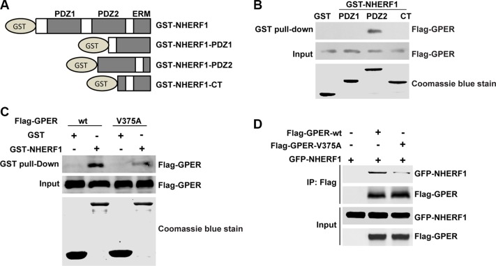
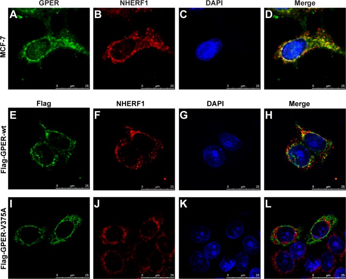
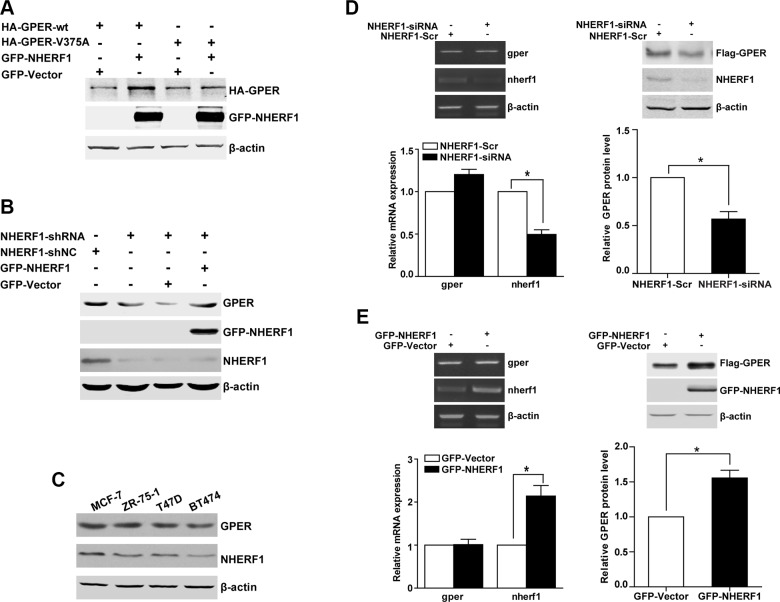
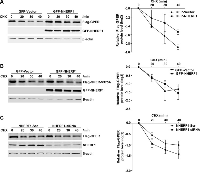
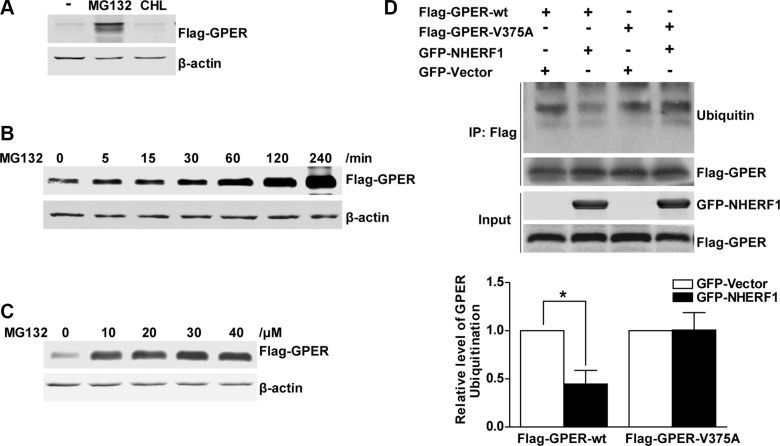
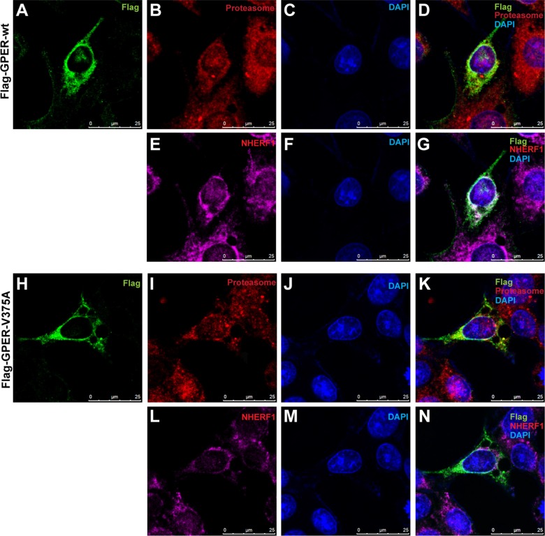
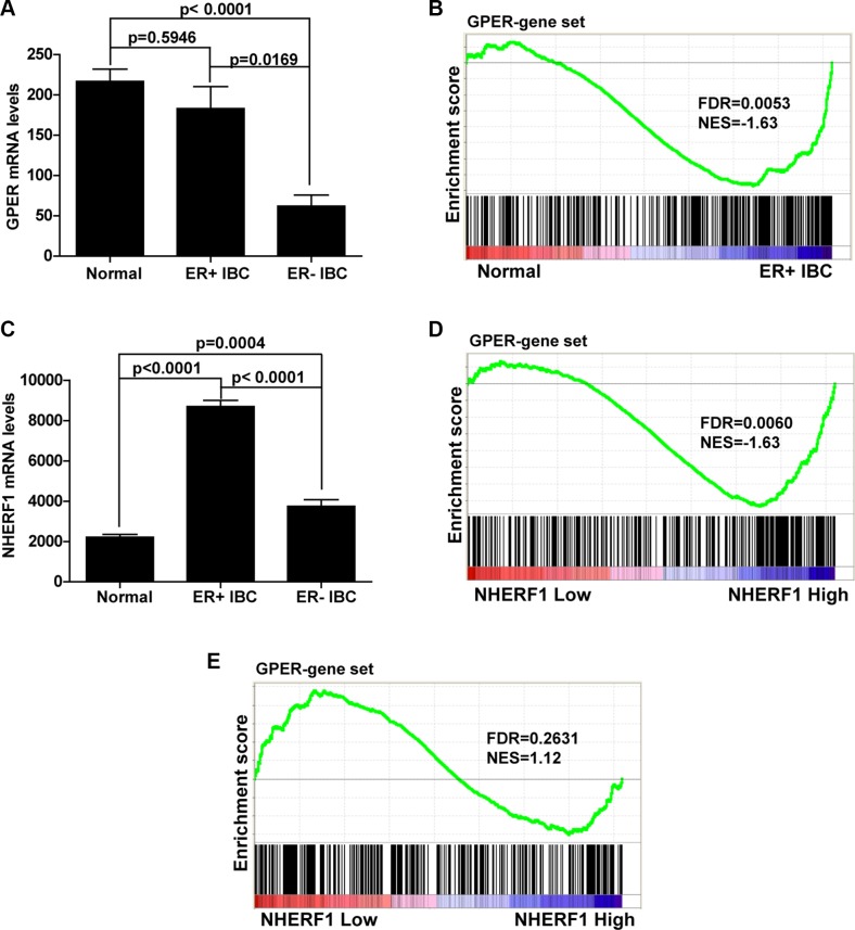
G protein-coupled estrogen receptor (GPER) plays an important role in mediating the effects of estradiol. High levels of GPER have been implicated to associate with the malignant progress of invasive breast cancer (IBC). However, the mechanisms by which GPER protein levels were regulated remain unclear. In this study, PDZ protein Na+/H+ exchanger regulatory factor (NHERF1) was found to interact with GPER in breast cancer cells. This interaction was mediated by the PDZ2 domain of NHERF1 and the carboxyl terminal PDZ binding motif of GPER. NHERF1 was demonstrated to facilitate GPER expression at post-transcriptional level and improve GPER protein stability by inhibiting the receptor degradation via ubiquitin-proteasome pathway in a GPER/NHERF1 interaction-dependent manner. In addition, GPER protein levels are positively associated with NHERF1 protein levels in a panel of estrogen receptor (ER)-positive breast cancer cells. Furthermore, analysis of clinical IBC data from The Cancer Genome Atlas (TCGA) showed no significant difference in GPER mRNA levels between ER-positive IBC and normal breast tissues. However, gene set enrichment analysis (GSEA) showed that GPER signaling is ultra-activated in ER-positive IBC when compared with normal and its activation is positively associated with NHERF1 mRNA levels. Taken together, our findings identify NHERF1 as a new binding partner for GPER and its overexpression promotes protein stability and activation of GPER in ER-positive IBC. Our data indicate that regulation of GPER stability by NHERF1 may contribute to GPER-mediated carcinogenesis in ER-positive IBC.

Keywords: EBP50; G protein-coupled receptor; carcinogenesis; protein degradation; protein-protein interaction.


Conflict of interest statement

CONFLICTS OF INTEREST

The authors declare that they have no conflicts of interest with the contents of this article.

Figures

Figure 1
Figure 1. NHERF1 is identified as a novel GPER-associated protein
(A) GPER interacts with NHERF1 in vitro. Lysates of HEK-293 cells that stably transfected with Flag-GPER (HEK-GPER) were incubated with GST or GST-NHERF1, followed by Western blotting using anti-Flag antibody. GST and GST-NHERF1 fusion proteins were stained by Coomassie blue dye (lower panel). (B) Cellular association of full-length Flag-GPER with GFP-NHERF1 in HEK-293 cells. HEK-293 cells were transfected with GFP-NHERF1 in the presence or absence of Flag-GPER. The cells were then lysed and immunoprecipitated using anti-Flag affinity gel. The immunoprecipitate samples (IP) and whole cell lysates (Input) were analyzed by Western blotting with anti-Flag or anti-GFP antibodies. (C) GPER associates with NHERF1 in MCF-7 cells. The lysates of MCF-7 cells were subjected to immunoprecipitation using anti-GPER antibody. Lysates and immunoprecipitated samples were probed with anti-GPER or anti-NHERF1 antibodies to detect the presence of GPER and NHERF1. Blots are representative of three to five independent experiments. Input, whole cell lysates; IP, immunoprecipitation.
Figure 2
Figure 2. Interaction of NHERF1 and GPER is mediated via the C-terminal of GPER and the PDZ2 domain of NHERF1
(A) Schematic diagram of GST-NHERF1 fusion proteins used in GST-pull down experiments. (B) GPER interacts specifically with the PDZ2 domain of NHERF1. Lysates of HEK-GPER were incubated with GST or GST fusion proteins containing different domains of NHERF1. Precipitates were subjected to Western blotting with anti-Flag antibody. Coomassie blue staining showed equal loading of the fusion proteins (bottom panels). (C) Association of NHERF1 and GPER via C-terminal of GPER. Equal amounts of purified GST or GST-NHERF1 fusion protein beads (lower panel) were used to pull down lysates from HEK-293 cells that transfected with Flag-GPER-wt or Flag-GPER-V375A respectively. Precipitates were detected by Western blotting using anti-Flag antibody. (D) The point mutation of GPER (GPER-V375A) abolishes the interaction between GPER and NHERF1 in cells. HEK-293 cells were either transfected with Flag-GPER-wt or Flag-GPER-V375A together with or without GFP-NHERF1. Cell lysates were then immunoprecipitated using anti-Flag affinity gel. Immunoprecipitated samples were subjected to Western blotting using anti-Flag or anti-GFP antibodies. The expression levels of Flag-GPER-wt and Flag-GPER-V375A were adjusted to similar levels through transfection with different amounts of the respective constructs. The experiments were repeated at least three times.
Figure 3
Figure 3. NHERF1 co-localizes with GPER in MCF-7 cells
(AD) Endogenous GPER and NHERF1 are co-localized in MCF-7 cells. MCF-7 cells were stained with anti-GPER (A) and anti-NHERF1 (B) antibodies followed by visualized using Alexa 488- and Alexa 594-conjugated secondary antibodies. Co-localization of GPER and NHERF1 is shown in yellow color in merged image (D). (EH) Wild type GPER co-localizes with NHERF1 in MCF-7 cells. MCF-7 cells were transfected with Flag-GPER-wt, and then incubated with anti-Flag (E) and anti-NHERF1 (F) primary antibodies followed by staining with Alexa 488- and Alexa 594-conjugated secondary antibodies. Nucleuses were stained with DAPI (G). Co-localization of Flag-GPER-wt and NHERF1 is shown in yellow color in panel (H). (IL) Mutation in the C-terminal of GPER abolishes its co-localization with NHERF1 in MCF-7 cells. MCF-7 cells were transfected with Flag-GPER-V375A and then stained using anti-Flag (I) and anti-NHERF1 (J) antibodies. Nuclei were detected with DAPI staining (K). Less co-localization of Flag-GPER-V375A and NHERF1 is detected following merging of the two individual images (L).
Figure 4
Figure 4. NHERF1 enhances the level of the GPER protein at the post-transcriptional level
(A) The protein level of GPER-wt is increased when NHERF1 is overexpressed. HEK-293 cells were transiently transfected with equal amount constructs of HA-GPER-wt or HA-GPER-V375A in the presence or absence of GFP-NHERF1. Protein levels of GPER and NHERF1 were detected by Western blotting using anti-HA and anti-GFP antibodies respectively. (B) NHERF1 enhances GPER protein level in breast cancer cells. MCF-7 cells were stably transfected with shNC or shNHERF1 (lane 1 and lane 2), and the expression of NHERF1 was rescued by transiently transfected the cells with GFP-Vector or GFP-NHERF1 (lane 3 and lane 4). The cell lysates were then tested by Western blotting using anti-GPER, anti-GFP or anti-NHERF1 antibodies. (C) Correlation of endogenous NHERF1 and GPER protein levels in ER-positive breast cancer cell lines. Lysates from four ER-positive breast cancer cell lines were analyzed by Western blotting by using anti-GPER or anti-NHERF1 antibodies. (D) Knock-down of NHERF1 expression decreases GPER expression at the post-transcriptional level. HEK-GPER cells were transiently transfected with siNHERF1 or scrambled sequence (Scr) respectively. Total RNA of the cells was isolated using Trizol reagent and the mRNA levels of GPER, NHERF1, and β-actin were then analyzed by RT-PCR (Left panel). Total cell lysates were subjected to Western blotting and detected with anti-Flag, anti-NHERF1, and anti-β-actin antibodies (Right panel). (E) Overexpression of NHERF1 dramatically increases GPER expression at post-transcriptional level. HEK-GPER cells were transiently transfected with GFP-NHERF1. The mRNA and protein levels of GPER, NHERF1, and β-actin were analyzed by RT-PCR (Left panel) and Western blotting (Right panel) as described in panel D, respectively. The experiments were repeated at least three times, with values within each experiment normalized with β-actin and analyzed by GraphPad Prism 5. Histogram represents average value of relative GPER protein levels (*p < 0.05). Data were presented as means ± SEM.
Figure 5
Figure 5. NHERF1 expression improves GPER protein stabilization
(A) Overexpression of NHERF1 reduces the turnover rate of Flag-GPER. HEK-GPER cells were transiently transfected GFP-Vector or GFP-NHERF1, then treated with CHX (20 μg/ml) and harvested at the indicated time points. The protein levels of GPER, NHERF1, and β-actin were analyzed by Western blotting. (B) NHERF1 has no effect on the protein stabilization of Flag-GPER-V375A. HEK-GPER-V375A cells were transiently transfected with GFP-Vector or GFP-NHERF1, then treated with CHX (20 μg/ml) at indicated time points prior to harvest. The protein levels of GPER, NHERF1, and β-actin were analyzed by Western blotting. (C) Knock-down of NHERF1 expression enhances Flag-GPER degradation. HEK-GPER cells were transiently transfected with siNHERF1 or scrambled sequence (Scr). Cells were then treated with CHX (20 μg/ml) for indicated time points. Western blotting was performed to detect the levels of GPER, NHERF1, and β-actin. The experiments were repeated at least three times, with values within each experiment normalized to those of β-actin and analyzed by GraphPad Prism 5. The plot shows relative decay rates of GPER after quantified by Log2. Data were presented as means ± SEM.
Figure 6
Figure 6. NHERF1 retards the ubiquitin–proteasome proteolysis of GPER protein
(A) The GPER protein is degraded via the proteasome pathway. HEK-GPER cells were treated with proteasome inhibitor MG132 or lysosome inhibitor chloroquine for 4 hours. Then the GPER protein levels were detected by Western blotting. (B) The time course of GPER protein expression is elevated by MG132. HEK-GPER cells were treated with MG132 (20 μM) at indicated time points. The levels of Flag-GPER were examined by Western blotting. (C) GPER protein levels are increased by MG132 in a dose-dependent manner. HEK-GPER cells were treated with different doses of MG132 for 4 hours prior to lysis and then GPER protein levels were examined by Western blotting. (D) Overexpression of NHERF1 inhibits GPER ubiquitination via GPER/NHERF1 association. HEK-293 cells were co-transfected with Flag-GPER-wt or Flag-GPER-V375A in the presence or absence of GFP-NHERF1. The cells were treated with MG132 (20 μM) for 4 hours and then the lysates were immunoprecipitated using anti-Flag affinity gel. The ubiquitin levels of Flag-GPER were probed using anti-Ubiquitin antibody in the precipitate fraction. The protein levels of total Flag-GPER and GFP-NHERF1 were detected with anti-Flag and anti-GFP antibodies (lower panels).
Figure 7
Figure 7. Association of NHERF1 inhibits GPER localization in the proteasome
(AG) With association of NHERF1, Flag-GPER-wt shows weak localization in proteasome. MCF-7 cells were transfected with Flag-GPER-wt. Following fixation with methanol, cells were incubated with mouse anti-Flag IgG1 (A), rabbit anti-proteasome 20 s α/β (B) and mouse anti-NHERF1 IgG2b (E) primary antibodies and then visualized using Alexa 488-conjugated anti-mouse IgG1, Alexa 594-conjugated anti-mouse IgG2b and Alexa 647-conjugated anti-rabbit IgG secondary antibodies. Nuclei were visualized with DAPI (C, F). No co-localization of proteasome and Flag-GPER-wt was detected in merged image (D). Co-localization of NHERF1 with Flag-GPER-wt was shown in panel (G). (HN) Flag-GPER robustly co-localizes with proteasome when the interaction of GPER and NHERF1 is abolished. MCF-7 cells were transfected with Flag-GPER-V375A and then stained as described in panel (A–G). A robust co-localization of proteasome and Flag-GPER-V375A was shown in merged image (K). A little co-localization of NHERF1 with Flag-GPER-V375A was detected in panel (N).
Figure 8
Figure 8. NHERF1 expression level positively associates with GPER activation in ER-positive breast cancer
(A) The mRNA levels of GPER are similar in ER-positive invasive breast cancer (IBC) and normal samples but significantly lower in ER-negative IBC. Clinical mRNA expression data of IBC are downloaded from Sage Synapse. Significance between the 3 groups was determined with a two tailed t-test assuming unequal variances. (B) Activation of GPER downstream gene set is found in ER-positive IBC. GSEA plot for target genes of GPER (340 genes) in subgroups (Normal and ER-positive IBC) showed that genes downstream of GPER target were abundantly activated in ER-positive IBC compared with that of normal tissues. (C) The mRNA levels of NHERF1 are significantly up-regulated both in ER-positive and negative IBC. The mRNA levels of NHER1 were compared in the same way as GPER in panel A. (D) GPER downstream gene set is positively associated with NHERF1 level in ER-positive IBC. ER-positive IBC samples were divided into high and low NHERF1 expression groups. GSEA plot for target genes of GPER showed that GPER was abundantly activated in NHERF1-High subgroup compared with that of NHERF1-Low subgroup. (E) GPER downstream gene set is not associated with NHERF1 level in ER-negative IBC. ER-negative IBC samples were divided into high and low NHERF1 expression groups. GSEA plot for target genes of GPER showed that there was no significant enrichment of GPER gene set in neither NHERF1-High nor NHERF1-Low subgroup. False discovery rate (FDR) gives the estimated probability that a gene set with a given normalized ES (NES) represents a false-positive finding; FDR < 0.05 is a widely accepted cutoff for the identification of biologically significant gene sets.

References

    1. Carmeci C, Thompson DA, Ring HZ, Francke U, Weigel RJ. Identification of a gene (GPR30) with homology to the G-protein-coupled receptor superfamily associated with estrogen receptor expression in breast cancer. Genomics. 1997;45:607–617. - PubMed
    1. Owman C, Blay P, Nilsson C, Lolait SJ. Cloning of human cDNA encoding a novel heptahelix receptor expressed in Burkitt's lymphoma and widely distributed in brain and peripheral tissues. Biochem Biophys Res Commun. 1996;228:285–292. - PubMed
    1. Prossnitz ER, Barton M. Estrogen biology: new insights into GPER function and clinical opportunities. Mol Cell Endocrinol. 2014;389:71–83. - PMC - PubMed
    1. Zimmerman MA, Budish RA, Kashyap S, Lindsey SH. GPER-novel membrane oestrogen receptor. Clin Sci (Lond) 2016;130:1005–1016. - PMC - PubMed
    1. Filardo EJ, Quinn JA, Frackelton AR, Jr, Bland KI. Estrogen action via the G protein-coupled receptor, GPR30: stimulation of adenylyl cyclase and cAMP-mediated attenuation of the epidermal growth factor receptor-to-MAPK signaling axis. Mol Endocrinol. 2002;16:70–84. - PubMed

MeSH terms