

Decreased Dp71 expression is associated with gastric adenocarcinoma prognosis
- PMID: 27449096
- PMCID: PMC5288215
- DOI: 10.18632/oncotarget.10724
Decreased Dp71 expression is associated with gastric adenocarcinoma prognosis
Abstract
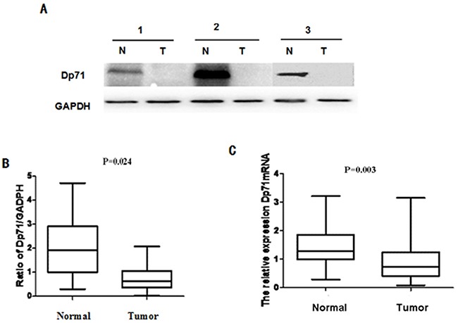
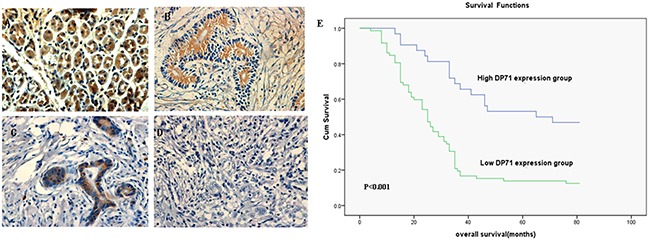
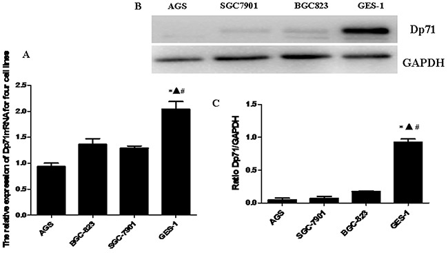
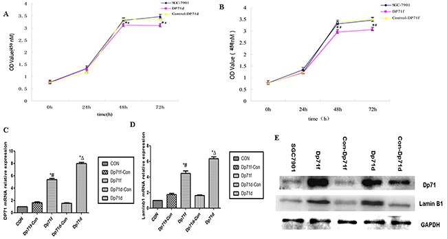
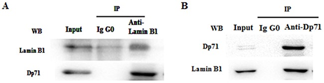
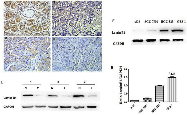
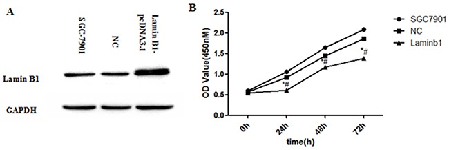
For the first time, dramatically decreased Dp71 protein and mRNA was found in 34 pairs of resected primary gastric adenocarcinoma. Immunohistochemistry identified Dp71 expression suppressed in 72.2% of 104 gastric cancer patients. The decreased Dp71 expression was significantly correlated with cancer differentiation (P=0.001) and lymph vascular invasion (p=0.041). Decreased Dp71 expression was associated with a poor gastric adenocarcinoma prognosis (P=0.001). Significantly less Dp71 mRNA and protein were found in BGC823, SGC7901, AGS compared with GES-1. Via increasing lamin B1 mRNA and protein, enforced Dp71d and Dp71f expression resulted in SGC7901 proliferation inhibition. Co-IP proved interaction of Dp71 with lamin B1 in GES-1 cells. Further expression characterization showed reduced lamin B1 in gastric cancer tissue and cancer cells. Increasing lamin B1 expression results in the growth inhibition of SGC7901, which suggests that Dp71-lamin B1 protein complex plays an important role for the newly identified tumor suppressive function of Dp71.
Keywords: Dp71; gastric cancer; gastric cancer cells; lamin B1; prognosis.
Conflict of interest statement
None.
Figures
Similar articles
-
Altered Biological Properties in Dp71 Over-Expressing HBE Cells.Cell Physiol Biochem. 2017;43(5):2022-2036. doi: 10.1159/000484181. Epub 2017 Oct 23. Cell Physiol Biochem. 2017. PMID: 29059680
-
Expression and prognostic impact of PRL-3 in lymph node metastasis of gastric cancer: its molecular mechanism was investigated using artificial microRNA interference.Int J Cancer. 2008 Sep 15;123(6):1439-47. doi: 10.1002/ijc.23643. Int J Cancer. 2008. PMID: 18561324
-
Expression of tumoral FOXP3 in gastric adenocarcinoma is associated with favorable clinicopathological variables and related with Hippo pathway.Int J Clin Exp Pathol. 2015 Nov 1;8(11):14608-18. eCollection 2015. Int J Clin Exp Pathol. 2015. PMID: 26823784 Free PMC article.
-
Overexpression of Ras-GTPase-activating protein SH3 domain-binding protein 1 correlates with poor prognosis in gastric cancer patients.Histopathology. 2015 Nov;67(5):677-88. doi: 10.1111/his.12695. Epub 2015 May 19. Histopathology. 2015. PMID: 25809930
-
Decreased Muc5AC expression is associated with poor prognosis in gastric cancer.Int J Cancer. 2014 Jan 1;134(1):114-24. doi: 10.1002/ijc.28345. Epub 2013 Jul 16. Int J Cancer. 2014. PMID: 23801416
Cited by
-
Dystrophin Dp71ab is monoclonally expressed in human satellite cells and enhances proliferation of myoblast cells.Sci Rep. 2020 Oct 13;10(1):17123. doi: 10.1038/s41598-020-74157-y. Sci Rep. 2020. PMID: 33051488 Free PMC article.
-
Schwann cell-specific Dp116 is expressed in glioblastoma cells, revealing two novel DMD gene splicing patterns.Biochem Biophys Rep. 2019 Nov 10;20:100703. doi: 10.1016/j.bbrep.2019.100703. eCollection 2019 Dec. Biochem Biophys Rep. 2019. PMID: 31737793 Free PMC article.
-
Dp71 depleted HBE cells displayed increased DNA damage and apoptosis induced by H2O2.Cell Mol Biol Lett. 2019 Jun 17;24:42. doi: 10.1186/s11658-019-0169-6. eCollection 2019. Cell Mol Biol Lett. 2019. PMID: 31236120 Free PMC article.
-
Comprehensive Genomic Analysis of Puerarin in Inhibiting Bladder Urothelial Carcinoma Cell Proliferation and Migration.Recent Pat Anticancer Drug Discov. 2024;19(4):516-529. doi: 10.2174/1574892819666230908110107. Recent Pat Anticancer Drug Discov. 2024. PMID: 37694778 Free PMC article.
-
Elevated FOXO6 expression correlates with progression and prognosis in gastric cancer.Oncotarget. 2017 May 9;8(19):31682-31691. doi: 10.18632/oncotarget.15920. Oncotarget. 2017. PMID: 28404958 Free PMC article.
References
-
- Jemal A, Bray F, Center MM, Ferlay J, Ward E, Forman D. Global cancer statistics. CA Cancer J Clin. 2011;61:69–90. - PubMed
-
- Hoffman EP, Brown RH, Jr, Kunkel LM. Dystrophin: the protein product of the Duchenne muscular dystrophy locus. Cell. 1987;51:919–28. - PubMed
-
- Austin RC, Howard PL, D'souza VN, Klamut HJ, Ray PN. Cloning and characterization of alternatively spliced isoforms of Dp71. Hum. Mol. Genet. 1995;4:1475–1483. - PubMed
MeSH terms
Substances
LinkOut - more resources
Full Text Sources
Other Literature Sources
Medical
Research Materials
