Case Reports
. 2016 Jul 22:16:120.
doi: 10.1186/s12886-016-0293-2.

Optical coherence tomography (OCT) angiography findings in retinal arterial macroaneurysms

Affiliations
Case Reports

Optical coherence tomography (OCT) angiography findings in retinal arterial macroaneurysms

Maged Alnawaiseh et al. BMC Ophthalmol. .

Abstract

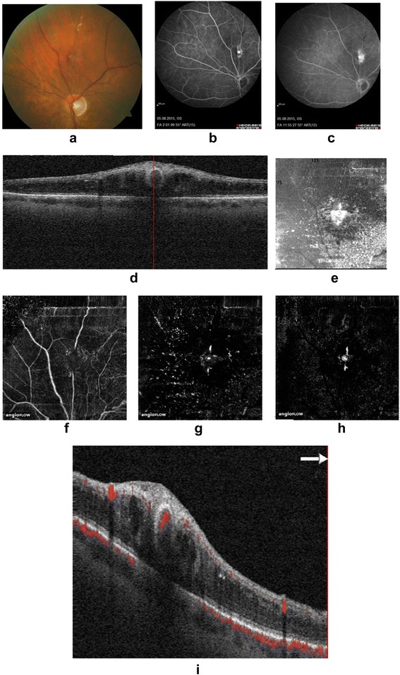
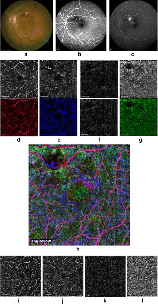
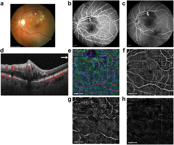
Background: Optical coherence tomography angiography is a novel imaging technique that allows dyeless in vivo visualization of the retinal and choroidal vasculature. The purpose of this study was to describe optical coherence tomography (OCT) angiography findings in patients with retinal arterial macroaneurysms (RAMs).

Methods: Three eyes of three patients with RAMs were retrospectively included. Fundus photography, OCT, fluorescein angiography (FA), and OCT angiography were performed. The entire imaging data was analyzed in detail.

Results: OCT angiography could detect the RAMs noninvasively without dye injection. By simultaneously observing the OCT scans, it was possible to determine the depth of the RAMs in the retina, to detect the exact localization in relation to the main vessel, and to determine the level of blood flow in the RAMs.

Conclusions: OCT angiography can clearly visualize RAMs without use of a dye. It also allows layer-specific observation of blood flow in each layer of the RAM. OCT angiography provides additional dynamic information on RAMs, which is not obtained with FA and facilitates a better understanding of its morphology and activity. This information in combination with ICG and fluorescein angiography can help to optimize direct laser treatment.

Keywords: Fluorescein angiography; OCT angiography; Retinal arterial macroaneurysms.


Figures

Fig. 1
Fig. 1
Fundus photography image a and early b and late c fluorescein angiogram showing a RAM superior to the optic disc. d Cross-sectional OCT. Sectional and e en face OCT image en face optical coherence tomography (OCT) images of the RAM. OCT angiograms of the different retinal layers showing an interruption of blood flow in the superficial OCT angiogram f and a significant flow in the deep OCT angiogram g and an OCT angiogram of the outer retina h. Cross-sectional OCT angiography visualizes blood flow in the RAMs i
Fig. 2
Fig. 2
Fundus photography image a and early b and late c fluorescein angiogram showing a superotemporal RAM. OCT angiograms, color-coded OCT angiograms d-g and composite OCT angiograms h of the different retinal layers: the RAM is best visualized in the deep OCT angiogram. i-l OCT angiograms of the different retinal layers two months after intravitreal anti-vascular endothelial growth factor therapy
Fig. 3
Fig. 3
Fundus photography image a early b and late c fluorescein angiogram showing the two main parts of a superotemporal RAM. OCT angiograms g-i and composite OCT angiograms of the RAM showing blood flow in the superficial f and deep OCT angiograms g. d Cross-sectional OCT angiography

References

    1. Pitkanen L, Tommila P, Kaarniranta K, Jaaskelainen JE, Kinnunen K. Retinal arterial macroaneurysms. Acta Ophthalmol. 2014;92:101–4. doi: 10.1111/aos.12210. - DOI - PubMed
    1. Bopp S, Joussen AM. Retinal Arterial Macroaneurysms (RAM)--pathology, differenzial diagnoses, and therapy. Klin Monbl Augenheilkd. 2014;231(9):890–900. doi: 10.1055/s-0034-1382953. - DOI - PubMed
    1. Meyer JC, Ahmad BU, Blinder KJ, Shah GK. Laser therapy versus observation for symptomatic retinal artery macroaneurysms. Graefes Arch Clin Exp Ophthalmol. 2015;253(4):537–41. doi: 10.1007/s00417-014-2730-3. - DOI - PubMed
    1. Cho HJ, Rhee TK, Kim HS, et al. Intravitreal bevacizumab for symptomatic retinal arterial macroaneurysm. Am J Ophthalmol. 2013;155(5):898–904. doi: 10.1016/j.ajo.2012.12.003. - DOI - PubMed
    1. Zweifel SA, Tönz MS, Pfenninger L, Becker M, Michels S. Intravitreal anti-VEGF therapy for retinal macroaneurysm. Klin Monbl Augenheilkd. 2013;230(4):392–5. doi: 10.1055/s-0032-1328377. - DOI - PubMed

Publication types