. 2016 Nov;37(11):1180-1189.
doi: 10.1002/humu.23055. Epub 2016 Aug 21.

Identification of a Large DNAJB2 Deletion in a Family with Spinal Muscular Atrophy and Parkinsonism

Affiliations

Identification of a Large DNAJB2 Deletion in a Family with Spinal Muscular Atrophy and Parkinsonism

Elena Sanchez et al. Hum Mutat. 2016 Nov.

Abstract

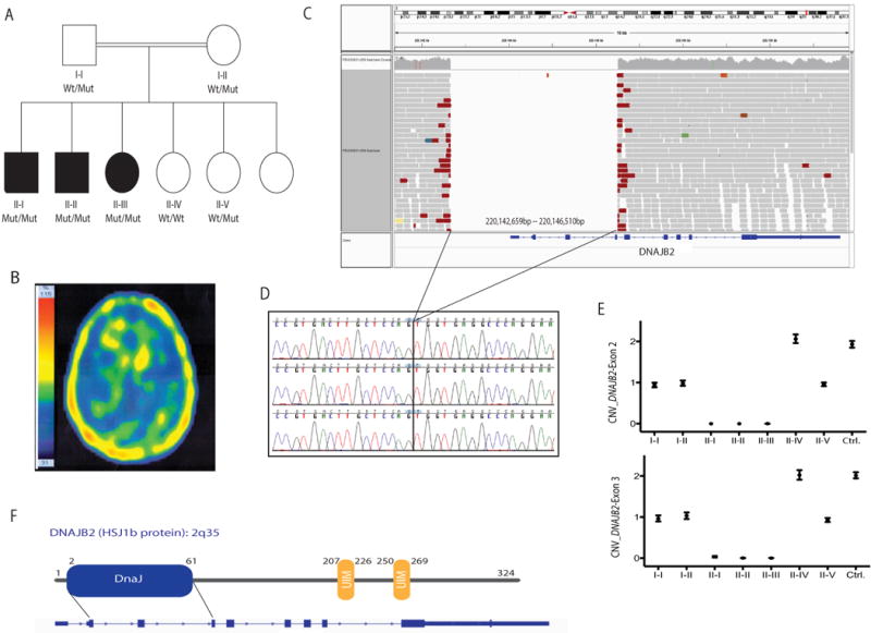
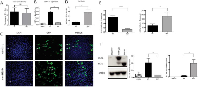
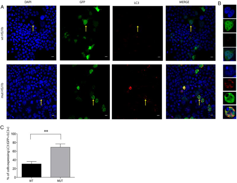
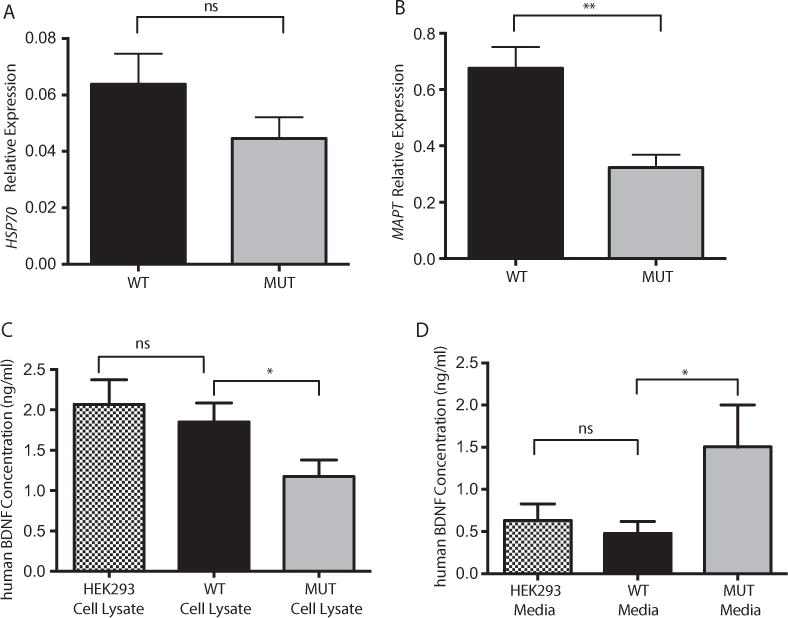
In this study, we described the identification of a large DNAJB2 (HSJ1) deletion in a family with recessive spinal muscular atrophy and Parkinsonism. After performing homozygosity mapping and whole genome sequencing, we identified a 3.8 kb deletion, spanning the entire DnaJ domain of the HSJ1 protein, as the disease-segregating mutation. By performing functional assays, we showed that HSJ1b-related DnaJ domain deletion leads to loss of HSJ1b mRNA and protein levels, increased HSJ1a mRNA and protein expressions, increased cell death, protein aggregation, and enhanced autophagy. Given the role of HSJ1 proteins in the degradation of misfolded proteins, we speculated that enhanced autophagy might be promoted by the elevated HSJ1a expression seen in HSJ1b-deficient cells. We also observed a significant reduction in both tau and brain-derived neurotrophic factor levels, which may explain the dopaminergic deficits seen in one of the affected siblings. We concluded that HSJ1b deficiency leads to a complex neurological phenotype, possibly due to the accumulation of misfolded proteins, caused by the lack of the DnaJ domain activity. We thus expand the phenotypic and genotypic spectrums associated with DNAJB2 disease and suggest relevant disease-associated mechanisms.

Keywords: DNAJB2; DnaJ domain deletion; Parkinsonism; WGS; spinal muscular atrophy.


Conflict of interest statement

Disclosure statement: The authors declare no conflict of interest.

Figures

Figure 1
Figure 1
A 3.8 kb deletion at the DNAJB2 locus (2q35) was identified as the disease-segregating mutation in a recessive family with SMA and Parkinsonism. A: Pedigree structure of a family presenting with SMA and Parkinsonism (one patient; II–I). Homozygous mutation carriers are represented as Mut/Mut, heterozygous carriers as Wt/Mut, and non-carriers as Wt/Wt. Dark squares (males) and circles (females) indicate neurological disease. B: Brain TRODAT SPECT imaging of affected sibling II–I showing decreased presynaptic dopamine activity in the striata, more prominent on the right side. C: WGS reads of an affected sibling showing the 3.8 kb chromosome 2 deletion identified in all affected members. WGS reads were visualized by using the IGV tool. D: Sanger chromatogram sequences of the three affected siblings confirming the presence of a homozygous deletion on chromosome 2q35. E: CNV plots of DNAJB2exons 2 and 3 obtained through ddPCR (QX100 system, Bio-rad). Two exon copies (homozygous wt allele) are represented with a CNV score of 2, one exon copy (heterozygous mutant allele) with a CNV score of 1, and no copies with a CNV score of 0. F: HSJ1b protein structure located at the 2q35 locus. The functional DnaJ domain expands from exons 2 through 4.
Figure 2
Figure 2
In vitro modeling of DNAJB2 exons 2 through 4 deletion leads to aggregations, cell death, and altered HSJ1 mRNA and protein expressions. A: Transfection efficiency. HEK293 cells were transfected in three independent experiments and only GFP+were considered for further analysis. B: Expression of HSJ1 b in non-transfected (black and white column), as well as wild type (black column) and mutant (solid gray column) transfected cells. Expression was evaluated by absolute quantification using the standard curve method. The y axis represents the mRNA expression (in number of copies) (P= 0.002, Mann-Whitney Test). C: HEK293 cells transfected with either wt HSJ1b-GFP (wt-HSJ1b) or mutant HSJ1b-GFP (mut-HSJ1b). Cells transfected with wt-HSJ1b do not show apoptotic nuclei and show small lysosomal deposits (white arrows). HEK293 cells transfected with Mut-DNAJB2 show apoptotic nuclei and aggregates (arrows). Scale bar: 50 μM. D: Quantification of HEK293 cells’ apoptotic nuclei in both wt and mutant HSJ1b cells. Cells transfected with mutant HSJ1b have a significantly higher number of apoptotic nuclei (Mean number of dead cells: WT = 3 and MUT = 24.50, P= 0.008, Mann-Whitney Test). E: HSJ1b (left) and HSJ1a (right) mRNA levels in wt (black column) and mutant (solid gray column) HSJ1b transfected cells. HSJ1b mRNA levels are highly decreased in mutant cells (left). F: Western blot analysis of HSJ1a, HSJ1b, and GAPDH proteins in cell lysatesfrom non-transfected cells (black and white columns), as well as wild type (black column) and mutant (solid gray column) HSJ1b transfected cells. HSJ1b or long isoform is of 36 kDa, HSJ1a or short isoform is of 31 kDa, and the GAPDH protein that was used as a loading control is of 37 kDa. Results are representative of four separate experiments. HSJ1b protein levels were significantly lower in mutant cells while HSJ1a were higher. Values represent the means ± SEM. *P< 0.05, **P< 0.01, ***P< 0.0001, ns = non-significant.
Figure 3
Figure 3
Increased co-localization of LC3 in mutant HSJ1b transfected cells versus wild type. A: LC3 localizes in both transfected (GFP+ cells) and non-transfected cells (GFP cells), being highly localized in cells presenting aberrant nuclei and big aggregates (yellow arrow, lower panel, mut-HSJ1b). However, transfected HEK293 cells with wild type HSJ1b have small aggregates and low levels of LC3 (yellow arrow, upper panel, wt-HSJ1b). B: Zoomed images of a sample cell with low levels of LC3 (upper panel, wt-HSJ1b) and a sample cell with high levels of LC3 (lower panel, mut-HSJ1b). C: Quantification of HEK293 transfected cells expressing LC3 (GFP+/LC3+) in wild type (solid black column) and mutant (solid gray column) HSJ1b (percentage of GFP+/LC3+ cells, wt-HSJ1b = 34.5% and mut-HSJ1b-Mut = 64.6%, P = 0.002, Mann–Whitney non-parametric test. Values represent the mean SEM. **P < 0.01. Scale bar = 50 μm.
Figure 4
Figure 4
HSJ1b deficiency does not alter Hsp70 mRNA levels but alters both MAPT and BDNF levels. A: Hsp70 mRNA expression levels are not significantly altered in wt (black column) versus mutant (solid gray column) transfected HEK293 cells (P= ns). B: MAPT (TAU) mRNA levels are significantly reduced in HSJ1b mutant transfected HEK293 cells (solid gray column) when compared with their wild type counterparts (black column) (P= 0.001).C: BDNF levels are reduced in cell lysatesof HSJ1b mutant cells (P= 0.05) but not in wtHSJ1b transfected cells (black column) (ns), which showed similar BDNF levels than non-transfected cells (ns). D: Overexpression of wild type HSJ1b (black column) did not increase the release of BDNF into the cell culture media when compared with non-transfected cells (black and white column) (ns). BNDF media release is increased in mutant HSJ1b transfected cells (solid gray column) (P = 0.03). Results are representative of two (A, B) or four (C, D) separate experiments. Values represent means ± SEM. *P≤ 0.05, **P< 0.001, ns = non-significant.

References

    1. Abisambra J, Jinwal UK, Miyata Y, Rogers J, Blair L, Li X, Seguin SP, Wang L, Jin Y, Bacon J, et al. Allosteric heat shock protein 70 inhibitors rapidly rescue synaptic plasticity deficits by reducing aberrant tau. Biol Psychiatry. 2013;74:367–374. - PMC - PubMed
    1. Arancio O, Chao MV. Neurotrophins, synaptic plasticity and dementia. Curr Opin Neurobiol. 2007;17:325–330. - PubMed
    1. Baquet ZC, Gorski JA, Jones KR. Early striatal dendrite deficits followed by neuron loss with advanced age in the absence of anterograde cortical brain-derived neurotrophic factor. J Neurosci. 2004;24:4250–4258. - PMC - PubMed
    1. Blumen SC, Astord S, Robin V, Vignaud L, Toumi N, Cieslik A, Achiron A, Carasso RL, Gurevich M, Braverman I, et al. A rare recessive distal hereditary motor neuropathy with HSJ1 chaperone mutation. Ann Neurol. 2012;71:509–519. - PubMed
    1. Borrell-Pages M, Canals JM, Cordelieres FP, Parker JA, Pineda JR, Grange G, Bryson EA, Guillermier M, Hirsch E, Hantraye P, et al. Cystamine and cysteamine increase brain levels of BDNF in Huntington disease via HSJ1b and transglutaminase. J Clin Invest. 2006;116:1410–1424. - PMC - PubMed

MeSH terms