Review
. 2016 Sep 1;8(9):a022061.
doi: 10.1101/cshperspect.a022061.

TGF-β Signaling from Receptors to Smads

Affiliations
Review

TGF-β Signaling from Receptors to Smads

Akiko Hata et al. Cold Spring Harb Perspect Biol. .

Abstract

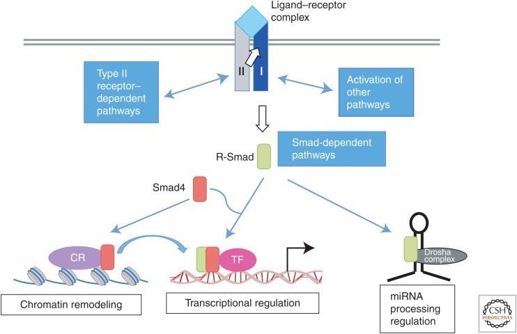
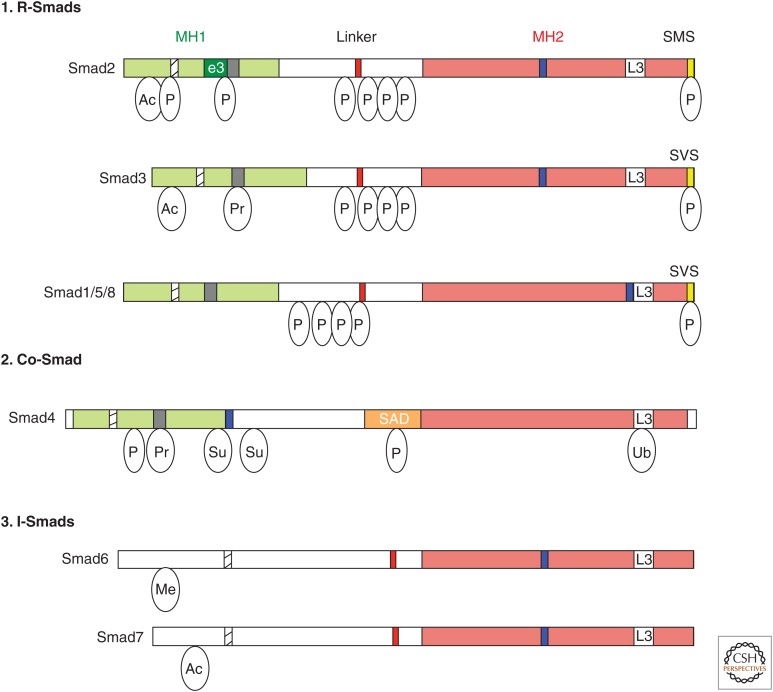
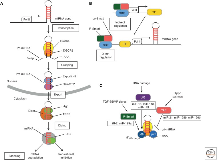
Transforming growth factor β (TGF-β) and related growth factors are secreted pleiotropic factors that play critical roles in embryogenesis and adult tissue homeostasis by regulating cell proliferation, differentiation, death, and migration. The TGF-β family members signal via heteromeric complexes of type I and type II receptors, which activate members of the Smad family of signal transducers. The main attribute of the TGF-β signaling pathway is context-dependence. Depending on the concentration and type of ligand, target tissue, and developmental stage, TGF-β family members transmit distinct signals. Deregulation of TGF-β signaling contributes to developmental defects and human diseases. More than a decade of studies have revealed the framework by which TGF-βs encode a context-dependent signal, which includes various positive and negative modifiers of the principal elements of the signaling pathway, the receptors, and the Smad proteins. In this review, we first introduce some basic components of the TGF-β signaling pathways and their actions, and then discuss posttranslational modifications and modulatory partners that modify the outcome of the signaling and contribute to its context-dependence, including small noncoding RNAs.


Figures

Figure 1.
Figure 1.
Transforming growth factor β (TGF-β) receptors and signal transducers. TGF-β family ligands, shown in light blue, transmit signals by assembling a heterotetrameric receptor complex with two type I receptors, shown in dark blue, and two type II receptors, shown in gray. Upon ligand binding, signaling is transmitted by a cytoplasmic kinase domain of type I receptors by phosphorylating receptor-regulated Smad proteins (R-Smad proteins, green box). This is considered as the “Smad signaling pathway.” Additionally, the receptor complex can activate “non-Smad signaling pathways” through type II receptor– and type I receptor–interacting proteins. TGF-β and bone morphogenetic protein (BMP) receptors can also activate mitogen-activated protein kinase (MAPK) and phosphoinositide-3-kinase (PI3K) pathways. Activated R-Smad proteins form a complex with the common-Smad, Smad4 (co-Smad, shown in red box), and, as a complex, translocate to the nucleus, where they regulate transcription of target genes together with cofactors (pink circle). R-Smads also form a complex with chromatin remodeling proteins (CR, purple circle) that recognizes certain histone modifications and promotes formation of active chromatin, which is a prerequisite for transcriptional activation by R-Smad/co-Smad complexes. Additionally, R-Smad proteins can participate in microRNA (miRNA) processing by the Drosha microprocessor complex (black circle) for the biogenesis of a subset of primary transcripts of miRNA (pri-miRNAs).
Figure 2.
Figure 2.
The Smad family. Schematic representations of the eight human Smad proteins divided into (1) receptor-regulated Smads (R-Smads), (2) common-Smad (co-Smad), and (3) inhibitory-Smads (I-Smads). The conserved amino-terminal Mad-homology 1 (MH1) and carboxy-terminal MH2 domains are indicated as green and red boxes, respectively. Highlighted are the nuclear localization signal (NLS, hatched box), the unique insert in Smad2-MH1 domain, which corresponds to exon 3 (e3, dark green box), the β-hairpin in the MH1 domain that binds DNA or the stem region of a subset of primary microRNAs (pri-miRNAs) (black box), the proline-tyrosine (PPXY) motif (red box) in the linker domain that is recognized by the WW domain of Smurf family proteins, the Smad activation domain (SAD, orange box) at the linker-MH2 border of Smad4, the nuclear export signal (NES, blue box), and the L3 loop of the MH2 domain (white box). The carboxy-terminal serine residues in the SXS motif that is phosphorylated by the type I receptor kinases are shown in the yellow box. Relative locations of different posttranslational modifications identified in Smad proteins are indicated. Ac, Acetylation; Ub, ubiquitylation; Pr, poly(ADP)ribosylation; Su, sumoylation; P, Ser or Thr phosphorylation.
Figure 3.
Figure 3.
MicroRNA (miRNA) biogenesis pathway and its regulation by Smad proteins. (A) The miRNA biogenesis pathway. The biogenesis of a miRNA is a stepwise process that includes (1) transcription of a primary transcript (pri-miRNA), (2) nuclear cropping to produce the precursor-miRNA (pre-miRNA), (3) export to the cytoplasm, and (4) cytoplasmic cropping to a double-stranded (ds) miRNA precursor. miRNA genes are generally transcribed by RNA polymerase II (Pol II) as long, 5′-capped and 3′-polyadenylated transcripts (pri-miRNAs) that are processed by the RNase III enzyme, Drosha, in the microprocessor complex to generate hairpin-loop RNAs, known as pre-miRNAs. Pre-miRNAs are recognized by the exportin 5 (Xpo5)/Ran-GTP transporter and exported to the cytoplasm, where another enzyme of the RNase III family, Dicer, catalyzes secondary processing (“dicing”) to produce miRNA/miRNA* duplexes. Dicer, TRBP, and Argonaute (Ago) proteins mediate the processing of pre-miRNAs and the assembly of the RNA-induced silencing complex (RISC) in mammalian cells. Ago proteins associate with Dicer in both the cropping and RISC assembly steps (Hata and Lieberman 2015). (From Hata and Lieberman 2015; adapted, with permission, from the authors.) (B) Transcription of miRNA genes can be regulated by TGF-β and BMP signaling pathways either by a direct binding of Smad complex (R-Smad/co-Smad) to Smad-binding element (SBE) in the promoter regions of miRNA genes or by transcriptional regulation of other transcription factors, which, in turn, modulate the transcription of miRNA genes. (C) Pri-miRNA to pre-miRNA processing of a subset of miRNAs catalyzed by Drosha and its cofactors p68 and p72 is positively regulated by transcription factors, such as R-Smad and p53, and negatively regulated by YAP, a signal transducer of the Hippo pathway. Specific binding of R-Smads to a dsRNA sequence motif located in the pre-miRNA provides specificity of this regulation. TF, Transcription factor. (From Hata and Lieberman 2015; adapted, with permission, from the authors.)

References

    1. Abdollah S, Macias-Silva M, Tsukazaki T, Hayashi H, Attisano L, Wrana JL. 1997. TβRI phosphorylation of Smad2 on Ser465 and Ser467 is required for Smad2–Smad4 complex formation and signaling. J Biol Chem 272: 27678–27685. - PubMed
    1. Agricola E, Randall RA, Gaarenstroom T, Dupont S, Hill CS. 2011. Recruitment of TIF1γ to chromatin via its PHD finger-bromodomain activates its ubiquitin ligase and transcriptional repressor activities. Mol Cell 43: 85–96. - PubMed
    1. Ahmed MI, Mardaryev AN, Lewis CJ, Sharov AA, Botchkareva NV. 2011. MicroRNA-21 is an important downstream component of BMP signalling in epidermal keratinocytes. J Cell Sci 124: 3399–3404. - PMC - PubMed
    1. Alarcon C, Zaromytidou AI, Xi Q, Gao S, Yu J, Fujisawa S, Barlas A, Miller AN, Manova-Todorova K, Macias MJ, et al. 2009. Nuclear CDKs drive Smad transcriptional activation and turnover in BMP and TGF-β pathways. Cell 139: 757–769. - PMC - PubMed
    1. Al-Salihi MA, Herhaus L, Macartney T, Sapkota GP. 2012. USP11 augments TGFβ signalling by deubiquitylating ALK5. Open Biol 2: 120063. - PMC - PubMed

Substances