Review
. 2017 Jan;367(1):125-140.
doi: 10.1007/s00441-016-2463-1. Epub 2016 Jul 23.

Mitochondrial lipids in neurodegeneration

Affiliations
Review

Mitochondrial lipids in neurodegeneration

Andreas Aufschnaiter et al. Cell Tissue Res. 2017 Jan.

Abstract

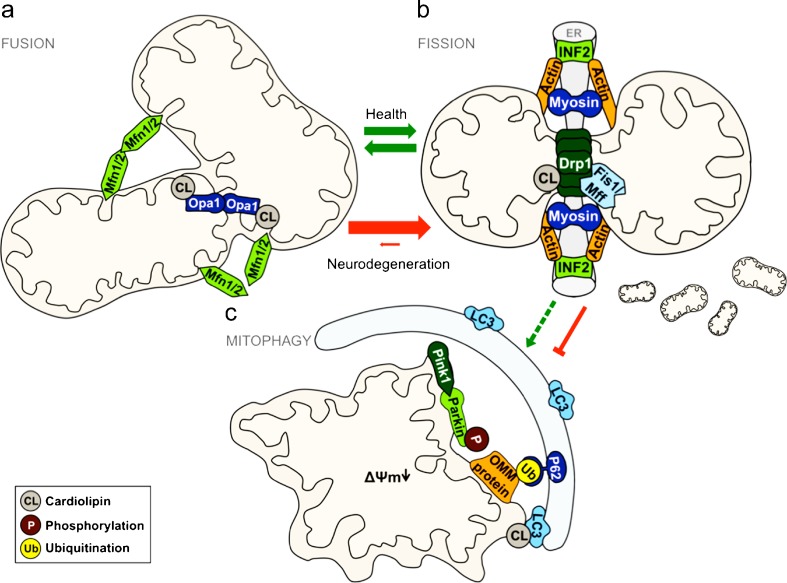
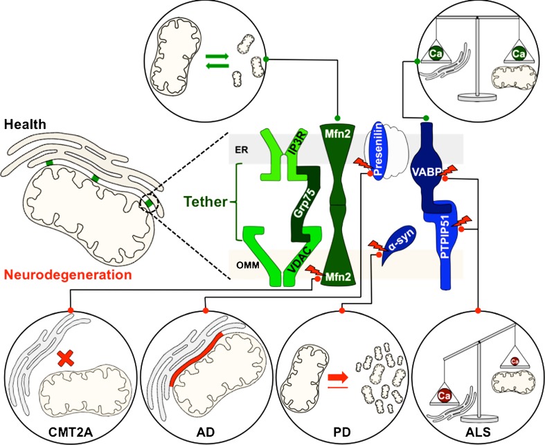
Mitochondrial dysfunction is a common feature of many neurodegenerative diseases, including proteinopathies such as Alzheimer's or Parkinson's disease, which are characterized by the deposition of aggregated proteins in the form of insoluble fibrils or plaques. The distinct molecular processes that eventually result in mitochondrial dysfunction during neurodegeneration are well studied but still not fully understood. However, defects in mitochondrial fission and fusion, mitophagy, oxidative phosphorylation and mitochondrial bioenergetics have been linked to cellular demise. These processes are influenced by the lipid environment within mitochondrial membranes as, besides membrane structure and curvature, recruitment and activity of different proteins also largely depend on the respective lipid composition. Hence, the interaction of neurotoxic proteins with certain lipids and the modification of lipid composition in different cell compartments, in particular mitochondria, decisively impact cell death associated with neurodegeneration. Here, we discuss the relevance of mitochondrial lipids in the pathological alterations that result in neuronal demise, focussing on proteinopathies.

Keywords: Lipids; Mitochondria; Mitochondria-associated membranes; Mitochondrial dynamics; Neurodegeneration.


Figures

Fig. 1
Fig. 1
Mitochondrial dynamics in health and neurodegenerative diseases. Under physiological conditions (indicated with green arrows), mitochondrial fusion (a) and fission (b) are balanced processes to adapt to the needs of a cell. In the wake of mitochondrial damage, mitophagy (c) acts as one of the protective mechanisms by degrading these organelles, thereby preventing oxidative stress and other deleterious consequences. However, in the pathogenesis of many neurodegenerative diseases (red arrows), the equilibrium of fission and fusion is shifted towards fission. This involves alterations of regulatory proteins and changes in the lipid composition of mitochondria. In such cases, cellular protection via mitophagy is severely impaired. Some key players in the molecular processes of mitophagy, for example, are also Parkinson’s disease-related proteins. For a detailed description of the pictured mechanisms, see main text
Fig. 2
Fig. 2
Mitochondria-associated membranes (MAMs) in health and neurodegenerative diseases. A close contact between the ER and mitochondria is crucial for mitochondrial function and morphology. This connectivity is maintained by a specific tethering-complex of MAMs (green). The proteins indicated in blue are additional components of these lipid raft-like microdomains, involved in Ca2+ homeostasis and balance of other cellular functions like mitochondrial fission/fusion processes. Mutations of indicated proteins (depicted with a red flash) are involved in the pathogenesis of specific neurodegenerative diseases, resulting in an imbalance of fission/fusion processes, gain or loss of ER–mitochondria contact area and impaired Ca2+ homeostasis. For a detailed description, see main text

References

    1. Abramovici H, Mojtabaie P, Parks RJ, Zhong XP, Koretzky GA, Topham MK, Gee SH. Diacylglycerol kinase zeta regulates actin cytoskeleton reorganization through dissociation of Rac1 from RhoGDI. Mol Biol Cell. 2009;20:2049–2059. doi: 10.1091/mbc.E07-12-1248. - DOI - PMC - PubMed
    1. Abrams AJ, Hufnagel RB, Rebelo A, Zanna C, Patel N, Gonzalez MA, Campeanu IJ, Griffin LB, Groenewald S, Strickland AV. Mutations in SLC25A46, encoding a UGO1-like protein, cause an optic atrophy spectrum disorder. Nat Genet. 2015;47:926–932. doi: 10.1038/ng.3354. - DOI - PMC - PubMed
    1. AhYoung AP, Jiang J, Zhang J, Khoi Dang X, Loo JA, Zhou ZH, Egea PF. Conserved SMP domains of the ERMES complex bind phospholipids and mediate tether assembly. Proc Natl Acad Sci U S A. 2015;112:E3179–E3188. doi: 10.1073/pnas.1422363112. - DOI - PMC - PubMed
    1. Alavi MV, Bette S, Schimpf S, Schuettauf F, Schraermeyer U, Wehrl HF, Ruttiger L, Beck SC, Tonagel F, Pichler BJ, Knipper M, Peters T, Laufs J, Wissinger B. A splice site mutation in the murine Opa1 gene features pathology of autosomal dominant optic atrophy. Brain. 2007;130:1029–1042. doi: 10.1093/brain/awm005. - DOI - PubMed
    1. Ardail D, Privat JP, Egret-Charlier M, Levrat C, Lerme F, Louisot P. Mitochondrial contact sites. Lipid composition and dynamics. J Biol Chem. 1990;265:18797–18802. - PubMed