Review
. 2016 Oct:104:87-103.
doi: 10.1016/j.biomaterials.2016.06.050. Epub 2016 Jul 9.

Superhydrophobic materials for biomedical applications

Affiliations
Review

Superhydrophobic materials for biomedical applications

Eric J Falde et al. Biomaterials. 2016 Oct.

Abstract

Superhydrophobic surfaces are actively studied across a wide range of applications and industries, and are now finding increased use in the biomedical arena as substrates to control protein adsorption, cellular interaction, and bacterial growth, as well as platforms for drug delivery devices and for diagnostic tools. The commonality in the design of these materials is to create a stable or metastable air layer at the material surface, which lends itself to a number of unique properties. These activities are catalyzing the development of new materials, applications, and fabrication techniques, as well as collaborations across material science, chemistry, engineering, and medicine given the interdisciplinary nature of this work. The review begins with a discussion of superhydrophobicity, and then explores biomedical applications that are utilizing superhydrophobicity in depth including material selection characteristics, in vitro performance, and in vivo performance. General trends are offered for each application in addition to discussion of conflicting data in the literature, and the review concludes with the authors' future perspectives on the utility of superhydrophobic biomaterials for medical applications.

Keywords: Biomaterials; Diagnostics; Drug delivery; High throughput assays; Polymers; Superhydrophobic; Tissue engineering.


Figures

Figure 1
Figure 1
Diagram of wetting states on rough materials and examples in nature. a) The Cassie-Baxter (CB) partially wetted state and c) the Wenzel complete wetting state. Examples of natural materials are b) lotus leaves, which are the canonical example of a natural superhydrophobic material, and d) the hydrophilic leaves of petunias in genus Ruellia. Photos are courtesy of Takashi Matsuzawa and Mark Swanson.
Figure 2
Figure 2
Superhydrophobic materials are of interest for a variety of medical applications including: a) control of the local release of drugs after tumor resection, b) patterned cell growth to study cellular communication (i.e., from a biopsy), c) reduced bacterial adhesion on implants such as hip replacements, or d) stabilization of droplets or drive flow in microfluidics and diagnostic assays. Photos are adapted with permission from [190] and [146].
Figure 3
Figure 3
Entrapped air at the superhydrophobic surface prevents protein binding (protein is shown in green) when incubated both with BSA or FBS (A,C). Removal of the air layer through sonication leads to protein binding in the regions that were once protected (B,D). Figure is reprinted with permission from [129].
Figure 4
Figure 4
Illustration of how protein fouling (biofouling) of superhydrophobic surfaces may lead to removal of the air layer. Proteins denature at the superhydrophobic/hydrophobic surface, leading to proteins binding and subsequent surface wetting due to formation of a more hydrophilic interface.
Figure 5
Figure 5
Superhydrophobic regions prevent cellular binding and growth, where superhydrophilic regions showed the opposite. a) Minimal binding of cells on superhydrophobic regions occurs while significant adhesion occurs on superhydrophilic surfaces, adapted with permission from [144]. b) Adjacent superhydrophilic spots can be seeded with droplets of different cell populations separated by superhydrophobic barriers, after which the entire surface can be wetted to study cell-cell communication, adapted with permission from [146].
Figure 6
Figure 6
(a) Standard hydrophobic ePTFE and (b) superhydrophobic PTFE showed no difference in neointima formation on the luminal side of the patch in a pig carotid circulation model after 4 weeks. No thrombus formation was observed. As shown by α-actin staining, the majority of neointima formation in (c) ePTFE and (d) superhydrophobic PTFE is smooth muscle, reprinted with permission from [155].
Figure 7
Figure 7
SEM images of P. aeruginosa PAO1 colonization on hydrophobic (a) and two types of superhydrophobic (b) (c) surfaces after 6 and 18 hours of incubation. Colonization occurred more slowly with a more stable superhydrophobic state (ab). Figure adapted with permission from [170].
Figure 8
Figure 8
Superhydrophobic surfaces allow deposition of molecules on a small number of pillars during evaporation (a, b, c). Concentration of non-volatile components at pillars allows detection of femto- to attomolar concentrations of biomolecules with an appropriate plasmonic structure on the pillars. Adapted with permission from [196].
Figure 9
Figure 9
The removal of air from superhydrophobic 3D materials controls the rate of drug release from the central layer of a tri-layered electrospun mesh. Each layer of an electrospun mesh acts as a new superhydrophobic surface to control the rate of air removal and penetration of a serum solution, imaged using μCT with a contrast agent. Figure adapted from [78].

References

    1. Marmur A. Superhydrophobic and superhygrophobic surfaces: from understanding non-wettability to design considerations. Soft Matter. 2013;9:7900–7904.
    1. Cassie ABD, Baxter S. Wettability of porous surfaces. Trans. Faraday Soc. 1944;40:546–551.
    1. Wenzel RN. Resistance of solid surfaces to wetting by water. J. Ind. Eng. Chem. 1936;28:988–994.
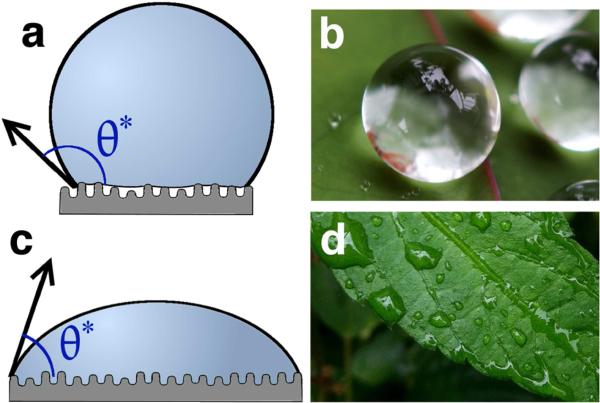
    1. Barthlott W, Neinhuis C. Purity of the sacred lotus, or escape from contamination in biological surfaces. Planta. 1997;202:1–8.
    1. Wagner P, Fürstner R, Barthlott W, Neinhuis C. Quantitative assessment to the structural basis of water repellency in natural and technical surfaces. J. Exp. Bot. 2003;54:1295–1303. - PubMed

Publication types

MeSH terms