

Prothymosin Alpha and Immune Responses: Are We Close to Potential Clinical Applications?
- PMID: 27450735
- PMCID: PMC7126549
- DOI: 10.1016/bs.vh.2016.04.008
Prothymosin Alpha and Immune Responses: Are We Close to Potential Clinical Applications?
Abstract
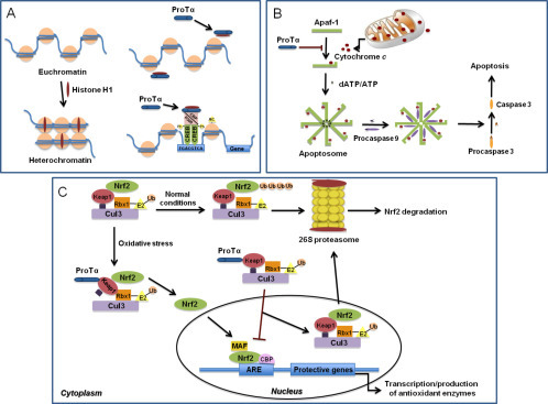
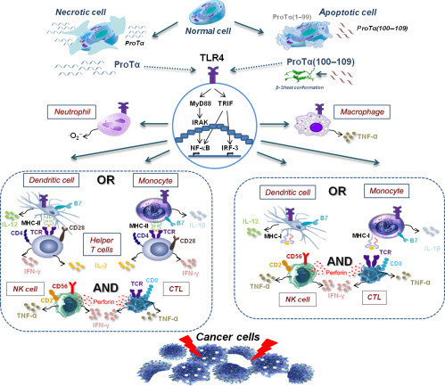
The thymus gland produces soluble molecules, which mediate significant immune functions. The first biologically active thymic extract was thymosin fraction V, the fractionation of which led to the isolation of a series of immunoactive polypeptides, including prothymosin alpha (proTα). ProTα displays a dual role, intracellularly as a survival and proliferation mediator and extracellularly as a biological response modifier. Accordingly, inside the cell, proTα is implicated in crucial intracellular circuits and may serve as a surrogate tumor biomarker, but when found outside the cell, it could be used as a therapeutic agent for treating immune system deficiencies. In fact, proTα possesses pleiotropic adjuvant activity and a series of immunomodulatory effects (eg, anticancer, antiviral, neuroprotective, cardioprotective). Moreover, several reports suggest that the variable activity of proTα might be exerted through different parts of the molecule. We first reported that the main immunoactive region of proTα is the carboxy-terminal decapeptide proTα(100-109). In conjunction with data from others, we also revealed that proTα and proTα(100-109) signal through Toll-like receptor 4. Although their precise molecular mechanism of action is yet not fully elucidated, proTα and proTα(100-109) are viewed as candidate adjuvants for cancer immunotherapy. Here, we present a historical overview on the discovery and isolation of thymosins with emphasis on proTα and data on some immune-related new activities of the polypeptide and smaller immunostimulatory peptides thereof. Finally, we propose a compiled scenario on proTα's mode of action, which could eventually contribute to its clinical application.
Keywords: Adjuvant; Alarmin; Cancer; DAMP; Immune response; Immunoenhancing peptide; ProTα(100–109); Prothymosin α; Thymic peptides.
© 2016 Elsevier Inc. All rights reserved.
Figures
Similar articles
-
Prothymosin Alpha: An Alarmin and More..Curr Med Chem. 2017;24(17):1747-1760. doi: 10.2174/0929867324666170518110033. Curr Med Chem. 2017. PMID: 28521686 Review.
-
The immunologically active site of prothymosin alpha is located at the carboxy-terminus of the polypeptide. Evaluation of its in vitro effects in cancer patients.Cancer Immunol Immunother. 2006 Oct;55(10):1247-57. doi: 10.1007/s00262-005-0108-4. Epub 2006 Feb 2. Cancer Immunol Immunother. 2006. PMID: 16453152 Free PMC article.
-
Prothymosin alpha immunoactive carboxyl-terminal peptide TKKQKTDEDD stimulates lymphocyte reactions, induces dendritic cell maturation and adopts a beta-sheet conformation in a sequence-specific manner.Mol Immunol. 2009 Feb;46(5):784-92. doi: 10.1016/j.molimm.2008.09.014. Epub 2008 Oct 30. Mol Immunol. 2009. PMID: 18976813
-
Prothymosin alpha: a ubiquitous polypeptide with potential use in cancer diagnosis and therapy.Cancer Immunol Immunother. 2012 May;61(5):599-614. doi: 10.1007/s00262-012-1222-8. Epub 2012 Feb 26. Cancer Immunol Immunother. 2012. PMID: 22366887 Free PMC article. Review.
-
Prothymosin α and a prothymosin α-derived peptide enhance T(H)1-type immune responses against defined HER-2/neu epitopes.BMC Immunol. 2013 Sep 22;14:43. doi: 10.1186/1471-2172-14-43. BMC Immunol. 2013. PMID: 24053720 Free PMC article.
Cited by
-
Proteomic profiling reveals engineered chitosan nanoparticles mediated cellular crosstalk and immunomodulation for therapeutic application in apical periodontitis.Bioact Mater. 2021 Oct 9;11:77-89. doi: 10.1016/j.bioactmat.2021.09.032. eCollection 2022 May. Bioact Mater. 2021. PMID: 34938914 Free PMC article.
-
Antitumor Reactive T-Cell Responses Are Enhanced In Vivo by DAMP Prothymosin Alpha and Its C-Terminal Decapeptide.Cancers (Basel). 2019 Nov 9;11(11):1764. doi: 10.3390/cancers11111764. Cancers (Basel). 2019. PMID: 31717548 Free PMC article.
-
Anti-β2glycoprotein I-induced neutrophil extracellular traps cause endothelial activation.Rheumatology (Oxford). 2025 Aug 1;64(8):4796-4805. doi: 10.1093/rheumatology/keaf204. Rheumatology (Oxford). 2025. PMID: 40238197 Free PMC article.
-
Evaluation of prothymosin alpha, trimethylamine-N-oxide, and ischemia-modified albumin in type 2 diabetes mellitus patients with dysregulated lipid profile.Qatar Med J. 2025 Jun 11;2025(2):39. doi: 10.5339/qmj.2025.39. eCollection 2025. Qatar Med J. 2025. PMID: 40792244 Free PMC article.
-
RNA-Seq-Based Gene Expression Pattern and Morphological Alterations in Chick Thymus during Postnatal Development.Int J Genomics. 2019 Apr 21;2019:6905194. doi: 10.1155/2019/6905194. eCollection 2019. Int J Genomics. 2019. PMID: 31179312 Free PMC article.
References
-
- Aisenberg A.C., Wilkes B. Partial immunological restoration of neonatally thymectomized rats with thymus-containing diffusion chambers. Nature. 1965;205:716–717. - PubMed
-
- Baxevanis C.N., Frillingos S., Seferiadis K., Reclos G.J., Arsenis P., Katsiyiannis A. Enhancement of human T lymphocyte function by prothymosin alpha: Increased production of interleukin-2 and expression of interleukin-2 receptors in normal human peripheral blood T lymphocytes. Immunopharmacology and Immunotoxicology. 1990;12:595–617. - PubMed
-
- Baxevanis C.N., Reclos G.J., Papamichail M. Prothymosin alpha restores depressed allogeneic cell-mediated lympholysis and natural-killer-cell activity in patients with cancer. International Journal of Cancer. 1993;53:264–268. - PubMed
Publication types
MeSH terms
Substances
LinkOut - more resources
Full Text Sources
Other Literature Sources
