Review
. 2016 Nov 1;419(1):99-113.
doi: 10.1016/j.ydbio.2016.07.014. Epub 2016 Jul 19.

Volvox: A simple algal model for embryogenesis, morphogenesis and cellular differentiation

Affiliations
Review

Volvox: A simple algal model for embryogenesis, morphogenesis and cellular differentiation

Gavriel Matt et al. Dev Biol. .

Abstract

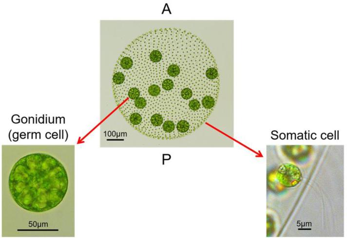
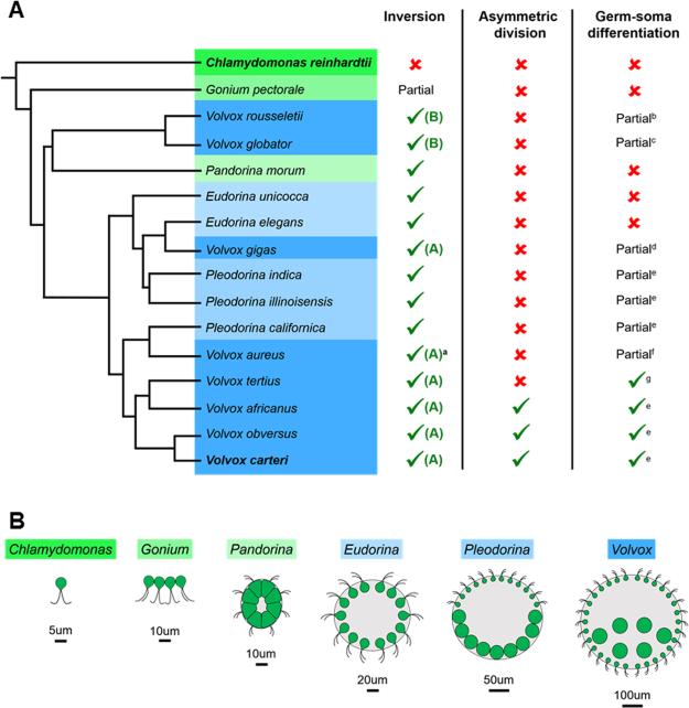
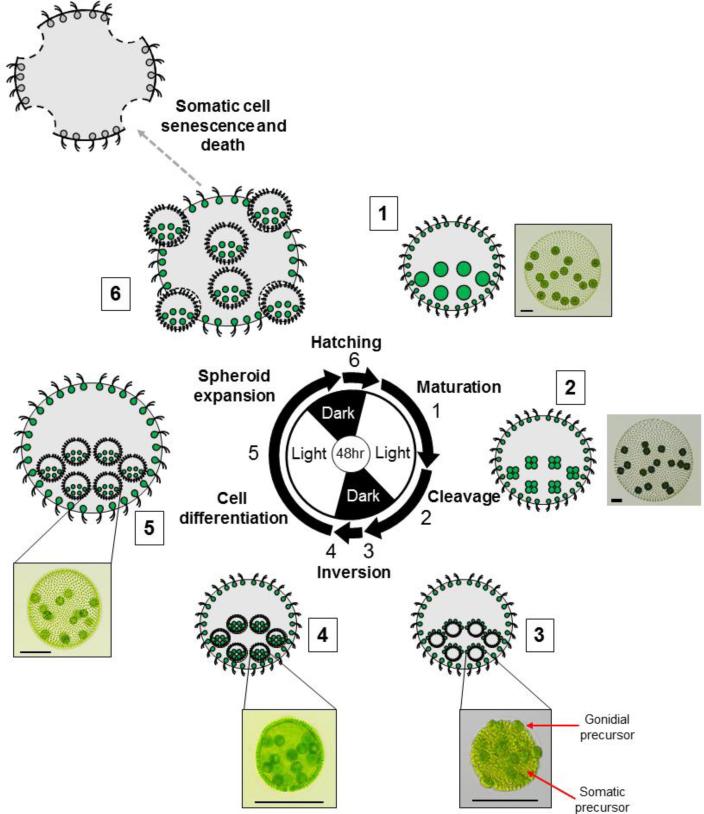
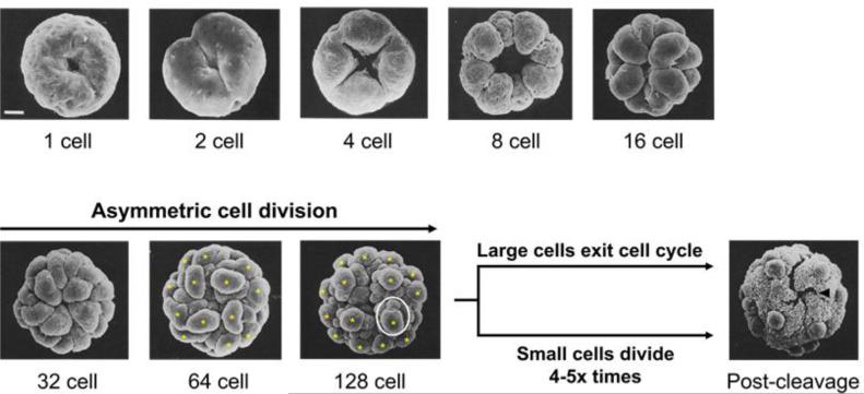
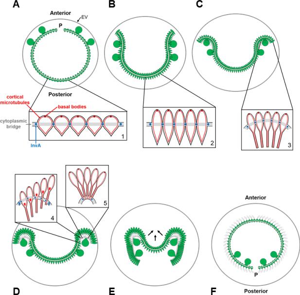
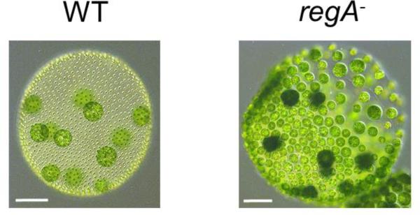
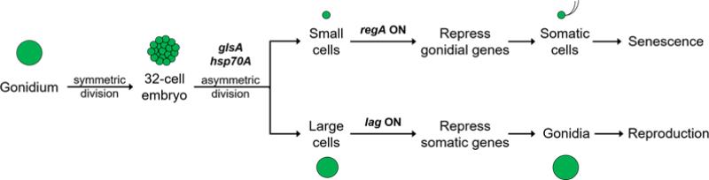
Patterning of a multicellular body plan involves a coordinated set of developmental processes that includes cell division, morphogenesis, and cellular differentiation. These processes have been most intensively studied in animals and land plants; however, deep insight can also be gained by studying development in simpler multicellular organisms. The multicellular green alga Volvox carteri (Volvox) is an excellent model for the investigation of developmental mechanisms and their evolutionary origins. Volvox has a streamlined body plan that contains only a few thousand cells and two distinct cell types: reproductive germ cells and terminally differentiated somatic cells. Patterning of the Volvox body plan is achieved through a stereotyped developmental program that includes embryonic cleavage with asymmetric cell division, morphogenesis, and cell-type differentiation. In this review we provide an overview of how these three developmental processes give rise to the adult form in Volvox and how developmental mutants have provided insights into the mechanisms behind these events. We highlight the accessibility and tractability of Volvox and its relatives that provide a unique opportunity for studying development.


Figures

Figure 1
Figure 1. Volvox carteri body plan and cell types
Center, an adult vegetative Volvox spheroid with two distinct cell types: ~2000 small, flagellated somatic cells (right inset) and ~16 large, aflagellate germ cells called gonidia (left inset). Somatic cells are on the outer surface of the spheroid with flagella oriented towards the exterior. Gonidia are just beneath the somatic cell layer in the posterior hemisphere. All cells are embedded within a clear, compartmentalized extracellular matrix. Anterior (A) and posterior (P) poles of the spheroid are labeled.
Figure 2
Figure 2. Grades of volvocine algal body plan complexity and polyphyletic origins of the genus Volvox
(A, left) Abbreviated volvocine phylogeny adapted from Herron and Michod (2008) and Herron et al. (2009). Different genera are highlighted by different colored boxes. Body plan schematics of each genus are shown in (B). The genus Volvox (dark blue highlighted species), which is characterized by spheroid size (typically >500 µm diameter), large cell number (>500), and composition of mostly terminally differentiated somatic cells (Coleman, 2012), is polyphyletic with at least three separate origins. Volvox carteri, the species that is the focus of this review, and Chlamydomonas reinhardtii, a unicellular outgroup for all multicellular volvocine species, are highlighted in bold. (A, right) Table indicating the presence or absence of developmental innovations that are discussed in this review. Complete inversion: inverted embryo forms a closed spheroid; partial inversion: inverted embryo reverses curvature but does not form a closed spheroid (Kirk, 2005). Type of inversion (A or B) refers to the spatial and temporal sequences of tissue and cell shape changes during inversion that differ between species (Hallmann, 2006). Presence/absence of asymmetric cell division as described in (Desnitski, 1995 and Herron et al., 2010). Complete germ-soma differentiation: germ and somatic cells have completely distinct fates; partial germ-soma differentiation: germ cell precursors first proceed through a flagellated somatic phase prior to acquiring a germ fate (Kirk, 2005 and Ransick, 1993). Note that asymmetric cell division and complete germ-soma differentiation are not universal traits of Volvox and are likely derived in V. carteri from a simpler ancestral program with no asymmetric division and partial germ-soma differentiation. (B) Cartoons illustrating volvocine body plan size and organization based on genus. Chlamydomonas, single celled; Gonium, discoidal body plan of 4–16 cells; Pandorina, spheroidal body plan with 16 cells; Eudorina, spheroidal body plan with 32–64 cells and expanded ECM; Pleodorina, spheroidal body plan with 32–128 cells, expanded ECM, and partial germ-soma differentiation; Volvox, spheroidal body plan with 500–50,000 cells, a small proportion of germ cells, and expanded ECM. aV. aureus was reported in Hallmann (2006) to undergo type B inversion; however its inversion appears closer to type A in (Darden, 1966). bPocock (1933a); cKirk et al.(1986); dPocock (1933b); eRansick (1993); fDarden (1966); gRansick (1993) noted that conclusive documentation for complete germ-soma differentiation in V. tertius is lacking, but Pocock (1938) reports that the gonidial initials of V. tertius are differentiated prior to the conclusion of embryogenesis implying that they do not have a transient somatic stage.
Figure 3
Figure 3. Asexual life cycle of Volvox
Stages of vegetative reproduction are depicted proceeding clockwise from upper right with cartoons and micrographs of selected stages. Center, two-day diurnal regime used to synchronize the Volvox life cycle with alternating light and dark periods of 16hrs and 8hrs respectively. Arrows represent the approximate duration of each labeled phase. Numbers next to cartoons and micrographs correspond to numbers next to arrows on diurnal diagram showing the stage depicted. 1, Mature adult spheroid with pre-cleavage gonidia (large green circles) and flagellated somatic cells (small green circles) embedded within extracellular matrix (grey). 2, Adult mother spheroid with four-cell stage embryos derived from gonidia. Note that adult somatic cells do not divide. 3, Adult mother spheroid with post-cleavage embryos prior to inversion. Note the large gonidial precursor cells on the exterior surface. 4, Adult mother spheroid with post-inversion juveniles. 5, Adult mother spheroid with differentiated and expanded juveniles. 6, Late stage juveniles hatching from their mother spheroid. The somatic cells from the mother spheroid undergo senescence and eventual death. Scale bars = 100μm.
Figure 4
Figure 4. Volvox embryogenesis
Scanning electron micrographs showing the anterior hemisphere of a cleaving gonidium at indicated stages. Yellow asterisks mark 16 large daughters produced by asymmetric division at the 6th cleavage cycle (32→64 cell stage) and 7th cleavage cycle (64→128 cell stage). Within the white circle at the 128 cell stage is a large cell undergoing visibly asymmetric cell division. The cross-shaped phialopore opening in the post-cleavage embryo before inversion is indicated by a black arrowhead. Scale bar, 10um. EM micrographs used with permission from (Green and Kirk, 1981) ©1981 Green and Kirk. Journal of Cell Biology. 91:743–755.
Figure 5
Figure 5. Stages of inversion and associated cell shape changes
A) Pre-inversion embryo. Presumptive gonidia (large cells) protrude from the exterior surface of the anterior hemisphere and flagellar ends of presumptive somatic cells (pointed ends of small pear-shaped cells) (inset 1) face the interior of the embryo. Cytoplasmic bridges (white band) connect cells at their midpoints and traverse the embryo except at the phialopore (P). A glycoprotein embryonic vesicle (gray circle labeled EV) surrounds the embryo and expands during inversion. Anterior and posterior poles of the embryo are labeled. B) Presumptive somatic cells transition to a spindle shape (inset 2) causing the embryo to shrink and allowing the phialopore lips to begin opening. C) Cells near the phialopore transition to an elongated flask shape with their cortical microtubules extending into their posterior ends (inset 3). D) The kinesin motor protein InvA that is anchored to the cytoplasmic bridges migrates along cortical microtubules in cells near the phialopore driving cell body movement relative to the cytoplasmic bridges (inset 4) to cause increased local curvature and acute bending near the phialopore lips (inset 5). A wave of InvA generated cell movement relative to bridges and local bending proceeds from the anterior region towards the equator. E) The bend reaches the equator and the posterior hemisphere contracts (arrows) allowing it to snap through the already inverted anterior hemisphere. Cells that have passed through the bend region undergo a third shape change to a compact columnar shape. F) Inversion is complete and the gonidia and phialopore are now positioned at the posterior pole of the juvenile spheroid.
Figure 6
Figure 6. The somatic regenerator (regA) mutant phenotype
Micrographs of a wild type (WT) adult spheroid (left) and a regA mutant (right). Note the cells that initially were somatic in the regA spheroid have re-differentiated as gonidia and enlarged, and will eventually undergo embyrogenesis. Scale bars, 100um.
Figure 7
Figure 7. Formal genetic pathway for germ-soma differentiation in V. carteri
During embryogenesis GlsA and Hsp70A are required for asymmetric divisions that give rise to small and large blastomeres. Cell size determines the fate of postembryonic cells with regA activated in small cells to repress germ cell fate, and lag genes required in large cells to partially suppress somatic fate. Modified from (Kirk, 2001).

References

    1. Arakaki Y, Kawai-Toyooka H, Hamamura Y, Higashiyama T, Noga A, Hirono M, Olson BJSC, Nozaki H. The simplest integrated multicellular organism unveiled. PLoS One. 2013;8 doi:10.1371/journal.pone.0081641. - PMC - PubMed
    1. Bell G. The origin and early evolution of germ cells as illustrated by the Volvocales. In: Halvorson H, Monroy A, editors. The Origin and Evolution of Sex. Wood's Hole Symposium. Liss, New York: 1985. pp. 221–256.
    1. Benfey PN. Essays on Developmental Biology, Part A. Elsevier Inc.; 2016. Defining the Path from Stem Cells to Differentiated Tissue; pp. 35–43. doi:10.1016/bs.ctdb.2015.12.002. - PubMed
    1. Bišová K, Zachleder V. Cell-cycle regulation in green algae dividing by multiple fission. J. Exp. Bot. 2014;65:2585–602. doi:10.1093/jxb/ert466. - PubMed
    1. Blumenstiel JP, Noll AC, Griffiths JA, Perera AG, Walton KN, Gilliland WD, Hawley RS, Staehling-Hampton K. Identification of EMS-induced mutations in Drosophila melanogaster by whole-genome sequencing. Genetics. 2009;182:25–32. doi:10.1534/genetics.109.101998. - PMC - PubMed

LinkOut - more resources