Review
. 2016 Jul;204(1):47-54.
doi: 10.1016/j.jss.2016.04.021. Epub 2016 Apr 22.

Indole-3-carbinol induces tumor cell death: function follows form

Affiliations
Review

Indole-3-carbinol induces tumor cell death: function follows form

Bryant W Megna et al. J Surg Res. 2016 Jul.

Abstract

Background: Even with colonoscopy screening and preventive measures becoming more commonplace, colorectal cancer (CRC) remains the third leading cause of oncologic death in the United States as of 2014. Many chemotherapeutics exist for the treatment of colorectal cancer, though they often come with significant side effect profiles or narrow efficacy ranges in terms of patient profile. Dietary phytochemicals such as glucobrassicin and its metabolite indole-3-carbinol (I3C) have been implicated in tumor prevention in many preclinical models across a variety of gastrointestinal tumors and represent an intriguing new class of natural chemotherapeutics for CRC. I3C has been identified as a ligand of the aryl hydrocarbon receptor (AHR), and we aimed to characterize this AHR activation in relation to its cytotoxic properties.

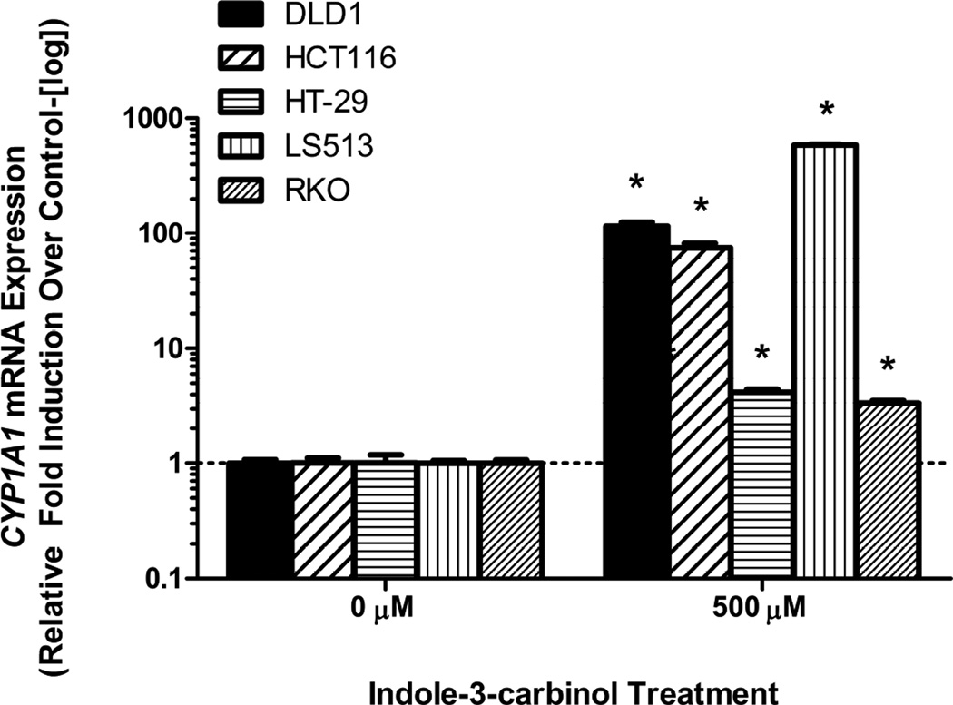
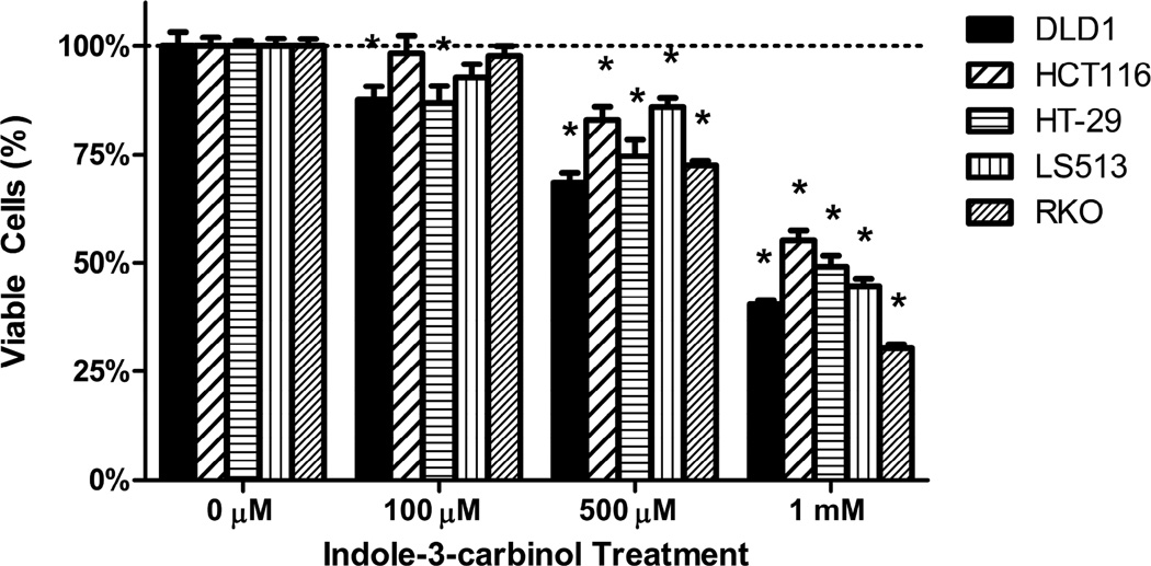
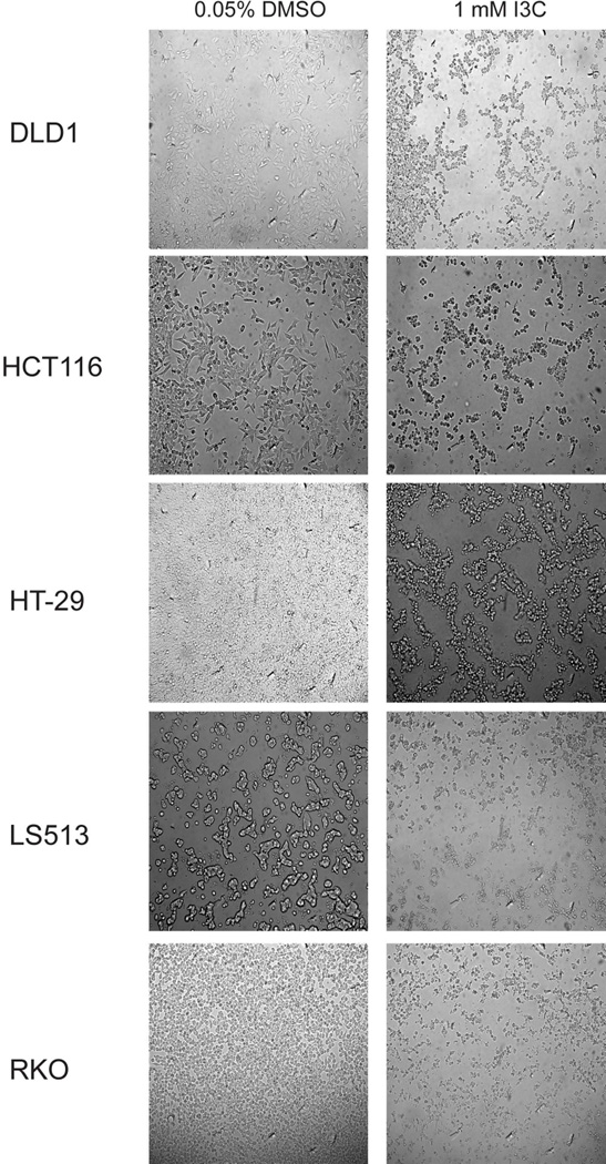
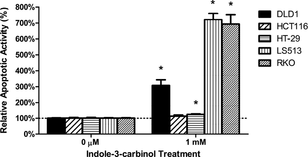
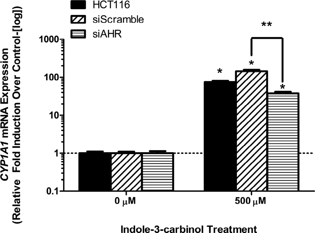
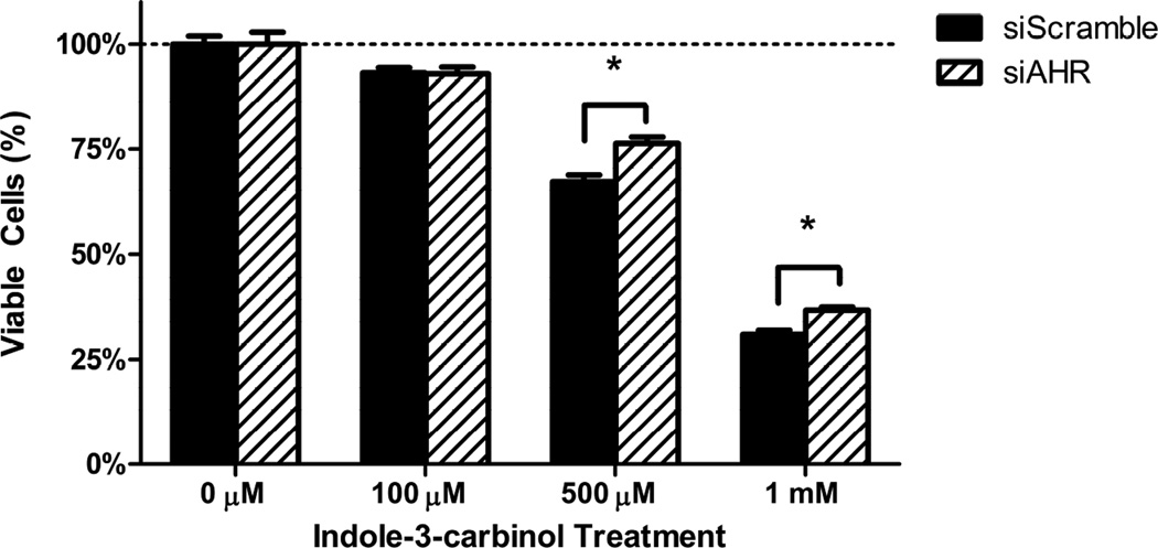
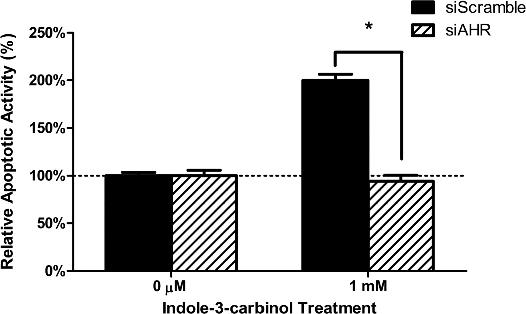
Methods: Human colorectal cancer cell lines DLD1, HCT116, HT-29, LS513, and RKO were treated with indole-3-carbinol or vehicle. Cell viability was assessed via a fluorescent product assay, and apoptotic activity was assessed via a luminescent signal tied to a ratio of caspase-3 and caspase-7 activity. Gene expression of AHR and CYP1A1 messenger ribonucleic acid (mRNA) was measured using quantitative real-time polymerase chain reaction. Small interfering RNA stable expression lines were established on a HCT116 background using a laboratory-developed transfection protocol as published elsewhere.

Results: Multiple colorectal cancer cell types express increased CYP1A1 mRNA levels (a specific marker of AHR-driven activity) after treatment with I3C, characterizing I3C treatment as agonistic of this pathway. Also, I3C induced a dose-dependent decrease in cell viability as well as inducing apoptosis. Furthermore, using small interfering RNA interference to knockdown AHR responsiveness generated a significant resistance to the chemotherapeutic actions of indole-3-carbinol regarding both cell viability and apoptotic activity.

Conclusions: Some degree of the cytotoxic and proapoptotic effects of indole-3-carbinol on colon cancer cells is dependent on activation of the aryl hydrocarbon receptor. This represents a novel mechanism for the molecular action of indole-3-carbinol and enhances our understanding of its effects in the context of colorectal cancer. Continued preclinical study of both indole-3-carbinol and the aryl hydrocarbon receptor pathway is warranted, which may one day lead to novel diet-derived colon cancer treatments that enlist the AHR.

Keywords: Aryl hydrocarbon receptor; Chemotherapy; Colorectal cancer; Indole-3-carbinol; Phytochemical.


Figures

Figure 1
Figure 1. Indole-3-carbinol treatment activates the aryl hydrocarbon receptor in colorectal cancer cells
A) DLD1, HCT116, HT-29, LS513, and RKO cells exhibited CYP1A1 mRNA upregulation following I3C treatment. With cell growth in logarithmic phase, cells were treated with either 500 μM I3C or 0.01% DMSO for 24 hours. Total RNA was harvested and reverse transcribed. Data are presented as relative fold induction of CYP1A1 mRNA of I3C treated cells over DMSO treated cells (controlled to internal levels of β-actin) ± standard error (representing at least three discrete experiments). “*” indicates statistical significance between I3C treated mRNA levels and vehicle (DMSO- μM) ) controls (p<0.05).
Figure 2
Figure 2. Effects of indole-3-carbinol (I3C) on colorectal cancer cell viability
A) I3C treatment of DLD1, HCT116, HT-29, LS513, and RKO cells lead to a dose-dependent decrease in cell viability. Cells were treated with I3C (100 μM, 500 μM, 1 mM) for 24 hours and cell viability was measured using a metabolically activated fluorogenic substrate. Data are depicted as mean viability percentage ± standard error, as normalized by DMSO treated control groups (representing at least three discrete experiments). “*” indicates statistical significance between I3C treated cell viability levels and vehicle (DMSO) controls in all cell types (p<0.05). B) Light microscope representation of 24 hour I3C treatment in all colorectal cancer cancer cell types. Images of monolayer cells are depicted with both vehicle (DMSO) and 1 mM I3C treatment at 24 hours. I3C treatment induced widespread pyknosis and fragmentation as compared to controls. C) I3C treatment stimulated apoptotic activity in all tested cell lines. Cells were treated with 1 mM I3C and luminescent caspase-3/7 activity was measured at 24 hours. Data are depicted as mean percentage of caspase-3/7 activity over DMSO treated controls ± standard error (representing at least three discrete experiments). “*” indicates statistical significance between I3C treated apoptotic activity levels and vehicle (DMSO) controls in all cell types (p<0.05).
Figure 3
Figure 3. Inhibition of AHR mRNA expression abrogates AHR activity induced by indole-3-carbinol (I3C)
Si-scramble cells display a more robust activation AHR signaling (in the form of CYP1A1 mRNA expression) than si-AHR cells following I3C treatment. Cells were treated with either 500 μM I3C or 0.01% DMSO for 24 hours. Wild type HCT116 mRNA response is depicted as an additional model. Data are presented as relative fold induction of CYP1A1 mRNA of I3C treated cells over DMSO treated cells (controlled to internal levels of β-actin) ± standard error (representing at least three discrete experiments). “*” indicates statistical significance between I3C treated mRNA levels and vehicle (DMSO) controls in all cell types (p<0.05). “**” with brackets indicate statistical significance of I3C treated mRNA level differences between si-scramble and si-AHR cells (p<0.05).
Figure 4
Figure 4. Inhibition of AHR expression blunts the cytotoxic action of indole-3-carbinol
A) I3C treatment of si-Scramble and si-AHR cells lead to a dose-dependent decrease in cell viability. This effect was more robust in si-Scramble cells. Cells were treated with I3C (100 μM, 500 μM, 1 mM) for 24 hours and cell viability was measured using a metabolically activated fluorogenic substrate. Data are depicted as mean viability percentage ± standard error, as normalized by DMSO treated control groups (representing at least three discrete experiments). “*” with brackets indicate statistical significance between the cell viability of si-scramble and si-AHR cells (p<0.05). B) I3C treatment stimulated apoptotic activity more so in si-Scramble cells than si-AHR cells. Cells were treated with 1 mM I3C and luminescent caspase-3/7 activity was measured at 24 hours. Data are depicted as mean percentage of caspase-3/7 activity over DMSO treated controls ± standard error (representing at least two discrete experiments). “*” with brackets indicates statistical significance between I3C treated levels of apoptotic activity between si-scramble and si-AHR cells (p<0.05).

References

    1. Siegel R, Ma J, Zou Z, Jemal A. Cancer statistics, 2014. CA Cancer J Clin. 2014;64:9–29. - PubMed
    1. Stintzing S. Management of colorectal cancer. F1000prime reports. 2014;6:108. - PMC - PubMed
    1. Braun MS, Seymour MT. Balancing the efficacy and toxicity of chemotherapy in colorectal cancer. Therapeutic advances in medical oncology. 2011;3:43–52. - PMC - PubMed
    1. Edwards MS, Chadda SD, Zhao Z, Barber BL, Sykes DP. A systematic review of treatment guidelines for metastatic colorectal cancer. Colorectal Dis. 2012;14:e31–e47. - PMC - PubMed
    1. Leavy O. Mucosal immunology: the 'AHR diet' for mucosal homeostasis. Nat Rev Immunol. 2011;11:806. - PubMed

Publication types

MeSH terms