. 2016 Oct 1;101(10):1285-1300.
doi: 10.1113/EP085768. Epub 2016 Sep 24.

OPN-a induces muscle inflammation by increasing recruitment and activation of pro-inflammatory macrophages

Affiliations

OPN-a induces muscle inflammation by increasing recruitment and activation of pro-inflammatory macrophages

Gina M Many et al. Exp Physiol. .

Abstract

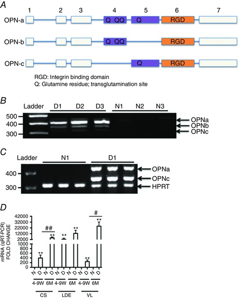
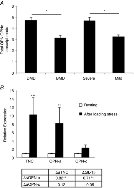
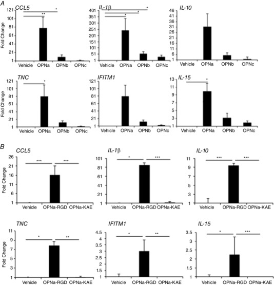
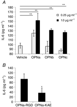
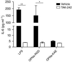
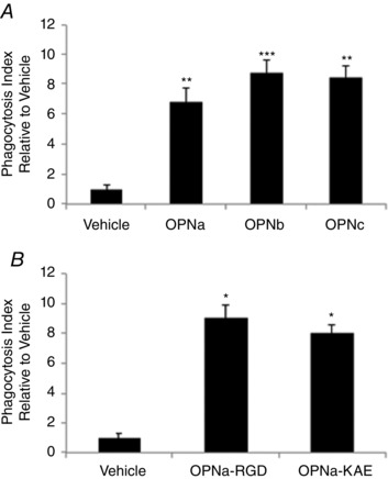
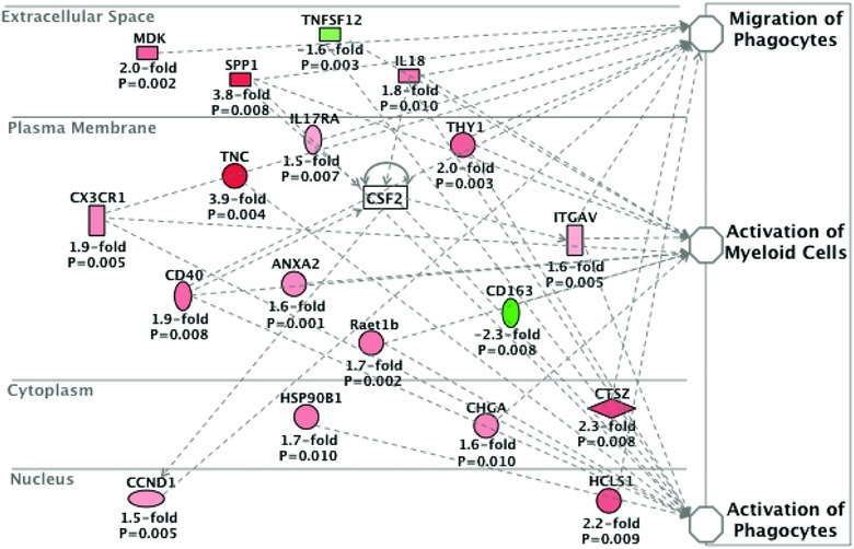
What is the central question of this study? What is the functional relevance of OPN isoform expression in muscle pathology? What is the main finding and its importance? The full-length human OPN-a isoform is the most pro-inflammatory isoform in the muscle microenvironment, acting on macrophages and myoblasts in an RGD-integrin-dependent manner. OPN-a upregulates expression of tenascin-C (TNC), a known Toll-like receptor 4 (TLR4) agonist. Blocking TLR4 signalling inhibits the pro-inflammatory effects of OPN-a, suggesting that a potential mechanism of OPN action is by promoting TNC-TLR4 signalling. Although osteopontin (OPN) is an important mediator of muscle remodelling in health and disease, functional differences in human spliced OPN variants in the muscle microenvironment have not been characterized. We thus sought to define the pro-inflammatory activities of human OPN isoforms (OPN-a, OPN-b and OPN-c) on cells present in regenerating muscle. OPN transcripts were quantified in normal and dystrophic human and dog muscle. Human macrophages and myoblasts were stimulated with recombinant human OPN protein isoforms, and cytokine mRNA and protein induction was assayed. OPN isoforms were greatly increased in dystrophic human (OPN-a > OPN-b > OPN-c) and dog muscle (OPN-a = OPN-c). In healthy human muscle, mechanical loading also upregulated OPN-a expression (eightfold; P < 0.01), but did not significantly upregulate OPN-c expression (twofold; P > 0.05). In vitro, OPN-a displayed the most pronounced pro-inflammatory activity among isoforms, acting on both macrophages and myoblasts. In vitro and in vivo data revealed that OPN-a upregulated tenascin-C (TNC), a known Toll-like receptor 4 (TLR4) agonist. Inhibition of TLR4 signalling attenuated OPN-mediated macrophage cytokine production. In summary, OPN-a is the most abundant and functionally active human spliced isoform in the skeletal muscle microenvironment. Here, OPN-a promotes pro-inflammatory signalling in both macrophages and myoblasts, possibly through induction of TNC-TLR4 signalling. Together, our findings suggest that specific targeting of OPN-a and/or TNC signalling in the damaged muscle microenvironment may be of therapeutic relevance.

Keywords: Duchenne muscular dystrophy; OPN-a; TNC; muscle inflammation; osteopontin; skeletal muscle.


Figures

Figure 1
Figure 1. Alternatively spliced osteopontin isoforms are expressed in dystrophic muscle
A, OPN spliced isoform transcript structure. The blocks in this figure represent exons and the lines represent introns. Here, OPN‐a is full length (top), OPN‐b is missing exon 5 (middle), and OPN‐c is missing exon 4 (bottom). B, osteopontin isoform transcript expression in muscle biopsies of three dystrophin‐deficient Duchenne muscular dystrophy (DMD) patients [D1–D3], and three dystrophin‐sufficient control subjects [N1–N3] as determined by RT‐PCR. Minimal expression is detected in dystrophin‐sufficient muscle, whereas all three isoforms are expressed in DMD muscle (full‐length OPN‐a, OPN‐b lacking exon 5, and OPN‐c lacking exon 4). C, dog dystrophin‐deficient muscle likewise shows no detectable expression in dystrophin‐sufficient littermates (N1), but high expression of OPN‐a and OPN‐c in dystrophin‐deficient golden retriever muscular dystrophy (GRMD) vastus lateralis muscle (D1). D, RT‐PCR analysis of muscle biopsies from three muscles and two age points (4‐9 W, 4–9 weeks old; and 6 M, 6 months old) in dystrophin‐sufficient (N) and ‐deficient (D) dogs shows elevated OPN mRNA in all dystrophic muscles at all age points. An increase in OPN‐a mRNA levels with age is seen in the three muscle groups tested (CS, cranial sartorius; LDE, long digital extensor; and VL, vastus lateralis). Here, OPN‐a mRNA levels were correlated with both age and severity of muscle involvement (CS, mildly affected; and LDE and VL, severely affected). Triplicates are shown per sample. Significant difference between dystrophic and non‐dystrophic littermates: ** P < 0.01. Significant differences with age per muscle group: # P ≤ 0.05 and ## P ≤ 0.01 (GRMD, n = 8; and dystrophin‐sufficient littermates, n = 4).
Figure 2
Figure 2. Human OPN‐a and OPN‐c isoform ratios change with dystrophic severity and mechanical loading
A, the expression ratio of total OPN (using primers specific to exon 7) to OPN‐c (specific to mRNA sequence spanning from exons 3–5, with exon 4 deleted) in Duchenne muscular dystrophy (DMD) and Becker's muscular dystrophy (BMD) vastus lateralis human muscle biopsies (n = 18; all male). Patients were grouped by histological severity prior to Nanostring mRNA molecule counting, as indicated by total fibre degeneration and regeneration and inflammatory foci, as assessed via Haematoxylin and Eosin staining. B, transcript fold changes (ΔΔC t) were quantified before and 24 h after mechanical loading (n = 22, matched for sex) by qRT‐PCR. IL‐1β and OPN‐c were undetectable in some subjects (n = 3 and n = 4, respectively) at either baseline or at 24 h and were excluded from analysis. The table below displays Pearson product–moment correlation coefficients between fold changes in transcripts (ΔΔC t values) in all subjects. * P < 0.05 and ** P < 0.01 between groups.
Figure 3
Figure 3. Human primary macrophages show a pro‐inflammatory response to recombinant OPN‐a in an RGD‐dependent manner
Shown are fold changes of immunoregulatory transcripts from primary human macrophages treated with 1 μg ml−1 OPN isoforms (A) or RGD‐sufficient (OPN‐a‐RGD) and RGD‐deficient [ΔRGD→KAE (OPN‐a‐KAE)] OPN (B). A, OPN‐a increased pro‐inflammatory transcripts, whereas OPN‐b and OPN‐c show less activity. Statistical significance is presented relative to vehicle control. B, macrophages stimulated with 2 μg ml−1 OPN‐a‐RGD or OPN‐a‐KAE show that the pro‐inflammatory activity of OPN‐a requires RGD‐mediated binding to integrins on macrophages. Statistical significance is compared between treatment conditions. Expression values after treatment with OPN isoforms are presented as the average fold change relative to vehicle [polymixin‐B (PMB)] treatment for n = 4 healthy male donors. * P < 0.05, ** P < 0.01 and *** P < 0.001 as assessed via Student's paired t tests (average donor response).
Figure 4
Figure 4. OPN isoforms differentially induce pro‐inflammatory cytokine production from human myoblasts in an RGD‐dependent manner
Immortalized human myoblasts were cultured for 48 h with recombinant human OPN proteins treated with 0.25 or 1.0 μg ml−1 human OPN‐a, OPN‐b or OPN‐c (A) or 1.0 μg ml−1 recombinant human OPNa‐RGD or OPNa‐KAE (B). Interleukin‐6 (IL‐6) concentrations in the secreted media were quantified after 48 h by enzyme‐linked immunosorbent assay. Samples were run in replicates of n = 5.
Figure 5
Figure 5. Pretreatment of human macrophages with TAK‐242, an inhibitor of Toll‐like receptor 4 (TLR4) signalling, inhibits human OPN‐a‐mediated IL‐6 production
Primary human macrophages were treated with TAK‐242 (100 nm) or vehicle (DMSO) for 1 h before stimulation with 1 μg ml−1 OPN‐a‐RGD and OPN‐a‐KAE or 10 ng ml−1 lipopolysaccharide (LPS; positive control). Interleukin‐6 concentrations in the secreted media were measured at 24 h by enzyme‐linked immunosorbent assay. The pro‐inflammatory activity of OPN‐a was largely blocked by TAK‐242. TAK‐242 had little effect on OPN‐a‐KAE activity. Samples were run in replicates of n = 4.
Figure 6
Figure 6. Human OPN‐mediated monocyte phagocytosis is isoform and RGD independent
After 30 min pretreatment with polymixin‐B (PMB), primary human monocytes were treated for 2 h with recombinant OPN proteins or vehicle (PMB) in the presence of fluorescently conjugated Escherichia coli particles. Cells were washed, and cellular fluorescence of non‐viable cells was quenched with Trypan Blue. Phagocytosis is presented as the fold increase in fluorescence from viable cells relative to vehicle control cells (PMB). All conditions were normalized for background fluorescence (E. coli particles only) before calculation of the fold change relative to vehicle control. A, phagocytosis induced by 0.5 μg ml−1 OPN spliced isoforms relative to vehicle (PMB). B, phagocytosis induced by 1.0 μg ml−1 OPN‐a‐RGD and OPN‐a‐KAE. All samples were run in replicates of n = 5, according to the manufacturer's instructions (Vybrant Phagocytosis Assay Kit; Life Technologies). Significance is presented relative to the vehicle control.
Figure 7
Figure 7. OPN‐a promotes pro‐inflammatory macrophage infiltration in vivo
BLA/J mice (n = 3) were injected with OPN‐a and an empty vector in contralateral tibialis anterior (TA) muscles. The TA muscles were harvested 7 days postinjection, and Illumina mRNA expression profiling was performed. Data are represented as the average transcript fold change by OPN‐a relative to empty vector control. Changes in all transcripts shown were statistically significant (P < 0.05). CSF2, granulocyte‐macrophage colony‐stimulating factor (GM‐CSF); SPP1, osteopontin; TNC, tenascin‐C.

References

    1. Arnold L, Henry A, Poron F, Baba‐Amer Y, van Rooijen N, Plonquet A, Gherardi RK & Chazaud B (2007). Inflammatory monocytes recruited after skeletal muscle injury switch into antiinflammatory macrophages to support myogenesis. J Exp Med 204, 1057–1069. - PMC - PubMed
    1. Bandopadhyay M, Bulbule A, Butti R, Chakraborty G, Ghorpade P, Ghosh P, Gorain M, Kale S, Kumar D, Kumar S, Totakura KV, Roy G, Sharma P, Shetti D, Soundararajan G, Thorat D, Tomar D, Nalukurthi R, Raja R, Mishra R, Yadav AS & Kundu GC (2014). Osteopontin as a therapeutic target for cancer. Expert Opin Ther Targets 18, 883–895. - PubMed
    1. Barfield WL, Uaesoontrachoon K, Wu CS, Lin S, Chen Y, Wang PC, Kanaan Y, Bond V & Hoffman EP (2014). Eccentric muscle challenge shows osteopontin polymorphism modulation of muscle damage. Hum Mol Genet 23, 4043–4050. - PMC - PubMed
    1. Bello L, Kesari A, Gordish‐Dressman H, Cnaan A, Morgenroth LP, Punetha J, Duong T, Henricson EK, Pegoraro E, McDonald CM, Hoffman EP & Cooperative International Neuromuscular Research Group Investigators (2015). Genetic modifiers of ambulation in the Cooperative International Neuromuscular Research Group Duchenne Natural History Study. Ann Neurol 77, 684–696. - PMC - PubMed
    1. Cavaillon JM & Haeffner‐Cavaillon N (1986). Polymyxin‐B inhibition of LPS‐induced interleukin‐1 secretion by human monocytes is dependent upon the LPS origin. Mol Immunol 23, 965–969. - PubMed

Publication types