Review
. 2016 Sep;12(9):519-33.
doi: 10.1038/nrneph.2016.101. Epub 2016 Jul 25.

Idiopathic hypercalciuria and formation of calcium renal stones

Affiliations
Review

Idiopathic hypercalciuria and formation of calcium renal stones

Fredric L Coe et al. Nat Rev Nephrol. 2016 Sep.

Abstract

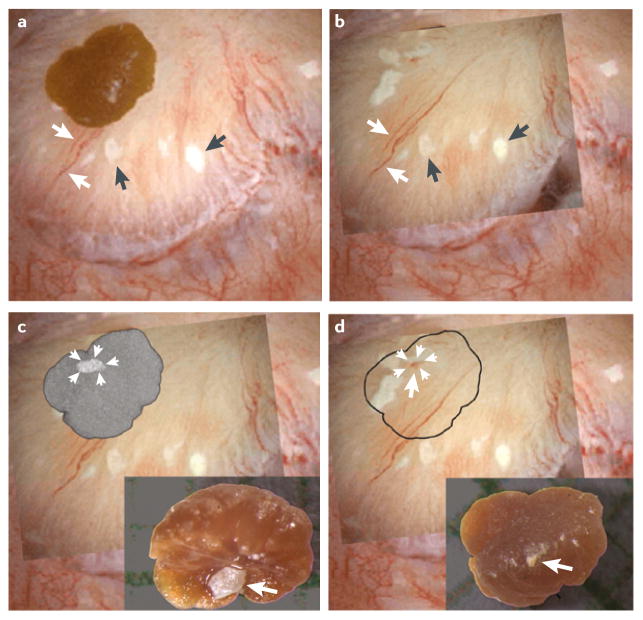
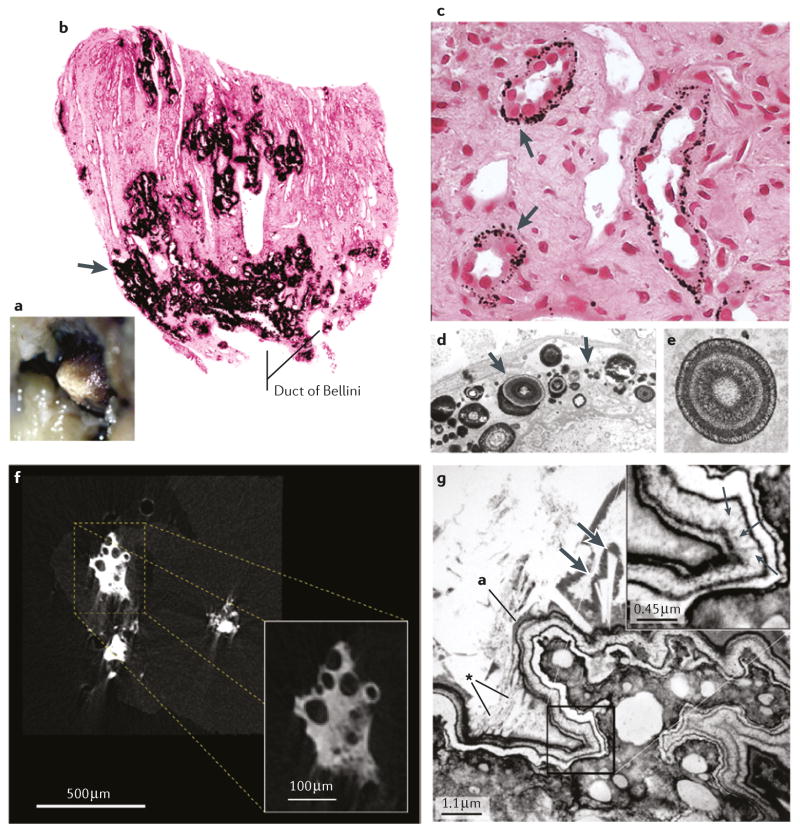
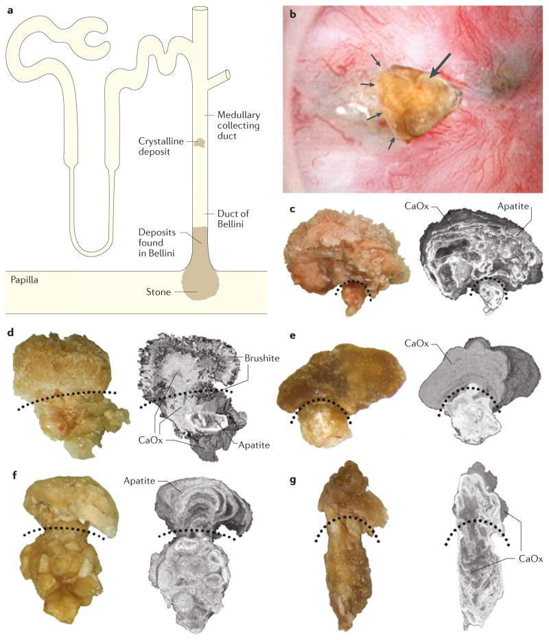
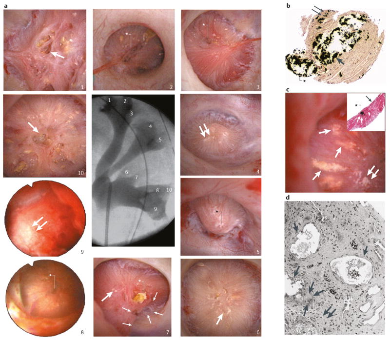
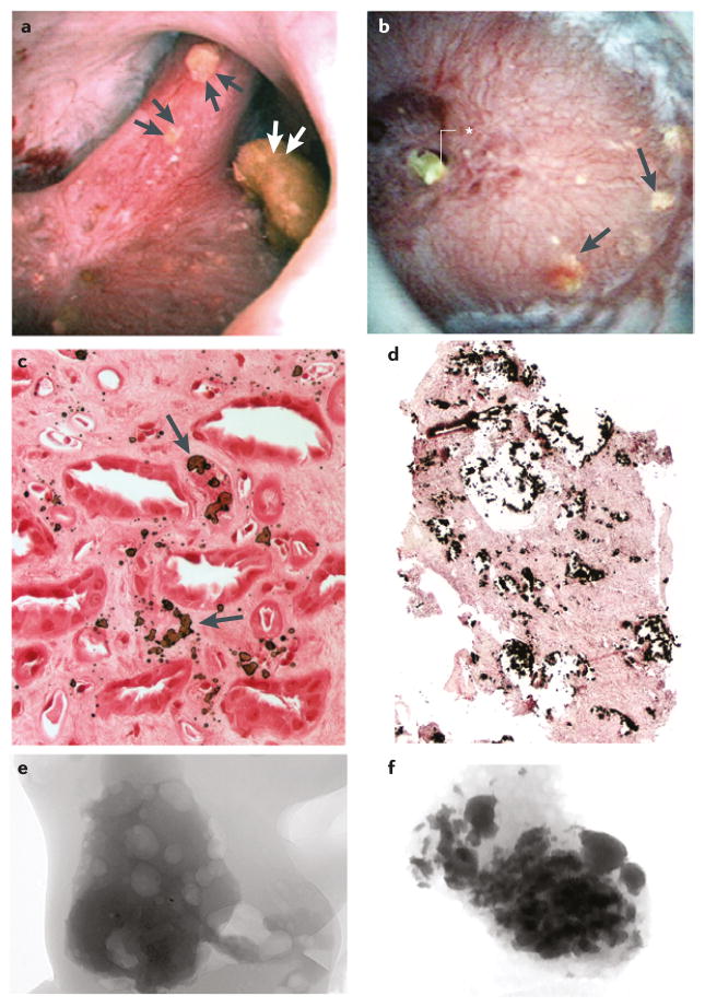
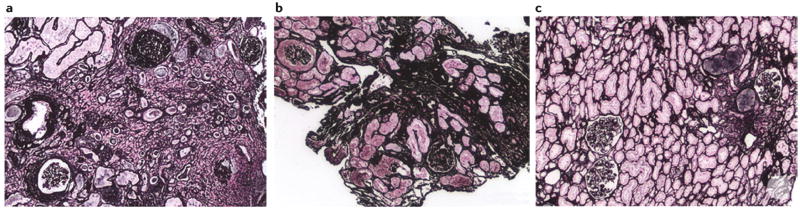
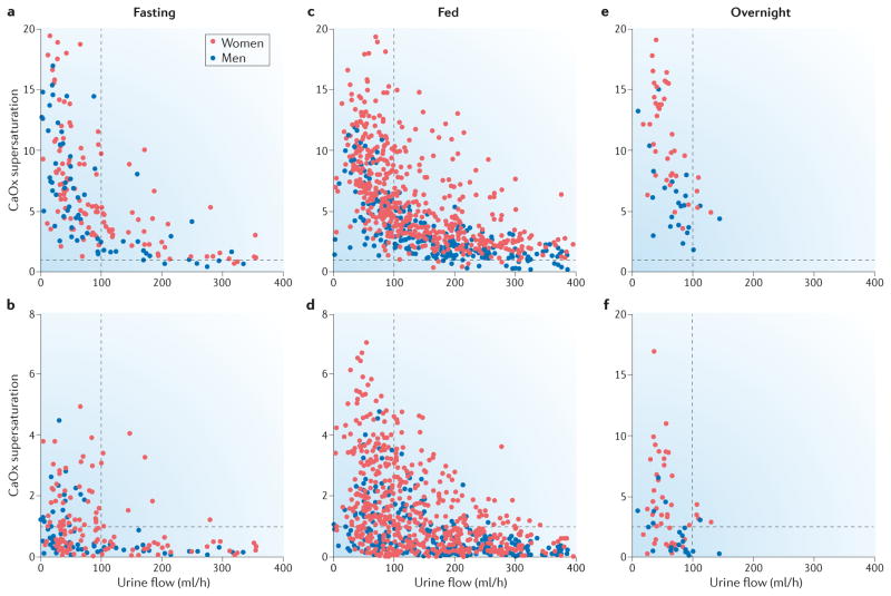
The most common presentation of nephrolithiasis is idiopathic calcium stones in patients without systemic disease. Most stones are primarily composed of calcium oxalate and form on a base of interstitial apatite deposits, known as Randall's plaque. By contrast some stones are composed largely of calcium phosphate, as either hydroxyapatite or brushite (calcium monohydrogen phosphate), and are usually accompanied by deposits of calcium phosphate in the Bellini ducts. These deposits result in local tissue damage and might serve as a site of mineral overgrowth. Stone formation is driven by supersaturation of urine with calcium oxalate and brushite. The level of supersaturation is related to fluid intake as well as to the levels of urinary citrate and calcium. Risk of stone formation is increased when urine citrate excretion is <400 mg per day, and treatment with potassium citrate has been used to prevent stones. Urine calcium levels >200 mg per day also increase stone risk and often result in negative calcium balance. Reduced renal calcium reabsorption has a role in idiopathic hypercalciuria. Low sodium diets and thiazide-type diuretics lower urine calcium levels and potentially reduce the risk of stone recurrence and bone disease.


Conflict of interest statement

Competing interests statement

The authors declare no competing interests.

Figures

Figure 1
Figure 1. Interstitial plaque and corresponding attachment site on a CaOx stone
a | Digital endoscopic image of a 3 mm CaOx stone attached to a papillum before stone removal by percutaneous nephrolithotomy. Several sites of interstitial plaque (arrowheads) are visible as well as blood vessels (arrows) that were used for image orientation. b | Following stone removal the papillum was reimaged — an overlay showing the sites of interstitial plaque and blood vessels after stone removal has been placed over the original endoscopic imagec | “Ghosted” CT image of the detached stone showing a site of calcium phosphate (arrowheads) on the papillary surface of the stone. The insert shows a light microscopic image of the papillary surface of the detached stone with the site of calcium phosphate (arrow). d | The site of calcium phosphate on the papillary surface of the stone (arrows) aligns with a region of interstitial plaque with a central blood spot (arrow) on the papillium, which is presumably the site of stone attachment. The insert shows the urinary surface of the detached stone.
Figure 2
Figure 2. Sites of interstitial plaque in idiopathic CaOx stone formers
a | Endoscopic image showing irregular whitish regions of interstitial plaque covering the papillary tip. b | A biopsy from this region showing black Yasue-stained material (arrow) within the interstitial space of the inner medulla. c | Light microscopy and d | transmission electron microscopy images showing regions of plaque in the basement membrane of the thin loops of Henle only, suggesting that plaque originates at this site. e | The plaque is composed of laminated spheres with up to nine alternating light (hydroxyapatite) and dark (matrix) layers. f | Micro CT image of plaque that has filled the interstitium and generated islands that encompass nearby tubules with no evidence of intraluminal deposits. g | Transmission electron microscopy imageshowing a multilayered ribbon-like structure separating a region of interstitial plaque (lower right) from an overgrowth of developing stone (upper left). In the thickest region of the white lamina of the ribbon (insert), tiny thin spicules run perpendicular to the surface adjacent to voids containing tightly packed crystals (small arrows). Numerous small crystals have grown into the outer border of the ribbon (asterisk) and merged with large crystals within the urinary space at the developing overgrowth. The double arrows indicate a large in-growing crystal.
Figure 3
Figure 3. Bellini duct plugs with overgrowths in patients who formed calcium stones
a | Sites of crystalline deposits that form plugged Bellini ducts (BD) with an overgrowth region protruding into the urinary space. b | Endoscopic image of a plug (arrow) protruding from a dilated BD (arrowheads). BD plugs with overgrowths visible by light microscopy and micro-CT from c | a patient with idiopathic hydroxyapatite stones, d | a patient with idiopathic brushite stones e | a patient with ileostomy, f | a patient with primary hyperparathyroidism and g | a medullary sponge kidney. The curved, dotted lines divide the BD plugs from the overgrowth regions.
Figure 4
Figure 4. Renal papilla of a patient who formed brushite stones
a | Endoscopic images of all ten papilla. The numbering corresponds to the x-ray image of the collection system. Papilla 1, 6, 7 and 10 show severe morphological changes including pitting (large arrows), flattening, plugs (black asterisk, papilla 7) and yellow plaque. Papilla 2, 3, 5 and 8 show small pits (white asterisks) whereas papilla 4, 7 and 9 show small sites of interstitial plaque (double arrows). b | Scattered inner medullary collecting ducts (IMCDs) and Bellini duct (BD) filled with yellow crystalline deposits that have dilated the tubular lumens (arrow) and protrude from the opening of the BD (asterisk). Small sites of interstitial plaque are also visible (double arrows). c | Sites of yellow plaque (arrows) in the lumens of IMCD (asterisk in insert) just beneath the urothelium (arrow in insert) and near sites of interstitial plaque (double arrows). d | showing extensive injury in the lining cells of IMCDs clogged with mineral deposits (arrows). Regions of extensive interstitial fibrosis surround the plugged IMCDs (double arrows) and entrapped and injured adjacent thin loops of Henle (asterisk). Giant cells (arrowheads) were occasionally found near damaged IMCDs.
Figure 5
Figure 5. Renal papilla of patients who formed idiopathic apatite stones
Patients who form apatite stones can be categorized into two groups on the basis of endoscopy images: those with a | normal appearing papillae and those with b | severe changes that resemble those of patients who form distal renal acidosis stones. Panel a | shows a papilla with three separate attached stones (double arrowheads) and no yellow plaque or Bellini duct plugs, whereas panel b | shows a papilla with numerous regions of yellow plaque (arrows) and a large BD plug (asterisk). Light microscopy images showing c | novel interstitial plaque structures (arrow) — a form of plaque that is unique to patients who form apatite stones — characterized by irregular, large and randomly distributed laminar structures of hydroxyapatite crystal and matrix, and d | numerous plugged inner medullary collecting ducts surrounding an area of extensive interstitial fibrosis in a papilla with severe changes. Micro-CT images of tubular deposits in biopsy samples from e | a normal appearing papilla with no deposits and f | a severely damaged papilla with numerous small deposits.
Figure 6
Figure 6. Renal cortical changes in patients who formed calcium stones
Histopathology images showing a | moderate changes in the cortex of a patient who formed brushite stones, b | patchy change in the cortex of a patient who formed apatite stones, and c | minimal interstitial fibrosis and glomerulosclerosis in a patient who formed idiopathic CaOx stones.
Figure 7
Figure 7. Effect of urine flow rate on calcium oxalate and calcium phosphate supersaturations
Urine supersaturations of calcium oxalate (CaOx) and calcium phosphate (CaP) varied inversely with urine flow rate in normal subjects (blue) and hypercalciuric idiopathic calcium stone formers (red) on the same diet. During fasting both a | CaOx and b |[ the figure is mislabeled, the y axis says CaOx, but should say CaP] CaP supersaturation values were generally high when urine flow was <100 ml/h. When urine flow was >100 ml/h many CaP supersaturation values were less than one, indicating that crystal formation will not occur. A much higher urine flow rate was required to reduce CaOx supersaturations to a similar level. When the participants were fed the distributions of c | CaOx and d | CaP supersaturations shifted to the right because of increased urinary excretion of calcium and oxalate. A urine flow rate of around 125 ml/h seems to be an appropriate clinical goal to prevent large increases in CaOx and CaP supersaturations. e and f | Although each participant provided only one urine sample overnight the general pattern of supersaturation and urine flow is similar to that seen during fasting. A goal of 100 ml/h urine flow overnight seems reasonable for stone prevention. Data from Bergsland et al. Am. J. Physiol Renal Physiol 297, F1017–F1023 (2009).

References

    1. Scales CD, Jr, Smith AC, Hanley JM, Saigal CS. Prevalence of kidney stones in the United States. Eur Urol. 2012;62:160–165. - PMC - PubMed
    1. Kirkali Z, Rasooly R, Star RA, Rodgers GP. Urinary Stone Disease: Progress, Status, and Needs. Urol. 2015;86:651–653. - PMC - PubMed
    1. Worcester EM, Coe FL. Nephrolithiasis. Prim Care. 2008;35:369–91. vii. - PMC - PubMed
    1. Worcester EM, Coe FL. New insights into the pathogenesis of idiopathic hypercalciuria. Semin Nephrol. 2008;28:120–132. - PMC - PubMed
    1. Parks JH, Worcester EM, Coe FL, Evan AP, Lingeman JE. Clinical implications of abundant calcium phosphate in routinely analyzed kidney stones. Kidney Int. 2004;66:777–785. - PubMed

Publication types